Se rum antigliadin antibo dy le ve ls
as a scre e ning crite rio n be fo re je junal
bio psy indicatio n fo r ce liac dise ase
in a de ve lo ping co untry
1Serviço de Gastroenterologia Pediátrica, Departamento de Pediatria,
Faculdade de Medicina, Universidade Federal de Minas Gerais, Belo Horizonte, MG, Brasil
2Centro de Pesquisas René Rachou, Belo Horizonte, MG, Brasil 3Departamento de Anatomia Patológica, Faculdade de Medicina,
Universidade Federal de Minas Gerais, Belo Horizonte, MG, Brasil M. Bahia1,
A. Rabello2,
G. Brasileiro Filho3
and F.J. Penna1
Abstract
The objective of the present study was to determine the efficacy of detection of antigliadin immunoglobulins G and A (IgG and IgA) for the diagnosis of celiac disease in a developing country, since other enteropathies might alter the levels of these antibodies. Three groups were studied: 22 patients with celiac disease (mean age: 30.6 months), 61 patients with other enteropathies (mean age: 43.3 months), and 46 patients without enteropathies (mean age: 96.9 months). Antigliadin IgG and IgA ELISA showed sensitivity of 90.9 and 95.5%, respec-tively. With the hypothetical values of prevalence ranging from 1:500 to 1:2000 liveborns, the positive predictive value varied from 8.5 to 2.3% for IgG and from 4.8 to 1.1% for IgA. Considering the patients without enteropathies, specificity was 97.8 and 95.7% for IgG and IgA, respectively. In patients with other enteropathies, specificity was 82.0 and 84.1%, respectively. When patients with and without other enteropathies were considered as a whole, specificity was 88.8 and 91.6%, respectively. The specificity of positive IgG or IgA was 93.5% in children without enteropathies and 78.7% in the presence of other enteropathies. The negative predictive value for hypothetical preva-lences varying from 1:500 to 1:2000 liveborns was 99.9%. Thus, even in developing countries where the prevalence of non-celiac enteropa-thies is high, the determination of serum antigliadin antibody levels is a useful screening test prior to the jejunal biopsy in the investigation of intestinal malabsorption.
Co rre spo nde nce
M. Bahia
Departamento de Propedêutica Complementar
Faculdade de Medicina, UFMG Av. Alfredo Balena, 190, 6º andar Belo Horizonte, MG
Brasil
Fax: + 55-31-248-9774
E-mail: magbahia@ medicina.ufmg.br Research supported by CNPq and FAPEMIG.
Received November 14, 2000 Accepted August 10, 2001
Ke y words
·Celiac disease
·Enteropathies
·Antigliadin antibodies
Intro ductio n
Currently, the criteria for the diagnosis of celiac disease require the demonstration of typical changes in the small intestinal (jeju-nal) biopsy followed by clinical
More-over, the total period of time for the diagnos-tic investigation is more than two years.
Valuable experience has been obtained with the quantitation of antigliadin antibod-ies as an additional diagnostic method in several developed countries (2-9). This method helps to prevent the lack of detection of cases of celiac disease and also to avoid many unnecessary jejunal biopsies. How-ever, despite important advances in the im-munological methods for the diagnosis of celiac disease, the high incidence of non-celiac enteropathies, that may induce the synthesis of antigliadin antibodies, may limit their feasibility in developing countries (10). Consequently, there is controversy about the usefulness of antibody detection in develop-ing countries.
The aim of the present study was to as-sess the profile and to determine the sensitiv-ity and specificsensitiv-ity of the enzyme-linked im-munosorbent assay (ELISA) for the determi-nation of antigliadin immunoglobulins G and A (IgG and IgA) antibodies in Brazilian children with celiac disease, with other en-teropathies and with no gastrointestinal symp-toms.
Patie nts and Me thods
Patie nts
During the 1991-96 period, three study groups were selected at the Gastroenterol-ogy Clinics of the University Hospital, Fed-eral University of Minas Gerais. The groups consisted of children with a clinical picture suggestive of intestinal malabsorption such as chronic gastroenterologic symptoms or low stature (below the 3rd percentile), who were submitted to jejunal biopsy and blood sampling as part of the routine clinical evalu-ation. Informed consent to participate in this research was obtained from the children’s parents. The project was approved by the Research Ethics Committee of the Federal University of Minas Gerais.
Study groups
Celiac disease group. Twenty-two
pa-tients with intestinal malabsorption, present-ing severe jejunal villous atrophy and cuboi-dal epithelial surface with many intraepithe-lial lymphocytes.
Other enteropathy group. Sixty-one
pa-tients with gastroenterologic symptoms such as abdominal distension or pain or chronic diarrhea, with normal or nonspecific changes in the intestinal mucosa.
Control group. Forty-six patients
sub-mitted to low stature evaluation, without hormonal or gastroenterologic symptoms and with a normal jejunal biopsy.
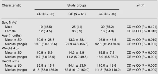
The general description of the patients is presented in Table 1. The mean age and consequently the weight and height were higher in the control group than in the other groups which were included in the study for the etiological diagnosis of low stature, usu-ally defined after three years of age.
Me tho ds
Antigliadin IgG and IgA detection.
100 µl per well of ABTS (2,2’-azinobis(3-ethylbenzthiazoline-6-sulfonic acid); Sigma). The reaction was interrupted after 10 min with 10% SDS (Sigma) and absorbance was read at 405 nm using an ELISA reader. After each of the above described steps, the plates were washed five times with PBS containing 5% Tween 20. The cutoff point was defined as the mean plus two standard deviations for a group of 20 normal children, being 0.022 for IgA (mean = 0.0065, SD = 0.0076) and 0.103 for IgG (mean = 0.0393, SD = 0.032).
Jejunal biopsy
Jejunal biopsies were obtained by the oral route using the pediatric Carey capsule placed at the angle of Treitz, and visualized by X-ray. The mucosal fragments were mounted on a Millipore filter with the vil-lous surface facing up, and immediately im-mersed in 10% formalin solution, embedded in paraffin, stained with hematoxylin-eosin and periodic acid Schiff and examined ac-cording to the method of Pereira et al. (12). The following aspects were considered for the definition of celiac pattern: absent or vestigial villi, cuboidal and basophilic epi-thelial surface containing many interepiepi-thelial lymphocytes (12).
Statistical analysis
Data were analyzed using the statistical EPI INFO v.6.0 and SPSS software (13). Proportions were compared by the chi-square test and means were compared by the Kruskal-Wallis test. Analysis of variance (ANOVA) was used to compare means and the Scheffé test was used to identify the groups responsible for the differences. The 95% confidence interval (95% CI) was de-fined for the determinations of sensitivity, specificity and predictive values. The level of significance was set at 5%.
For the calculation of the predictive val-ues, hypothetical incidences of 1:500, 1:1000 and 1:2000 were used based on literature data from other countries (14-17).
Re sults
Antigliadin antibodie s
Antigliadin IgA antibodies were detected in 21 of 22 patients with celiac disease, resulting in a sensitivity of 95.5% (95% CI: 75.1 to 99.8%). Specificity was 88.5% when considering patients from the other enter-opathy group, since 7 of 61 patients pre-sented positive antigliadin IgA antibodies.
Table 1. Sex, age, w eight and height distribution of patients in the groups w ith celiac disease (CD), other enteropathies (OE) and control (CO).
Characteristic Study groups c2 (P)
CD (N = 22) OE (N = 61) CO (N = 46)
Sex, N (% )
M ale 10 (45.5) 25 (41) 30 (65.2) CD vs CO (P = 0.121)
Female 12 (54.5) 36 (59) 16 (34.8) OE vs CO (P = 0.013)
Age (months)
M ean ± SD 30.6 ± 28.8 43.3 ± 38.1 96.9 ± 48.5 CD vs CO (P = 0.010)
M edian (range) 19.3 (6.0-135.6) 27.9 (4.8-156.5) 92.6 (12.2-170.8) OE vs CO (P = 0.000) Weight (kg)
M ean ± SD 10.9 ± 5.9 14.3 ± 8.9 19.5 ± 7.3 CD vs CO (P = 0.000)
M edian (range) 9.7 (6.0-35.0) 11.2 (5.0-49.5) 18.9 (6.5-36.7) OE vs CO (P = 0.001) Height (cm)
M ean ± SD 85.8 ± 16.1 94.1 ± 23.0 110.0 ± 19.6 CD vs CO (P = 0.000)
When these antibodies were evaluated in the control group, only 2 of 46 patients were positive, with a specificity of 95.7% (95% CI: 84.0 to 99.2%). When the other enter-opathy and control groups were considered together, 9 of the 107 patients were positive for antigliadin IgA antibodies, with an over-all specificity of 91.6%.
Antigliadin IgG antibodies were positive in 20 of 22 patients with celiac disease, resulting in a sensitivity of 90.9% (95% CI: 69.4 to 98.4%). The specificity was 82% when considering patients from the other enteropathy group, since 11 of 61 patients presented positive antigliadin IgG. When these antibodies were evaluated in the con-trol group, only one of 46 patients was posi-tive, resulting in a specificity of 97.8% (95% CI: 87.0 to 99.9%). Taken together, 12 of the 107 patients that comprised the other enter-opathy and control groups had positive anti-gliadin IgG, with an overall specificity of 88%. The absorbance values for antigliadin IgG and IgA are shown in Table 2. A signifi-cant difference was observed in the mean absorbance values recorded for the three study groups for IgA (Figure 1) and IgG (Figure 2) using ANOVA. Using the Scheffé test, the difference was attributed to the dif-ferences between the control group and other enteropathy and celiac disease groups ver-sus control. No difference was observed be-tween the other enteropathy and control groups.
Predictive positive and negative values were calculated considering hypothetical values of incidence of celiac disease ranging from 1:500 to 1:2000 liveborns. The positive predictive values were very low, ranging from 8.5 to 2.3% and from 4.8 to 1.1% for IgG and IgA, respectively. However, a high negative predictive value of 99.9% was found for IgG and IgA (Table 3).
D iscussio n
The main objective of this study was to
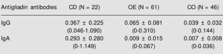
Table 2. Serum antigliadin IgG and IgA in the groups w ith celiac disease (CD), other enteropathies (OE) and control (CO).
Antigliadin antibodies CD (N = 22) OE (N = 61) CO (N = 46)
IgG 0.367 ± 0.225 0.065 ± 0.081 0.039 ± 0.032
(0.046-1.090) (0-0.310) (0-0.144)
IgA 0.293 ± 0.280 0.009 ± 0.015 0.007 ± 0.008
(0-1.149) (0-0.067) (0-0.036)
Data are reported as means ± SD and range of ELISA absorbance values. IgG antibod-ies: ANOVA: F = 81.69; P = 0.0000. Scheffé test (P<0.05): CD>OE; CD>CO. IgA antibodies: ANOVA: F = 56.24; P = 0.0000. Scheffé test (P<0.05): CD>OE; CD>CO.
Table 3. Positive (PPV) and negative predictive values (NPV) reported as percent of antigliadin IgG and IgA antibodies based on hypothetical val-ues of prevalence.
Prevalence IgG IgA
PPV NPV PPV NPV
1:500 8.5 99.9 4.8 99.9
1:1000 4.4 99.9 2.0 99.9
1:2000 2.3 99.9 1.1 99.9
Figure 1. ELISA absorbance val-ues of antigliadin IgG antibodies in celiac disease (CD, N = 22), control (CO, N = 46) and other ent eropat hy (OE, N = 61) groups. The dotted line repre-sents the cutoff value. The solid lines represent the mean of the absorbance values f or each
group. Abs
o
rb
a
n
c
e
a
t
4
0
5
n
m
1.2
1.0
0.8
0.6
0.4
0.2
0.0
CD CO OE
Figure 2. ELISA absorbance val-ues of antigliadin IgA antibodies in the celiac disease (CD, N = 22), control (CO, N = 46) and other enteropathy (OE, N = 61) groups. The dotted line repre-sents the cutoff value. The solid lines represent the mean of the absorbance values f or each
group. A
b
s
o
rb
a
n
c
e
a
t
4
0
5
n
m
1.2
1.0
0.8
0.6
0.4
0.2
0.0
evaluate the usefulness of antigliadin anti-body quantitation in the screening for jejunal biopsy indication during the investigation of the causes of enteropathies and low stature, especially in those children with ambient enteropathies.
In the last two decades, the determination of serum antibodies has proved to be a useful tool for diagnosis and follow-up of patients on a gluten-free diet (2,3,18-21). Nevertheless, most of the studies refer to populations from developed countries, raising questions about the utility of these tests in nondeveloped coun-tries, where the high frequency of so-called ambient enteropathies may produce false-posi-tive results for antigliadin antibodies (10).
In the present study, high sensitivity for the IgG and IgA antigliadin antibody deter-mination, 90.9 and 95.5%, respectively, was observed in children subjected to jejunal biopsy on the basis of routine clinical indica-tions, with typical pathological findings of celiac disease.
Two other tests represent alternative se-rological tools for the celiac disease diag-nostic screening test. The determination of endomysium antibodies (22-24) and anti-tissue transglutaminase (25,26), both psenting high sensitivity and specificity, re-quires more in-depth studies in order to es-tablish its usefulness in developing coun-tries. Grodzinski (27) suggests that antiglia-din antibodies should be used as a screening test and the anti-endomysium antibodies as a confirmatory test before the intestinal bi-opsy. However, several investigators have pointed out the possibility of false-negative antibody detection (9,28,29). The advantages of the detection of antigliadin antibodies are the feasibility of the ELISA technique and
the low cost.
Khoshoo et al. (30), in India, reported that IgG and IgA antigliadin antibodies were sig-nificantly higher in children with celiac dis-ease than in children with other enteropathies. In Brazil, antigliadin antibodies were in-vestigated in children with celiac disease, showing a sensitivity of 90.4% for IgG and of 64.2% for IgA and a specificity of 87.0% for IgG and 92.1% for IgA. The authors consider that the positive antibodies associ-ated with the characteristic histological find-ings confirm the diagnosis of celiac disease. However, a small number of patients were tested for IgA by Medeiros (31): 14 of 29 patients with celiac disease, 49 of 106 with other enteropathies, 27 of 45 with protracted diarrhea, and 26 of 56 control patients.
Many studies have used groups of adults as control, some of them without a jejunal biopsy (4,32-36). In general, antigliadin IgA has proven to be more specific for celiac disease. Tucker et al. (36) observed a higher specificity for IgG, in agreement with the present results. Although the frequency of celiac dis-eases in our country has not been estimated, the hypothetical values permit us to con-clude that if a child has no IgG and no IgA antigliadin antibodies he has a 99.9% chance of not having celiac disease, with no need to be submitted to a jejunal biopsy.
Ackno wle dgm e nts
We are grateful to the staff of the Pediat-ric Gastroenterology Service, University Hos-pital, Federal University of Minas Gerais, for referring the patients and to the staff of the Centro de Pesquisas René Rachou for technical assistance.
Re fe re nce s
1. M cNeish AS & Anderson CM (1974). Coeliac disease; the disorder in childhood. Clinical Gastroenterology, 3: 127-144. 2. Kilander AF, Nilsson LA & Gillberg R
(1987). Serum antibodies to gliadin in
coeliac disease after gluten w ithdraw al. Scandinavian Journal of Gastroenterology, 22: 29-34.
3. Stenhammar L, Kilander AF, Nilsson LA, Stromberg L & Tarkow ski A (1984).
Se-rum gliadin antibodies for detection and control of childhood coeliac disease. Acta Paediatrica Scandinavica, 73: 657-663. 4. Volta U, Lenzi M , Lazzari R, Cassani F,
Anti-bodies to gliadin detected by immuno-fluorescence and a micro-ELISA method: markers of active childhood and adult coeliac disease. Gut, 26: 667-671. 5. Lindberg T, Nilsson LA, Borulf S, Cavell B,
Fallstrom SP, Jansson U, Stenhammar LS & Stintzing G (1985). Serum IgA and IgG gliadin antibodies and small intestinal mu-cosa damage in children. Journal of Pedi-atric Gastroenterology and Nutrition, 4: 917-922.
6. Stahlberg M R, Savilahti E & Viander M (1986). Antibodies to gliadin by ELISA as a screening test for childhood celiac dis-ease. Journal of Pediatric Gastroenterol-ogy and Nutrition, 5: 726-729.
7. Vitoria JC, Arrieta A, Astigarraga I, Garcia-M asdevall D & Rodriguez-Soriano J (1994). Use of serological markers as a screening test in family members of pa-tients w ith celiac disease. Journal of Pedi-atric Gastroenterology and Nutrition, 19: 304-309.
8. Altuntas B, Kansu A, Ensari A & Girgin N (1998). Celiac disease in Turkish short-statured children and the value of antiglia-din antibody in diagnosis. Acta Paediatrica Japonica, 40: 457-460.
9. Chartrand LJ, Agulnik J, Vanounou T, Russo PA, Baehler P & Seidm an EG (1997). Effectiveness of antigliadin anti-bodies as a screening test for celiac dis-ease in children. Canadian M edical Asso-ciation Journal, 157: 527-533.
10. Walker-Smith JA, Guandalini S, Schmitz J, Shmerling DH & Visakorpi JK (1990). Revised criteria for diagnosis of coeliac disease. Archives of Disease in Childhood, 65: 909-911.
11. Huff JC, Weston WL & Zirker DK (1979). Wheat protein antibodies in dermatitis herpetiformis. Journal of Investigative Dermatology, 73: 570-574.
12. Pereira DR, Weinstein WM & Rubin CE (1975). Symposium on pathology of the gastrointestinal tract. Part II. Small intesti-nal biopsy. Human Pathology, 6: 157-217. 13. Dean AG, Dean JA, Coulom bier D, Bremde KA, Smith DC, Burton AH, Dicker RC, Sullivan K, Fagan RF & Arner TG (1994). Epi Info, Version 6: A Word Pro-cessing, Database and Statistic Program for Epidemiology on M icrocomputers. Centers for Disease Control and Preven-tion, Atlanta, GA.
14. M ylotte S, Egan M itchell B, M cCarthy CF & M cNicholl B (1973). Incidence of celiac disease in the West of Ireland. British M edical Journal, 1: 703-705.
15. Rossipal E (1975). Incidence of coeliac disease in children in Austria. Zeitschrift für Kinderheilkunde, 119: 143-149. 16. Hed J, Lieden G, Ottosson E, Strôm M ,
Walan A, Groth O, Sjôgren F & Franzen L (1984). IgA antigliadin antibodies and jeju-nal mucosal lesions in healthy blood do-nors. Lancet, 2: 215.
17. Stevens FM , Egan-M itchell B, Cryan E, M cCarthy CF & M cNicholl B (1987). De-creasing incidence of coeliac disease. Ar-chives of Disease in Childhood, 62: 465-468.
18. Kilander AF, Dotevall G, Fallstrom SP, Gillberg RE, Nilsson LA & Tarkow ski A (1983). Evaluation of gliadin antibodies for detection of coeliac disease. Scandinavian Journal of Gastroenterology, 18: 377-383. 19. Sbolgi P, M angiante G, Ruffa G, Caprioli F, Bartolozzi G, Cinigolani M & Gemme G (1991). Gli anticorpi antigliadina nella diagnosi e nel follow -up della malattia celiaca. M inerva Pediatrica, 43: 783-788. 20. Bodé S, W eile B, Krasilnikof f PA &
Gudmand-Hoyer E (1993). The diagnostic value of the gliadin antibody test in celiac disease in children: a prospective study. Journal of Pediatric Gastroenterology and Nutrition, 17: 260-264.
21. Carroccio A, Iacono G, M ontalto G, Cava-t aio F, Soresi M , Kazm ierska I & Notarbartolo A (1993). Immunologic and absorptive tests in celiac disease: can they replace intestinal biopsies? Scandinavian Journal of Gastroenterology, 28: 673-676. 22. Burgin-Wolff A, Gaze H, Hadziselimovic F, Huber H, Lentze M J, Nusslé D & Reymond-Berthet C (1991). Antigliadin and antiendomysium antibody determina-tion for coeliac disease. Archives of Dis-ease in Childhood, 66: 941-947. 23. Lerner A, Kumar V & Iancu TC (1994).
Immunological diagnosis of childhood co-eliac disease: comparison betw een anti-gliadin, antireticulin and antiendomysial antibodies. Clinical and Experimental Im-munology, 95: 78-82.
24. Lindquist BL, Rogoziniski T, M oi H, Danielsson D & Olcên P (1994). Endomy-sium and gliadin IgA antibodies in children w ith coeliac disease. Scandinavian Jour-nal of Gastroenterology, 29: 452-456. 25. Dieterich W, Ehnis T, Bauer M , Donner P,
Volta U, Riecken EO & Schuppan D (1997). Identification of tissue transglutaminase as the autoantigen of coeliac disease. Na-ture M edicine, 3: 797-801.
26. Dieterich W, Laag E, Schôpper H, Volta U, Fergunson A, Gillet H, Riecken EO &
Schuppan D (1998). Autoantibodies to tis-sue transglutaminase as predictors of ce-liac disease. Gastroenterology, 115: 1317-1321.
27. Grodzinsky E (1996). Screening for coeliac disease in apparently healthy blood do-nors. Acta Paediatrica, 412 (Suppl): 36-38. 28. Rostami AM , Sater RA, Bird SJ, Galetta S, Farber RE, Kamoun M , Silberg DH, Gross-man RI & Pfohl D (1999). A double-blind, placebo-controlled trial of extracorporeal photopheresis chronic progressive mul-tiple sclerosis. M ulmul-tiple Sclerosis, 5: 198-203.
29. Altuntas B, Gul H, Yarali N & Ertan U (1999). Etiology of chronic diarrhea. In-dian Journal of Pediatrics, 66: 657-661. 30. Khoshoo V, Bhan M K, Puri S, Jain R,
Jayashree S, Bhatnagar RS, Kumar R & Stintzing G (1989). Serum antigliadin anti-body profile in childhood protracted diar-rhoea due to coeliac disease and other causes in a developing country. Scandina-vian Journal of Gastroenterology, 24: 1212-1216.
31. M edeiros EHGR (1992). Anticorpo sérico antigliadina no diagnóstico e seguimento da doença celíaca. Doctoral thesis, Escola Paulista de M edicina, São Paulo, SP, Bra-zil.
32. Taylor KB, Truelove SC, Thompson DL & Wright R (1961). An immunological study of coeliac disease and idiopathic steator-rhoea; serological reactions to gluten and milk proteins. British M edical Journal, 2: 1727-1731.
33. Walia BNS, Sidhu JK, Tandon BN, Ghai AP & Bhargava S (1966). Coeliac disease in North Indian children. British M edical Journal, 2: 1233-1234.
34. Burgin-Wolff A, Bertele RM , Berger R, Gaze H, Harms HK, Just M , Khanna S, Schurmann K, Signer E & Tomovic D (1983). A reliable screening test for child-hood celiac disease: fluorescent immuno-sorbent test for gliadin antibodies; a pro-spective multicenter study. Journal of Pe-diatrics, 102: 655-660.
35. Husby S, Foged N, Oxelius VA & Svehag SE (1986). Serum IgG subclass antibodies to gliadin and other dietary antigens in children w ith coeliac disease. Clinical and Experimental Immunology, 64: 526-535. 36. Tucker NT, Barghuthy FS, Prihoda TJ,