CASE REPORT
J of Evidence Based Med & Hlthcare, pISSN- 2349-2562, eISSN- 2349-2570/ Vol. 2/Issue 5/Feb 2, 2015 Page 592
A RARE CASE OF OMENTAL HEMANGIOPERICYTOMA: A CASE
REPORT
G. Kiran Kumar1, V. Manmadharao2, K. B. Muralidhar3, K. Prakash4, K. V. A. Devara5
HOW TO CITE THIS ARTICLE:
G. Kiran Kumar, V. Manmadharao, K. B. Muralidhar, K. Prakash, K. V. A. Devara. ”A Rare Case of Omental Hemangiopericytoma: A Case Report”. Journal of Evidence based Medicine and Healthcare; Volume 2, Issue 5, February 2, 2015; Page: 592-596.
ABSTRACT: A hemangiopericytoma is a rare, soft-tissue tumor of vascular origin derived from a pericyte of Zimmerman, which is a modified smooth muscle cell that surrounds the small blood vessels. Hemangiopericytomas can occur wherever there are vascular capillaries. However hemangiopericytoma from omentum is very rare. We describe a case of a hemangiopericytoma found in the lesser omentum. A 53-year-old man presented with pain abdomen, lump abdomen. Preoperative imaging indicated that the tumor was in the lesser sac without invasion of other organs. A complete resection was performed. The histological pattern supported the diagnosis of a hemangiopericytoma. A complete resection is the best way to treat a hemangiopericytoma. Recognizing the presence of a hemangiopericytoma in the lesser omentum requires appropriate surgery.
KEYWORDS: omentum, hemangiopericytoma, computed tomography.
INTRODUCTION: A hemangiopericytoma is a rare, soft-tissue tumor of vascular origin derived from a pericyte of Zimmerman, which is a modified smooth muscle cell that surrounds the small blood vessels. This type of tumor was first described by Stout and Murray in 1942.[1] It represents approximately 5% of all sarcomatous tumors, and can occur anywhere, but more usually in the musculature of the extremities, retroperitoneum, pelvis (uterus, ovary, and urinary bladder), head, neck and lungs.[2] Hemangiopericytoma in the lesser omentum is a very rare entity. Since the recommended treatment for a hemangiopericytoma is wide excision, due to high local recurrence,[3,4] it is important to recognize the presence of this malignant tumor in the abdomen where various tumors occur. We describe a rare case of a hemangiopericytoma in the lesser omentum.
CASE REPORT
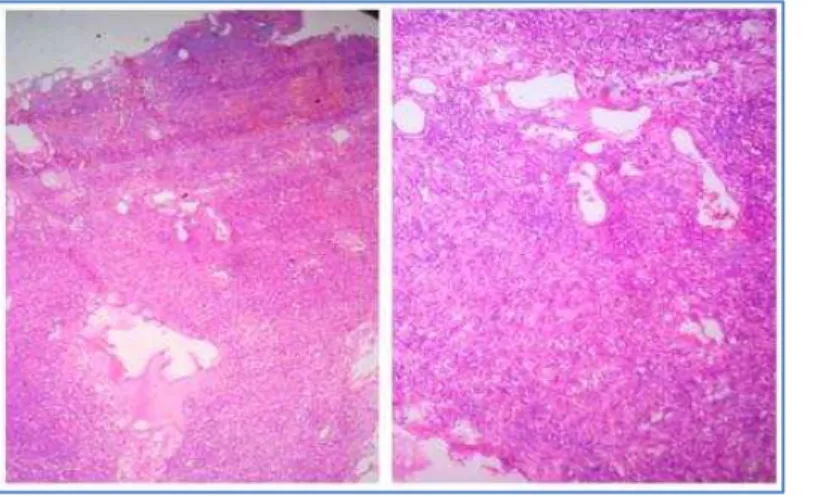
J of Evidence Based Med & Hlthcare, pISSN- 2349-2562, eISSN- 2349-2570/ Vol. 2/Issue 5/Feb 2, 2015 Page 593 The pre-operative image indicated that the tumor was not derived from the pancreas. A biopsy was avoided due to the risk of needle track seeding. The patient underwent a complete resection via midline laparotomy incision without pancreatectomy because pre-operative CT revealed that the pancreas was intact. The tumor was completely removed (Figure 2). The excised tumor was 16 × 10 × 8cm in diameter with a capsule. Its cut surface was mostly multilocular cystic grayish-white and partially reddish. Histopathological features of the hematoxylin and eosin staining revealed that the tumor contained spindle-shaped cells arranged in fascicular pattern and small clusters, predominantly around blood vessels. Individual tumor have ill-defined cell boarder, eosinophilic cytoplasm, elongated to oval vescicular nucleus. There is 3 to 4 mitotic figures/10hpf. Extensive areas of hyalinisation, dilated congested blood vessels, areas of hemorrhages and foci of calcification seen. These features suggestive of hemangiopericytoma (Figure 3).
Fig. 1: Computed tomography scan showing a large mass in the lesser omentum
CASE REPORT
J of Evidence Based Med & Hlthcare, pISSN- 2349-2562, eISSN- 2349-2570/ Vol. 2/Issue 5/Feb 2, 2015 Page 594 We didn’t perform immunohistological staining for this tumor. No necrotic lesion was observed in our patient's tumor. The tumor was pathologically diagnosed as a hemangiopericytoma.
DISCUSSION: Little has been published about hemangiopericytoma, a rare, soft-tissue tumor. It can occur anywhere vascular capillaries are found. The tumors most commonly occurs in the musculature of the extremities, retroperitoneum, pelvis (uterus, ovary, and urinary bladder), head, neck and lungs.[3,4]
The pathological diagnosis of a hemangiopericytoma, in comparison to other mesenchymal tumors such as solitary fibrous tumors, can be difficult.[5] Bcl-2 and CD99 immunohistochemistry were used to distinguish a hemangiopericytoma from a solitary fibrous tumor because a solitary fibrous tumor is positive for Bcl-2[6] and CD99.[7] We diagnosed a hemangiopericytoma following an examination of the structural features of the mass. Spindle-shaped cells surrounding the endothelial-lined vascular spaces were observed by hematoxylin and eosin staining. Making a differential diagnosis between a solitary fibrous tumor and a hemangiopericytoma is particularly difficult and controversial,[8] and a novel molecular marker for distinguishing between the two close entities is required.
Radiotherapy and chemotherapy are not generally effective for the treatment of a hemangiopericytoma.[9] Some have advocated the use of adjuvant radiotherapy in response to the locally aggressive nature of hemangiopericytomas but, due to tumor radioresistance, no differences in local disease control were observed between treatment with and without adjuvant radiotherapy.[10] Spitz et al. reported that hemangiopericytomas showed a poor response to chemotherapy. They treated six patients with pre-operative chemotherapy, and only one of them responded to anthracycline-based chemotherapy.[3] Therefore, complete surgical resection is the only effective therapy for hemangiopericytoma.
CASE REPORT
J of Evidence Based Med & Hlthcare, pISSN- 2349-2562, eISSN- 2349-2570/ Vol. 2/Issue 5/Feb 2, 2015 Page 595 Spitz et al. also reported that 5-year and 10-year survival rates of patients with a hemangiopericytoma were 71% and 54%, respectively. In addition, they noted that the survival rate differed between a curative and a non-curative resection. The 5-year survival rate in patients treated with curative resection and non-curative resection was 79% and 50%, respectively.[3] These data indicate that a complete resection is necessary to improve patients' survival rates.
CONCLUSION: This report presented a rare case of a hemangiopericytoma in the lesser omentum. A thorough differential diagnosis and complete resection without piecemeal excision must always be performed in the management of this type of malignant tumor.
REFERENCES:
1. Hart S, Sisely L: Haemangiopericytoma of the presacral space. Br J Surg 1973, 60:583-584. 2. Vennarecci G, Boschetto A, Esposito A, Giovannelli L, Buscaglia F, Corazza V, Santoro R, Mancini P, Lorusso R, Marino M, Ettorre GM: Malignant haemangiopericytoma of the mesorectum. ChirItal 2004, 56: 865-868.
3. Spitz FR, Bouvet M, Pisters PW, Pollock RE, Feig BW: Hemangiopericytoma: a 20-year single-institution experience. Ann SurgOncol 1998, 5: 350-355.
4. Goldman SM, Davidson AJ, Neal J: Retroperitoneal and pelvic hemangiopericytomas: clinical, radiologic, and pathologic correlation. Radiology 1988, 168: 13-17.
5. Enzinger FM, Smith BH: Hemangiopericytoma: an analysis of 106 cases. Hum Pathol 1976, 7: 61-82.
6. Fukunaga M, Naganuma H, Ushigome S, Endo Y, Ishikawa E: Malignant solitary fibrous tumour of the peritoneum. Histopathology 1996, 28: 463-466.
7. Cristi E, Perrone G, Battista C, Benedetti-Panici P, Rabitti C: A rare case of solitary fibrous tumour of the presacral space: morphological and immunohistochemical features. In Vivo 2005, 19: 777-780.
8. Gengler C, Guillou L: Solitary fibrous tumour and haemangiopericytoma: evolution of a concept. Histopathology 2006, 48: 63-74.
9. Del Rosario ML, Saleh A: Preoperative chemotherapy for congenital hemangiopericytoma and a review of the literature. J PediatrHematolOncol 1997, 19:247-250.
CASE REPORT
J of Evidence Based Med & Hlthcare, pISSN- 2349-2562, eISSN- 2349-2570/ Vol. 2/Issue 5/Feb 2, 2015 Page 596 AUTHORS:
1. G. Kiran Kumar 2. V. Manmadharao 3. K. B. Muralidhar 4. K. Prakash 5. K. V. A. Devara
PARTICULARS OF CONTRIBUTORS:
1. Assistant Professor, Department of Surgery, Andhra Medical College, Visakhapatnam.
2. Associate Professor, Department of Surgery, Andhra Medical College, Visakhapatnam.
3. Post Graduate, Department of Surgery, Andhra Medical College, Visakhapatnam. 4. Post Graduate, Department of Surgery,
Andhra Medical College, Visakhapatnam.
5. Chief Interventional Radiologist,
Department of Radio-Diagnosis, Medall, Visakhapatnam.
NAME ADDRESS EMAIL ID OF THE CORRESPONDING AUTHOR:
Dr. G. Kiran Kumar, Assistant Professor, Department of Surgery, Andhra Medical College, King George Hospital, Visakhapatnam.
E-mail: surgeonkiran@yahoo.co.in

