113
CaSE REPoRtRenal myxoma: a case report
Mixoma renal: relato de caso
Carlos Henrique C Souza1; Kennedy S. Carneiro2; Katia R. M. Leite3; Alcendino A. Junior4; Fredwilson S. Costa1
1. Medical Student. 2. Universidade Federal de São Paulo (UNIFESP); Faculdade de Medicina da Universidade Severino Sombra (USS), Vassouras-RJ. 3. UNIFESP; Faculdade de Medicina da Universidade de São Paulo (FMUSP). 4. Universidade do Estado do Rio de Janeiro (UERJ);
Faculdade de Medicina da Universidade Severino Sombra, Vassouras-RJ.
First submission on 05/06/14; last submission on 11/02/15; accepted for publication on 19/02/15; published on 20/04/15
aBStRaCt
Myxomas are rare tumors that can appear in many anatomical locations. There are only 14 cases of renal involvement documented in the literature. This article reports a case of renal myxoma in an elderly woman with recurrent cystitis. After ive years of follow-up, the computed tomography (CT) revealed a large solid tumor mass in the left kidney. Tumor resection was performed preserving the affected kidney with histopathological diagnosis of renal myxoma. The objective of this study is to report a rare case of renal myxoma, emphasizing the importance of the differential diagnosis from other benign and malignant mesenchymal tumors.
Key words: renal myxoma; kidney; myxoma; renal neoplasm.
J Bras Patol Med Lab, v. 51, n. 2, p. 113-116, April 2015
intRoDuCtion
Myxomas are uncommon benign neoplasms, extremely rare in the kidney. They can be found in the skin, soft tissues, bones, joint spaces, paranasal sinuses, maxillary antrum, viscera(1),
and intramuscular(2, 3). The affected viscera are eyes(4), heart(5),
ovaries(6) and kidneys(2, 3, 7-15).
Only 14 cases of patients with renal myxomas have been reported in the literature(3, 16), none preceded by renal cyst. The
tumor is well-circumscribed and is composed of spindle-shaped cells, scattered in a myxoid stroma, showing immunoreactivity for vimentin and negative reactivity for S-100 protein, epithelial membrane antigen (EMA), pancytokeratin and smooth muscle actin(2, 7-15). There is still no description of this type of renal
tumor in the literature. The importance of the recognition of its existence is to prevent diagnostic errors with malignant and benign neoplasms that present secondary myxoid features, which may involve the kidney. This paper reports the second case of renal myxoma in which was performed tumor excision only, and the affected kidney was preserved.
CaSE REPoRt
The patient was a white woman, aged 73 years, showing recurrent cystitis since 2003. In 2005, after new bouts of cystitis, an ultrasonography (US) was performed and bilateral renal cysts were detected, they were 4.2 cm in the left kidney and 1.4 cm in the right kidney. In 2007, she complained of right-sided cramp-like lumbar pain, irradiating anteriorly, with nausea, negative renal ist percussion, and right-sided lank pain at palpation. Computed tomography (CT) was ordered, and showed kidneys with reduced volume, lobulated contour, preserved parenchymal thickness, bilateral renal cortical with rounded and hypodenses formations, (cysts) without signiicant enhance after contrast medium, measuring 7.4 × 6.8 cm in the middle third of the left
kidney (Figure 1). After six months, the control CT scanning
showed, besides the cortical cysts, the presence of a solid tumor mass, enhanced by contrast medium, measuring 11.9 × 10.1 cm, with well-deined edges in the left lank, suggesting neoplasia
(Figure 2). There were no other changes in biochemical and
urinalysis tests.
114
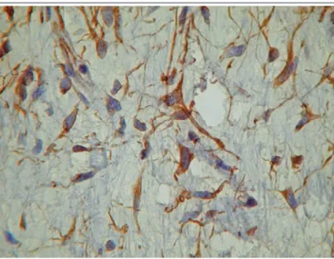
The patient underwent partial nephrectomy due to the exophytic tumor found with gelatinous and well-defined aspect, with narrow pedicle, without lymphadenopathy. The histopathologic diagnosis was myxoma. It showed short spindle-shaped cells with small, oval, delicate nuclei, without hyperchromasia or mitotic activity. Cytoplasm was reticular diffuse and eosinophilic. The cells were involved in a myxoid stroma highly vascularized. Immunohistochemistry showed unique positive tumor cells for vimentin. However, the de S-100 protein, smooth muscle actin, cytokeratin, and murine double minute 2 (MDM2) expression were negative (Figures 3 and 4).
figuRE 1 − Kidneys normally located, reduced volume, lobulated contour, cortical
parenchymal thickness preserved with rounded and hypodenses formations without significant enhancement after contrast medium, measuring 7.4 × 6.8 cm the largest cm in the middle third of the left kidney
figuRE 3 − Benign mesenchymal neoplasm consists of scattered spindle cells in myxoid
stroma. No mitotic activity or pleomorphism is observed 3A): HE, 100×; 3B) HE, 400×.
HE: hematoxylin and eosin.
figuRE 4 − Immunohistochemistrystudy showing only positive tumor cells for vimentin
(400×)
figuRE 2 − Presence of large tumor mass, solid, enhanced by iodinated contrast
medium, not calcified, irregular contour, projected on the left flank, anteromedial and exophytic flank in relation to the left kidney, measuring 119.7 × 101.9 mm, suggesting renal neoplasm. Identification of renal cortical cysts
Renal myxoma: a case report
DiSCuSSion
It was Virchow(17) who irst introduced the term myxoma,
and Stout(18) established that the myxoma is a tumor composed
of stellate or spindle-shaped cells, with myxoid stroma containing mucopolysaccharide, through which delicate reticular ibers run in various directions. We also examined and determined criteria for distinguishing myxomas from sarcoma variations and mesenchymal neoplasms.
Currently, there is no records of speciic clinical presentation
for renal myxoma(16) and, due to its rarity, renal myxoma is
often confused with other malignant lesions(16). It is known so
115
Carlos Henrique C Souza; Kennedy S. Carneiro; Katia R. M. Leite; Alcendino A. Junior; Fredwilson S. Costa
renal myxoma case, the patient had concomitant kidney cysts, and in a recent article published, there was also, hemorrhagic cysts concomitant with myxoma(16). Its differential diagnosis
includes a variety of benign and malignant mesenchymal tumors that may show occasionally prominent myxoid
features(2), namely: perineurioma, myxoid neuroibroma, myxoid
leiomyoma, myxolipoma and myxoid variant of malignant ibrous histiocytoma, leiomyosarcoma(19), rhabdomyosarcoma
and extraskeletal chondrosarcoma(3, 7, 8), low-grade ibromyxoid
sarcoma(20, 21), and solitary ibrous tumor(22). There is still no
description of renal myxomas in the literature(3).
Macroscopically and microscopically, renal myxomas resemble the primitive mesenchyme. The tumor may be found in different locations in the body, the most common is intramuscular(2, 3). It is believed that
it is originated in ibroblasts that have lost the ability to polymerize collagen(3), however, there are controversies in the literature(2, 10, 15).
Some authors believe that myxoma is a myxoid change of some mesenchymal tumors, such as leiomyoma and degenerative changes seen in adipose tissue in brown atrophy of the heart(2, 10, 15).
Macroscopically, the tumor has gelatinous aspect and is well-deined(8). Histopathologically, it is composed of thin
ibroblasts-like spindle-shaped cells, scattered in an abundant myxoid stroma, closely resembling primitive mesenchyme and myxomas of other
sites in the body(3). It is considered a benign ibroblastic tumor
because mitotic activity and cell pleomorphism are not present(8).
In a wide literature review in 1994, Melamed et al. assert
that only ive case reports were truly renal myxomas, including their two cases. The remaining cases exhibit features of sarcoma, ibroepithelial, polyp or myxolipoma(2).
The immunohistochemical indings of previous cases of renal myxoma, tumor cells stained positive for vimentin, but
negative for S-100 protein, EMA, pancytokeratin and smooth muscle actin(2, 7-15), except in one case, that was focally positive
for smooth muscle actin(3).
The imaging exams demonstrate the renal myxoma as a
large heterogeneous mass, predominantly hyperechoic on US and hypodense on CT scan, with more homogeneous signal on magnetic resonance imaging (MRI), which presents with low signal intensity on T1 and hyperintense on T2. Its contours are relatively regular, discreetly multilobulated, and its interface is well-deined with the adjacent renal parenchyma, only shifting the structures without invading them(23).
Radical nephrectomy is considered the treatment of choice, showing no recurrence or metastasis in any case(8). In one case
only tumor enucleation was performed(24). Our case is the second
in which there was kidney preservation, and the second that had renal cysts. As it is a benign tumor, we agree that, under the guidance of imaging examination, percutaneous biopsy may be a better option in the future for renal preservation, enabling better operational planning with maximum preservation of the affected kidney(16).
The patient is currently being monitored, free of recurrence
and metastasis.
In conclusion, renal myxoma is a rare tumor with good prognosis and has its origin in ibroblasts(3). The differential
diagnosis is important to avoid confusing it with a variety of malignant and benign mesenchymal tumors such as sarcomatoid carcinoma, which may show secondary myxoid
features(2). The best treatment option would be, when conditions
are favorable, tumor enucleation with the preservation of the affected kidney.
RESuMo
Mixomas são tumores raros que podem ser encontrados em muitas localizações anatômicas. Na literatura, há apenas 14 casos de acometimento renal. Neste artigo, é relatado um caso de mixoma renal em mulher idosa com cistites de repetição. Após cinco anos de acompanhamento, a tomografia computadorizada (TC) evidenciou grande massa tumoral sólida em rim esquerdo. Realizou-se exérese do tumor preservando o restante do rim afetado com diagnóstico histopatológico de mixoma renal. O objetivo deste trabalho é relatar um caso raro de mixoma renal, enfatizando a importância do diagnóstico diferencial de outros tumores mesenquimais benignos e malignos.
116
Renal myxoma: a case reportMaiLing aDDRESS
Carlos Henrique Câmara de Souza
Universidade Severino Sombra, Centro de Ciências da Saúde, Faculdade de Medicina;Av. Expedicionário Oswaldo de Almeida Ramos, 280; Centro; CEP:
27700-000; Vassouras-RJ, Brazil; e-mail: carloshenrique.camara@hotmail.com.
REfEREnCES
1. Allen PW. Myxoma is a not single entity: a review of the concept of myxoma. Ann Diagn Pathol. 2000; 4: 99-123.
2. Melamed J, Reuter VE, Erlandson RA, Rosai J. Renal myxoma: a report of two cases and review of the literature. Am J Surg Pathol. 1994; 18: 187-94. 3. Yildirim U, Erdem H, Kayikçi A, Uzunlar AK, Tekin A, Kuzey MA. Myxoma of the renal sinus: case report and literature review. Turk J Pathol. 2012; 28(1): 76-9.
4. Patrinely JR, Green WR. Conjunctival myxoma: a clinicopathologic study of four cases and a review of the literature. Arch Ophthalmol. 1983; 101: 1416-20.
5. Acebo E, Val-Bernal JF, Gómez-Román JJ, Revuelta JM. Clinicopathologic study and DNA analysis of 37 cardiac myxomas: a 28-year experience. Chest. 2003; 123: 1379-85.
6. Kumar R, Dey P, Nijhawan R. Myxoma of ovary: an uncommon entity. Arch Gynecol Obstet. 2011; 284(5): 1317-9.
7. Bolat F, Turunç T, Kayaselçuk F, Usulan S, Bal N. Primary renal myxoma. Turk J Pathol. 2007; 23: 160-3.
8. Hakverdi S, Görür S, Yaldiz M, Kuper AN. Renal myxoma: case report and review of the literature. Turk J Urol. 2010; 36(3): 318-21.
9. Koike H, Hayasi Y, Imanishi M. Case of the renal myxoma. Acta Urol Jpn. 2004; 50: 128-9.
10. Kundu AK, Chakraborty AK, Chakraborty S. Myxoma of the kidney. J Indian Med Assoc. 1995; 93: 462-4.
11. Nishimoto K, K, Fukushima M, Hatachi K, Yonehara S, Horiguchi J. Case of the renal myxoma. Jpn J Urol Surg. 1996; 9: 1075-8.
12. Nishimoto K, Sumimoto M, Kakoi N, Asano T, Hayakawa M. Case of renal myxoma. Int J Urol. 2007; 14(3):242-4.
13. Owari Y, Konda R, Omori So, Seo Takashi, Suzuki Kaoro, Fujioka T. Myxoma of the kidney. Int J Urol. 2006; 13: 987-9.
14. Shenansky JH, Gillenwater JY. Myxoma of the kidney. Urology. 1973; 1: 240-2.
15. Val-Bernal JF, Aguilera C, Villagrá NT, Correas MA. Myxoma of the renal capsule. Pathol Res Pract. 2005; 200: 835-40.
16. Shah A, Sun W, Cao D. Myxoma of the kidney associated with hemorrhage. Indian J Surg. 2013: 75 (Suppl. 1): S480-3.
17. Virchow R. Die krankhaften geschwülste. vol. 1. Hischwad VVA, editor. Berlim; 1863.
18. Stout AP. Myxoma, the tumor of primitive mesencyhme. Ann Surg. 1948; 127: 706-19.
19. Sobral APV, Nascimento GJF, Soubhia AMP, Pinto Jr DS, Araújo NS. Leiomiossarcoma de boca: estudo histoquímico e imuno-histoquímico de dois casos clínicos. J Bras Patol Med Lab. 2004; 40(5): 358-63.
20. Papp S, Dickson BC, Chetty R. Low-grade ibromyxoid sarcoma mimicking solitary ibrous tumor: a report of two cases. Virchows Arch. 2004; 1-6.
21. Kim M, Song TJ, Kang SD, et al. A case of low-grade ibromyxoid sarcoma of the colon. Korean J Gastroenterol. 2014; 64(6): 375-9. 22. Batista GR, D’Ippolito G, Szejnfeld J, Menasce S, Fischman MTI, Moraes Jr RL. Tumor ibroso solitário do rim: descrição de caso. Radiol Bras. 2005; 38(4): 313-15.
23. Ribeiro SM, Ajzen AS, Trindade JCS. Comparação dos métodos de imagem no diagnóstico dos tumores renais e calciicações nestas neoplasias. Rev Assoc Med Bras. 2004; 50(4): 403-12.
