of the cervix and its association with p53,
Ki-67 and CD31
Expressão do PTEN em pacientes com carcinoma de colo
uterino e sua associação com p53, Ki-67 e CD31
Abstract
PURPOSE: To investigate protein expression and mutations in phosphatase and tensin homolog (PTEN) in patients with stage IB cervical squamous cell carcinoma (CSCC) and the association with clinical-pathologic features, tumor p53 expression, cell proliferation and angiogenesis. METHODS: Women with stage IB CSCC (n=20 – Study Group) and uterine myoma (n=20 – Control Group), aged 49.1±1.7 years (mean±standard deviation, range 27–78 years), were prospectively evaluated. Patients with cervical cancer were submitted to Piver-Rutledge class III radical hysterectomy and pelvic lymphadenectomy and patients in the Control Group underwent vaginal hysterectomy. Tissue samples from the procedures were stained with hematoxylin and eosin for histological evaluation. Protein expression was detected by immunohistochemistry. Staining for PTEN, p53, Ki-67 and CD31 was evaluated. The intensity of PTEN immunostaining was estimated by computer-assisted image analysis, based on previously reported protocols. Data were analyzed using the Student’s t-test to evaluate signiicant differences between the groups. Level of signiicance was set at p<0.05. RESULTS: The PTEN expression intensity was lower in the CSCC group than in the Control (benign cervix) samples (150.5±5.2 versus 204.2±2.6; p<0.001). Our study did not identify any mutations after sequencing all nine PTEN
exons. PTEN expression was not associated with tumor expression of p53 (p=0.9), CD31 (p=0.8) or Ki-67 (p=0.3) or clinical-pathologic features in patients with invasive carcinoma of the cervix. CONCLUSIONS: Our indings demonstrate that the PTEN protein expression is signiicantly diminished in CSCC.
Resumo
OBJETIVO: O objetivo do estudo foi investigar a expressão e mutações do PTEN em pacientes com Carcinoma de Células Escamosas (CCE) de Colo do Útero com estadiamento IB e sua associação com fatores prognósticos, expressão do p53, proliferação celular e angiogênese. MÉTODOS: Mulheres com diagnóstico de CCE de colo uterino em estágio IB (n=20) (casos) e mioma uterino (n=20) (controle) com idade de 49.1±1.7 foram acompanhadas. As pacientes com câncer de colo do útero foram submetidas a histerectomia Piver-Rutledge classe III associada a linfadenectomia pélvica e aquelas com mioma uterino a histerectomia vaginal. Amostras de tumor e colo normal foram retiradas para avaliação histológica e marcação histoquímica das proteínas PTEN, p53, ki-67 e CD 3. A intensidade imuno-histoquímica do PTEN foi estimada por processamento de imagem digital a partir de protocolos pré-estabelecidos. Os dados foram analisados através do teste de qui - quadrado (χ2). O nível de signiicância foi considerado quando p < 0,05. RESULTADOS: A expressão do PTEN estava diminuída no grupo de pacientes com CCE em comparação ao grupo controle (150.5±5.2 versus 204.2±2.6; p<0.001). Nenhuma mutação no seqüenciamento genético dos nove exons do PTEN foi encontrada. Não houve associação estatisticamente signiicativa entre a expressão do PTEN e a expressão do p53 (p=0,969), Ki-67 (p=0.283) e CD 31 (p=0.817) ou fatores prognósticos anátomo-clínicos nas pacientes com carcinoma invasor do colo uterino. CONCLUSÕES: Este estudo demonstrou que o PTEN estava signiicativamente diminuído nas pacientes com CCE.
Hospital das Clínicas da Universidade Federal de Minas Gerais – UFMG – Belo Horizonte (MG), Brazil.
1Department of Obstetrics and Gynecology, Universidade Federal de Juiz de Fora – UFJF – Juiz de Fora (MG), Brazil. 2Department of Obstetrics and Gynecology, Universidade Federal de Minas Gerais – UFMG – Belo Horizonte (MG), Brazil. 3Department of Pathology, Universidade Federal de Minas Gerais – UFMG – Belo Horizonte (MG), Brazil.
4Clinical Hospital, Faculdade de Medicina, Universidade Federal de Minas Gerais – UFMG – Belo Horizonte (MG), Brazil. 5Department of Surgery, Universidade Federal de Minas Gerais – UFMG – Belo Horizonte (MG), Brazil.
6Department of Obstetrics, Gynecology and Mastology, Faculdade de Medicina de Botucatu, Universidade Estadual Paulista “Júlio de
Mesquita Filho” – UNESP – Botucatu (SP), Brazil. Conlicts of interests: none.
Keywords Uterine cervical neoplasms Immunohistochemistry Gene expression Antigens, neoplasm
Palavras-chave Neoplasias do colo do útero Imunohistoquímica Expressão gênica Antígenos de neoplasias
Correspondence
Agnaldo Lopes Silva-Filho Department of Obstetrics and Gynecology, Universidade Federal de Minas Gerais Avenida Professor Alfredo Balena, 190 – Santa Eigênia CEP: 30130-100 Belo Horizonte (MG), Brazil
Received
03/09/2014
Accept with modiications
04/23/2014
Original Article
PauLa Vieira teixeira VidigaL
Mariana ataydes Leite seaBra4
Luiz arMando cunhade Marco5
Introduction
Cervical cancer is a worldwide public health problem, with an incidence of 530,232 new cases and 275,008 deaths every year1. Most cases occur in undeveloped coun-tries where no effective screening systems are available2. In Brazil, 17,540 new cases were estimated to occur in 2012, making it the third most common malignancy
and the fourth leading cause of death among women3.
Persistent infection with human papillomavirus (HPV) plays a critical role in cervical carcinogenesis. However, HPV infection alone is not suficient to induce malignant transformation, and additional genetic or epigenetic changes in tumor cells are required4,5. The development and pro-gression of cervical squamous cell carcinoma (CSCC) are likely to be associated with the loss of growth suppression, increased cell growth rates, and angiogenesis6,7. These combinations of genetic abnormalities generate cells that divide more rapidly or evade cell death, thus liberating them from growth control and cell cycle checkpoints.
Phosphatase and tensin homolog (PTEN) is a tumor suppressor gene localized on chromosome 10 (10q23.3) in a region that is often related to loss of heterozygosis and consequent predisposition to carcinogenesis in a number of malignancies8-10. Genetic, epigenetic and protein expression alterations in PTEN have been described in several types of tumors such as brain, prostate, breast, thyroid and endome-trial tumors11-14. PTEN phosphatase is a negative regulator of the Akt/PKB survival pathway, which is over-expressed in CSCC15. Mutations are common in many human cancers, but the loss of heterozygosity, a genomic deletion and punctual mutations in PTEN and their associations with squamous cells carcinoma are controversial15-19.
The role of PTEN in cervical carcinogenesis has not been clearly deined and there is no general agreement on the mechanism related to the reduction of PTEN expres-sion in cervical cancer20. Therefore, the purpose of this study was to investigate protein expression and muta-tions in PTEN in patients with stage IB CSCC and the association with clinical-pathologic features, tumor p53 expression, cell proliferation and angiogenesis.
Methods
Women with stage IB CSCC (n=20 – Study Group) and uterine myoma (n=20 – Control Group), aged 49.1±1.7 years (mean±standard deviation, range 27–78 years) were prospectively evaluated. The study was performed in accordance with the Ethical Committee for Research in Human Beings guidelines of the institu-tion. Informed consent was obtained from all included patients and the research was conducted in accordance with the Declaration of Helsinki revised in 2008.
Patients with CSCC underwent class III Piver-Rutledge radical hysterectomy and pelvic lymphadenectomy. This was the primary treatment for all patients because none had previously been treated with radiotherapy and/or chemo-therapy. The clinical stage was deined preoperatively by pelvic examination under general anesthesia according to the International Federation of Gynecology and Obstetrics
(FIGO) recommendations21,22. Vaginal hysterectomy was
performed for uterine myomas according to the modiied Heaney technique.
Tissue samples were ixed in 10% neutral-buffered formalin, embedded in parafin, and stained with hema-toxylin and eosin for histological evaluation. Histological specimens were analyzed by the same pathologists accord-ing to the recommendations of the American Society of Pathologists23. Clinical-pathologic characteristics, such as tumor size, differentiation grade, lymphatic vascular space invasion (LVSI), parametrial involvement and pelvic lymph node status, were recorded.
Immunohistochemistry
Tissue sections from CSCC and normal mucosa were stained with PTEN, p53, Ki-67 and CD31 antiserum. Briely, 4 µm parafin-embedded sections were deparafinized in xylene and hydrated with graded ethanol solutions. Endogenous peroxidase activity was blocked with 3% H2O2 in water for 10 min. Heat-induced epitope retrieval was performed with 1 mM EDTA buffer (pH=8.0) for 30 min in a steamer at 96°C. Primary polyclonal rabbit antisera were used at a 1:100 dilution for PTEN, p53, and Ki-67 and a 1:40 dilution for CD31 antiserum for 18 h at 4°C. This was followed by incubation with a labeled streptavidin-biotin kit, the NovoLinkTM Max Polymer Detection System (Novocastra, United Kingdom). Peroxidase activity was developed with DAB (Sigma, St Louis, MI) with timed monitoring using a positive control sample. Sections were then counterstained with hematoxylin, dehydrated and mounted.
traced areas. For each ield, one nucleus with an evident nucleolus was randomly chosen for measurement, and the next nine consecutive nuclei were quantiied. Out-of-focus and altered-form nuclei were not considered. Background intensity was determined by tracing an unlabeled area adjacent to the measured cells. The inal pixel intensity was calculated by subtracting the values detected in the labeled nuclei from the background (Figure 1).
Phosphatase and tensin homolog sequencing
Genomic DNA was isolated from CSCC tissue samples according to a proteinase K-based protocol. After DNA isolation, exons 1 through 9 of PTEN were ampliied by PCR with speciic primers for each region. For the PCR reactions, 2 µL of DNA at 30 ng/µL were mixed with 2.5 µL of 10X IIB Buffer (40 mM NaCl;
10 mM TrisHCl pH=8.4; 0.1% Triton X-100; 1.5 mM
MgCl2), 2.5 µL of 0.2 mM dNTPs, 0.5 µL of each primer
at 10 pmol/µL and 0,25 µL of Taq polymerase (0,625 U), for a inal volume of 25 µL. Samples were ampliied
us-ing an Eppendorf Mastercycler® (Hamburg, Germany)
gradient thermocycler at 94°C for 3 minutes followed by 35 cycles of 94°C for 30 seconds, 55°C for 30 seconds, 72°C for 30 seconds and a inal extension time at 72°C for 5 minutes. PCR products were puriied using Illustra GFX PCR DNA and Gel Band Puriication Kit (GE Healthcare), following the manufacturer’s protocol, and were visualized on a silver-stained 6,5% polyacrylamide gel. Sequences were obtained using an ABI 3130 Genetic Analyzer (Applied Biosystems). Bidirectional sequence data were analyzed with Sequencher 4.9 software, and the analysis was followed by a manual review.
Analysis of p53, Ki-67, and CD31 staining
All slides were examined under light microscopy. Staining for p53, Ki-67, and CD31 were evaluated accord-ing to the number of positively stained cells by a saccord-ingle pathologist who was blinded to the clinical data of the patients. This was performed by classifying the protein expression into 4 categories for statistical purposes as follows: grade 1 – 0 to 25% expression; grade 2 – 26 to 50% expression; grade 3 – 51 to 75% expression; and grade 4 – greater than 75% expression.
Statistical analyses
Statistical analyses were performed with Statistical Package for the Social Sciences (SPSS) 18.0 software (SPSS Inc., Chicago, IL, USA). Data were analyzed using Student’s t-test to evaluate signiicant differences between
the groups. The level of signiicance was set at p<0.05. Power calculations showed that the sample size (n=20) allowed a minimal detectable difference of 35% between the 2 prevalence rates, with a power of 80% and a type I error of 5%.
Results
The clinical stage (FIGO) was IB1 in 14 patients (70%) and IB2 in 6 patients (30%). The tumors were well differentiated (G1) in 1 (5%) patient, moderately differentiated (G2) in 15 (75%) patients and poorly differentiated in 4 (20%) patients. Lymphatic vascular invasion was present in 4 patients (20%).
The PTEN expression intensity was lower in the CSCC group than in the benign cervix samples (150.5±5.2 versus
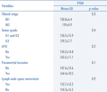
204.2±2.6; p<0.001) (Figure 2). No associations were identiied between tumor PTEN expression and tumor stage (p=0.3), grade of differentiation (p=0.4), presence of lymphatic vascular invasion (p=0.2), parametrium
A
B
involvement (p=0.6) or pelvic lymph node metastasis (p=0.9) in patients with invasive carcinoma of the cervix (Study Group) (Table 1).
All 9 PTEN exons were sequenced, and we were unable to ind modiications in the PTEN DNA se-quence in the 20 CSCC samples included in the study or in the normal cervical squamous epithelial sample used as control.
PTEN expression was not associated with tumor ex-pression of p53 (p=0.9), CD31 (p=0.8) or Ki-67 (p=0.3) in patients with invasive carcinoma of the cervix (Figure 3).
Discussion
PTEN genetic alterations occur in multiple types of cancer, such as brain, prostate, breast, thyroid and endo-metrial tumors. Thus, PTEN inactivation may play an important role in the pathogenesis of a variety of human
malignancies24. The present study found PTEN protein
signiicantly diminished in CSCC compared to control. This suggests that the loss of PTEN expression plays a role in cervical carcinogenesis. Previous reports have also demonstrated that PTEN expression is progressively reduced along a continuum from normal epithelium to squamous cell carcinoma25,26.
Our study did not identify mutations after sequenc-ing all 9 PTEN exons, even in the hot spot in exon 5. Structural changes of PTEN in cervical carcinomas do not appear to be common. Previous reports have not identiied mutations in the PTEN gene in this type of cancer8,19. However, Poetsch et al.27 demonstrated PTEN mutations in 23% of head and neck SCC tumor samples,
and Kurose et al.28 found intragenic PTEN mutations in
15% (3/20) of cervical tumors. PTEN mutations were frequently found in cancers arising from the endome-trium29,30, brain31 and prostate32. Rashmi et al.33 found results with activating PIK3CA (E545K, E542K) and inactivating PTEN (R233) mutations were identiied in human cervical cancer. An analysis of PTEN gene in squamous cell carcinomas from other sites also found that PTEN is not frequently mutated in the lung34, cer-vix14, skin35, head and neck36 or esophagus37. Although numerous somatic mutations have been localized in the PTEN gene, these only occur in a minority of tumors, which indicates that alternative mechanisms of PTEN inactivation, both genetic and non-genetic, must exist.
Table 1. Association of tumor size, grade of differentiation, presence of lymphatic vascular invasion, parametrium involvement and pelvic lymph node metastasis with tumor phosphatase and tensin homolog expression in patients with squamous cell carcinoma of the cervix
Variables PTEN
Mean±SD p-value
Clinical stage 0.3
IB1 150.8±6.4
IB2 159±4.9
Tumor grade 0.4
G1 and G2 150.5±15.9
G3 159.2±7.7
LVSI 0.2
No 150.2±14.8
Yes 163.2±11.1
Parametrial invasion 0.1
No 147.6±13.6
Yes 164.4±10.5
Lymph node space metastasis 0.9
Yes 152.7±13.2
No 154.3±16.5
PTEN: phosphatase and tensin homolog; SD: standard deviation
250
200
150
100
PTEN expression
50
SCC group Control p<0.001
0
PTEN: phosphatase and tensin homolog
Figure 2. Expression of PTEN intensity in benign cervix (control group) and in the tumor (study group)
250
200
150
100
PTEN expression
50
Grade 1 Grade 2 Grade 3 CD31 expression
p=0.817
0
1. International Agency for Research on Cancer. World Health Organization. GLOBOCAN 2012: estimated cancer incidence, and mortality and prevalence worldwide in 2012 [Internet]. Lyon: IARC; 2013 [cited 2014 Mar 20]. Available from: http://globocan.iarc.fr/ Default.aspx
2. Waggoner SE. Cervical cancer. Lancet. 2003;361(9376): 2217-25.
3. Brasil. Ministério da Saúde. Instituto Nacional de Câncer José Alencar Gomes da Silva. Coordenação Geral de Ações Estratégicas. Coordenação de Prevenção e Vigilância. Estimativa 2012: incidência de câncer no Brasil [Internet]. Rio de Janeiro: INCA; 2011 [citado 2012 Maio 15]. Disponível em: http:// portal.saude.sp.gov.br/resources/ses/peril/gestor/homepage/ estimativas-de-incidencia-de-cancer-2012/estimativas_incidencia_ cancer_2012.pdf
4. Castellsagué X. Natural history and epidemiology of HPV infection and cervical cancer. Gynecol Oncol. 2008;110(3 Suppl 2):S4-7. 5. Giarnieri E, Zanesi N, Bottoni A, Alderisio M, Lukic A, Vecchione A, et al. Oncosuppressor proteins of fragile sites are reduced in cervical cancer. Cancer Lett. 2010;289(1):40-5.
6. Hanahan D, Weinberg RA. The hallmarks of cancer. Cell. 2000;100(1):57-70.
7. Hahn WC, Weinberg RA. Rules for making human tumor cells. N Engl J Med. 2002;347(20):1593-603.
8. Cheung TH, Lo KW, Yim SF, Chan LK, Heung MS, Chan CS, et al. Epigenetic and genetic alternation of PTEN in cervical neoplasm. Gynecol Oncol. 2004;93(3):621-7.
9. Tamguney T, Stokoe D. New insights into PTEN. J Cell Sci. 2007;120(Pt 23):4071-9.
References
Our study did not focus on the role of epigenetic changes of PTEN in the development of squamous cell carcinoma. The future challenge is to further understand the roles of these possible epigenetic mechanisms of PTEN alteration and their biological relevance is a chal-lenge for the future. PTEN inactivation via epigenetic mechanisms was irst demonstrated in prostate cancer cell lines38 and later found in prostate cancer and mela-noma39. Ojesina et al.40 implicated somatic mutations in PIK3CA, PTEN, TP53, STK11 and KRAS as well as several copy-number alterations in the pathogenesis of cervical carcinomas and reported whole-exome sequencing analysis of 115 cervical carcinoma-normal paired samples, transcriptome sequencing of 79 cases and whole-genome sequencing of 14 tumour-normal pairs. Previously un-known somatic mutations in 79 primary squamous cell carcinomas include recurrent E322K substitutions in the MAPK1 gene (8%), inactivating mutations in the HLA-B gene (9%), and mutations in EP300 (16%), FBXW7 (15%), NFE2L2 (4%), P53 (5%) and ERBB2 (6%). The association of poor prognosis with PTEN aberration has also been reported previously in glioma41
and tongue carcinoma42. Whether PTEN methylation
contributes to the development of cervical cancer and affects the prognosis has not been elucidated. Cheung
et al.8 found that PTEN methylation was signiicantly
associated with reduced total and disease-free survival and suggested that tumors without normal PTEN functioning may be more aggressive.
In the present report, we analyzed p53, CD31 and Ki-67 expression and their association with PTEN ex-pression in CSCC. We believe that our study is the irst report describing PTEN expression in a benign cervix and cervical cancer and its association with those biomarkers. The roles of p53 and PTEN in cervical carcinogenesis have been addressed, but little is known about the interaction
of these proteins in cervical cancer. PTEN has been shown to directly associate with p53, increasing its stability, protein levels, and transcriptional activity43. The expres-sion of Ki-67 and CD31 was assessed in various grades of cervical intraepithelial neoplasia (CIN) to evaluate their potential to predict the extent of possible damage to the epithelium and CIN progression. Ki-67, a non-histone protein with short half-life, is expressed in the nuclei of proliferating cells, becoming a marker of cell prolifera-tion44,45. Angiogenesis is correlated with the potential for solid tumor metastasis. CD31 expression is related to neovascularization and may be associated with the clinical course of the cervical tumor46. No signiicant association was found between PTEN and other biomarkers, sug-gesting that the loss of PTEN expression is suficient to facilitate tumorigenesis, but different mechanisms may explain cervical carcinogenesis.
We were not able to identify any clinical or patho-logic factors associated with PTEN expression in CSCC specimens. Given the limited number of samples inves-tigated, our ability to detect prognostic associations of modest size was limited. The association between PTEN expression and disease outcome warrants further inves-tigation in a larger study cohort along with the analysis of prognostic factors.
In conclusion, our indings demonstrate that the PTEN protein is signiicantly diminished in CSCC. Therefore, we suggest that PTEN plays an important role in carcinogenesis in the uterine cervix.
10. Vazquez F, Ramaswamy S, Nakamura N, Sellers WR. Phosphorylation of the PTEN tail regulates protein stability and function. Mol Cell Biol. 2000;20(14):5010-8.
11. Alvarez-Nuñez F, Bussaglia E, Mauricio D, Ybarra J, Vilar M, Lerma E, et al. PTEN promoter methylation in sporadic thyroid carcinomas. Thyroid. 2006;16(1):17-23.
12. Li J, Yen C, Liaw D, Podsypanina K, Bose S, Wang SI, et al. PTEN, a putative protein tyrosine phosphatase gene mutated in human brain, breast, and prostate cancer. Science. 1997;275(5308):1943-7. 13. Mutter GL, Lin MC, Fitzgerald JT, Kum JB, Baak JP, Lees JA, et al. Altered PTEN expression as a diagnostic marker for the earliest endometrial precancers. J Natl Cancer Inst. 2000;92(11):924-30. 14. Tashiro H, Blazes MS, Wu R, Cho KR, Bose S, Wang SI, et al.
Mutations in PTEN are frequent in endometrial carcinoma but rare in other common gynecological malignancies. Cancer Res. 1997;57(18):3935-40.
15. Sansal I, Sellers WR. The biology and clinical relevance of the PTEN tumor suppressor pathway. J Clin Oncol. 2004;22(14):2954-63.
16. Gasparotto D, Vukosavljevic T, Piccinin S, Barzan L, Sulfaro S, Armellin M, et al. Loss of heterozygosity at 10q in tumors of the upper respiratory tract is associated with poor prognosis. Int J Cancer. 1999;84(4):432-6.
17. Harima Y, Sawada S, Nagata K, Sougawa M, Ostapenko V, Ohnishi T. Mutation of the PTEN gene in advanced cervical cancer correlated with tumor progression and poor outcome after radiotherapy. Int J Oncol. 2001;18(3):493-7.
18. Henderson YC, Wang E, Clayman GL. Genotypic analysis of tumor suppressor genes PTEN/MMAC1 and p53 in head and neck squamous cell carcinomas. Laryngoscope. 1998;108(10):1553-6. 19. Su TH, Chang JG, Perng LI, Chang CP, Wei HJ, Wang NM,
et al. Mutation analysis of the putative tumor suppressor gene PTEN/MMAC1 in cervical cancer. Gynecol Oncol. 2000;76(2):193-9.
20. Vázquez-Ulloa E, Lizano M, Avilés-Salas A, Alfaro-Moreno E, Contreras-Paredes A. Abnormal distribution of hDlg and PTEN in premalignant lesions and invasive cervical cancer. Gynecol Oncol. 2011;122(3):663-8.
21. Benedet JL, Bender H, Jones H 3rd, Ngan HY, Pecorelli S. FIGO staging classiications and clinical practice guidelines in the management of gynecologic cancers. FIGO Committee on Gynecologic Oncology. Int J Gynaecol Obstet. 2000;70(2):209-62.
22. Shepherd JH. Cervical and vulva cancer: changes in FIGO deinitions of staging. Br J Obstet Gynaecol. 1996;103(5):405-6. 23. Kamura T, Shigematsu T, Kaku T, Shimamoto T, Saito T, Sakai K,
et al. Histopathological factors inluencing pelvic lymph node metastases in two or more sites in patients with cervical carcinoma undergoing radical hysterectomy. Acta Obstet Gynecol Scand. 1999;78(5):452-7.
24. Nakanishi A, Kitagishi Y, Ogura Y, Matsuda S. The tumor suppressor PTEN interacts with p53 in hereditary cancer (Review). Int J Oncol. 2014;44(6):1813-9.
25. Lee JS, Choi YD, Lee JH, Nam JH, Choi C, Lee MC, et al. Expression of PTEN in the progression of cervical neoplasia and its relation to tumor behavior and angiogenesis in invasive squamous cell carcinoma. J Surg Oncol. 2006;93(3):233-40.
26. Qi M, Anderson AE, Chen DZ, Sun S, Auborn KJ. Indole-3-carbinol prevents PTEN loss in cervical cancer in vivo. Mol Med. 2005;11(1-12):59-63.
27. Poetsch M, Lorenz G, Kleist B. Detection of new PTEN/MMAC1 mutations in head and neck squamous cell carcinomas with loss of chromosome 10. Cancer Genet Cytogenet. 2002;132(1):20-4.
28. Kurose K, Zhou XP, Araki T, Eng C. Biallelic inactivating mutations and an occult germline mutation of PTEN in primary cervical carcinomas. Genes Chromosomes Cancer. 2000;29(2):166-72. 29. Kong D, Suzuki A, Zou TT, Sakurada A, Kemp LW, Wakatsuki S, et al.
PTEN1 is frequently mutated in primary endometrial carcinomas. Nat Genet. 1997;17(2):143-4.
30. Husseinzadeh N, Husseinzadeh HD. mTOR inhibitors and their clinical application in cervical, endometrial and ovarian cancers: a critical review. Gynecol Oncol. 2014;133(2):375-81.
31. Boström J, Cobbers JM, Wolter M, Tabatabai G, Weber RG, Lichter P, et al. Mutation of the PTEN (MMAC1) tumor suppressor gene in a subset of glioblastomas but not in meningiomas with loss of chromosome arm 10q. Cancer Res. 1998;58(1):29-33. 32. Feilotter HE, Nagai MA, Boag AH, Eng C, Mulligan LM. Analysis
of PTEN and the 10q23 region in primary prostate carcinomas. Oncogene. 1998;16(13):1743-8.
33. Rashmi R, Deselm C, Helms C, Bowcock A, Rogers BE, Rader J, et al. AKT inhibitors promote cell death in cervical cancer through disruption of mTOR signaling and glucose uptake. PLoS One. 2014;9(4):e92948. 34. Petersen S, Rudolf J, Bockmühl U, Gellert K, Wolf G, Dietel M, et al. Distinct regions of allelic imbalance on chromosome 10q22-q26 in squamous cell carcinomas of the lung. Oncogene. 1998;17(4):449-54. 35. Kubo Y, Urano Y, Hida Y, Arase S. Lack of somatic mutation in the
PTEN gene in squamous cell carcinomas of human skin. J Dermatol Sci. 1999;19(3):199-201.
36. Okami K, Wu L, Riggins G, Cairns P, Goggins M, Evron E, et al. Analysis of PTEN/MMAC1 alterations in aerodigestive tract tumors. Cancer Res. 1998;58(3):509-11.
37. Hu YC, Lam KY, Tang JC, Srivastava G. Mutational analysis of the PTEN/MMAC1 gene in primary oesophageal squamous cell carcinomas. Mol Pathol. 1999;52(6):353-6.
38. Wang L, Wang WL, Zhang Y, Guo SP, Zhang J, Li QL. Epigenetic and genetic alterations of PTEN in hepatocellular carcinoma. Hepatol Res. 2007;37(5):389-96.
39. Zhou XP, Gimm O, Hampel H, Niemann T, Walker MJ, Eng C. Epigenetic PTEN silencing in malignant melanomas without PTEN mutation. Am J Pathol. 2000;157(4):1123-8.
40. Ojesina AI, Lichtenstein L, Freeman SS, Pedamallu CS, Imaz-Rosshandler I, Pugh TJ, et al. Landscape of genomic alterations in cervical carcinomas. Nature. 2014;506(7488):371-5. 41. Lin H, Bondy ML, Langford LA, Hess KR, Delclos GL, Wu X, et
al. Allelic deletion analyses of MMAC/PTEN and DMBT1 loci in gliomas: relationship to prognostic signiicance. Clin Cancer Res. 1998;4(10):2447-54.
42. Lee JI, Soria JC, Hassan KA, El-Naggar AK, Tang X, Liu DD, et al. Loss of PTEN expression as a prognostic marker for tongue cancer. Arch Otolaryngol Head Neck Surg. 2001;127(12):1441-5. 43. Freeman DJ, Li AG, Wei G, Li HH, Kertesz N, Lesche R, et al.
PTEN tumor suppressor regulates p53 protein levels and activity through phosphatase-dependent and -independent mechanisms. Cancer Cell. 2003;3(2):117-30.
44. Shiohara S, Shiozawa T, Miyamoto T, Feng YZ, Kashima H, Kurai M, et al. Expression of cyclins, p53, and Ki-67 in cervical squamous cell carcinomas: overexpression of cyclin A is a poor prognostic factor in stage Ib and II disease. Virchows Arch. 2005;446(6):626-33. 45. Korolenkova LI, Stepanova EV, Ermilova VD, Baryshnikov AIu, Briuzgin VV.
[Ki-67 expression, thymidine phosphorylase and PTEN in intraepithelial cervical carcinoma]. Vopr Onkol. 2011;57(2):199-203. Russian. 46. Sauer G, Deissler H. Angiogenesis: prognostic and therapeutic