ACUTE APPENDICITIS: COMPUTED TOMOGRAPHY FINDINGS
– AN ICONOGRAPHIC ESSAY*
Marcelo Eustáquio Montandon Júnior1
, Cristiano Montandon1
, Gustavo Ribeiro Fiori2 , Carlos Alberto Ximenes Filho3
, Fernanda Coelho Barbosa da Cruz4
Acute appendicitis is the most important cause of abdominal pain requiring surgical intervention in the Western world. The early diagnosis of this disease is of paramount relevance for minimizing its morbidity. Imaging methods have represented a huge progress in the diagnosis of this entity, which used to be based essen-tially on clinical history, physical examination and laboratory tests results, considering that 20% to 33% of patients present with atypical symptoms. Diagnostic difficulty is higher in children, the elderly, and women in childbearing age. The main imaging methods for evaluation of acute appendicitis are ultrasound and com-puted tomography. The present study is aimed at describing the disease physiopathology, commenting main computed tomography technical aspects, demonstrating and illustrating tomographic findings, and describ-ing main differential diagnoses.
Keywords: Acute appendicitis; Vermiform appendix; Computed tomography.
Apendicite aguda: achados na tomografia computadorizada – ensaio iconográfico.
A apendicite aguda é a causa mais comum de dor abdominal aguda que requer intervenção cirúrgica no mundo ocidental. O diagnóstico precoce é essencial para minimizar a morbidade da doença. O uso dos métodos de imagem significou grande avanço no diagnóstico desta entidade, até então avaliada apenas com base na história clínica, exame físico e dados laboratoriais, haja vista que 20% a 33% dos pacientes apresentam sintomas atípicos. O diagnóstico é mais difícil nas crianças, nos idosos e nas mulheres em idade fértil. Os principais métodos de imagem para sua avaliação são a ultra-sonografia e a tomografia computadorizada. Os objetivos deste trabalho são: descrever a fisiopatologia da doença, comentar os principais aspectos téc-nicos da tomografia computadorizada, demonstrar e ilustrar os achados tomográficos e citar os principais diagnósticos diferenciais.
Unitermos: Apendicite aguda; Apêndice vermiforme; Tomografia computadorizada. Abstract
Resumo
* Study developed at Clínicas da Imagem, Multimagem, São Camilo and São Mateus, and Hospital e Maternidade Jardim América, Goiânia, GO, Brazil.
1. Titular Members of Colégio Brasileiro de Radiologia e Diag-nóstico por Imagem (CBR), MDs, Radiologists at Clínicas da Imagem and Multimagem, and Hospital e Maternidade Jardim América, Goiânia, GO, Brazil.
2. Titular Member of Colégio Brasileiro de Radiologia e Diag-nóstico por Imagem (CBR), MD, Radiologist at Clínicas da Ima-gem and São Camilo, Goiânia, GO, Brazil.
3. Titular Member of Colégio Brasileiro de Radiologia e Diag-nóstico por Imagem (CBR), MD, Radiologist at Clínicas da Ima-gem and São Mateus, Goiânia, GO, Brazil.
4. Graduate Student (6th year) at Faculdade de Medicina da Universidade Federal de Goiás (UFG), Goiânia, GO, Brazil.
Mailing address: Dr. Marcelo Eustáquio Montandon Júnior. Avenida Ismerino S. Carvalho, 775, Setor Aeroporto. Goiânia, GO, Brazil, 74075-040. E-mail: [email protected]
Received May 12, 2006. Accepted after revision September 26, 2006.
INTRODUCTION
Acute appendicitis is the most impor-tant cause of abdominal pain requiring sur-gical intervention in the Western world(1,2).
The early diagnosis of this disease is of paramount relevance for minimizing its morbidity. Imaging methods have repre-sented a huge progress in the diagnosis of this entity, which used to be based
essen-tially on clinical history, physical examina-tion and laboratory tests results, consider-ing that 20% to 33% of patients present with atypical symptoms(2).
Diagnostic difficulty is higher in chil-dren, the elderly, and women in childbear-ing age. The disease may occur at any age range, with higher incidence in the second decade of life(1).
The main imaging methods for acute appendicitis evaluation are ultrasonogra-phy (US) and computed tomograultrasonogra-phy (CT). Patients presenting with typical clinical and laboratory signs may be directly referred for surgery and can dispense with imaging methods(1). However, imaging methods
become essential when patients present with atypical symptoms, in retrocecal ap-pendicitis, in obese patients, an in case of complications of the disease.
The choice between US and CT is ex-tremely variable, depending on some fac-tors such as preference and experience of the institution, age, sex and biotype of the patient.
Advantages of US include short acqui-sition time, non-invasiveness, low-cost be-sides not requiring preparation of the pa-tients or contrast agent administration; however, is extremely operator-depen-dent(3). Considering the lack of ionizing
ra-diation, and the fact of representing a good method for evaluation of acute gynecologi-cal conditions, US is recommended as the initial imaging test in women of childbear-ing age, pregnant women and children. CT represents an excellent diagnostic alterna-tive for all the other cases, especially obese patients and in the complications of the disease (appendix perforation).
The present study is aimed at describing the disease physiopathology; commenting main CT technical aspects; demonstrating and illustrating tomographic findings; and describing main differential diagnoses.
PHYSIOPATHOLOGY
tion may occur. The appendicolith may migrate towards other sites of the abdomi-nal cavity, determining collections forma-tion(1,2).
Other possible complications are infec-tion disseminainfec-tion to the abdominal wall, ureteral obstruction, venous thrombosis (portal system) and hepatic abscesses(1,2).
Indiscriminate use of antibiotics may change the disease progress, difficulting an early diagnosis and increasing the morbid-ity. A delayed surgical intervention in-creases the risk for complications(1,2).
EXAMINATION TECHNIQUE
Notwithstanding the advantages of he-lical CT over the conventional CT (sequen-tial, transverse sections), with shorter ac-quisition time and possibility of images re-construction with thinner slices, in our ex-perience they present similar final results. Transverse multidetector CT followed by coronal reconstruction may improve the characterization of the appendix, but its sensitivity is the same only with the utili-zation of transverse sections(4).
The images acquisition must cover the whole abdomen, from the xiphoid appen-dix to the pubic symphysis, since the ap-pendix localization is highly variable and distant complications may coexist. Besides, the possibility of other differential diagno-sis should be considered(3,5,6).
In conventional CT equipment, the col-limation (slice thickness) may range be-tween 5 mm and 10 mm, possibly requir-ing thicker slices. We consider the evalua-tion of the whole abdomen with 10 mm col-limation followed by thin slices (5 mm) on the right iliac fossa or on the suspicious region as sufficient. Many times, these thin slices are performed on the topography of the painful area indicated by the patients, facilitating the inflammatory process iden-tification. In institutions where helical equipment is available, 5 mm-thick-slices are performed at 8 mm-intervals, followed by 5 mm-reconstruction, according to Lane et al.(6).
Intravenous contrast agent is not rou-tinely utilized(7,8), although it may be quite
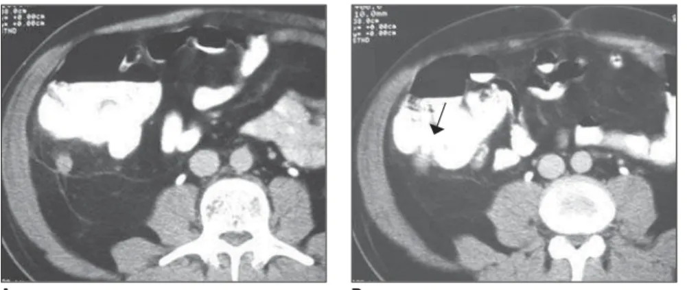
useful, especially in case of complications (perforated appendicitis), in young and thin patients (with paucity of peritoneal fat), in Figure 2. Computed tomography. Signs of appendicitis (parietal thickening and increase in the
mesen-teric fat density) in the right flank, located more anteriorly than usual (arrow).
A B
➝
Figure 1. Normal appendix. Opaque enema (A) and computed tomography (B – arrow).
A B
➝
from the medial posterior wall of the cecum, about 3 cm below the ileocecal valve. The base is at a constant location, whereas the position of the tip of the ap-pendix varies and may occupy several re-gions inside de abdominal cavity (Figure 2), including the pelvic region(1), the left
iliac fossa, or even inside the inguinal ca-nal. Clinical presentation is highly influ-enced by this wide variation in the topog-raphy of the appendix(1).
Obstruction of the appendiceal lumen due to the presence of fecalith (the most frequent one), lymphoid hyperplasia, for-eign body or tumor(1). Appendicolith, a
cal-cified fecalith, is less frequent but is asso-ciated with perforation and abscess forma-tion(1,2). The obstruction of the lumen there
is secretion accumulation leading to an in-crease in the intraluminal pressure, and de-termining stimulation of afferent visceral fibers between T8 and T10, with perium-bilical epigastric pain as a consequence(1).
This pain is not intense and usually poorly localized, with 2–5-hour duration. Anor-exia, nausea and emesis may be present in this phase. The gradual increase in the in-traluminal pressure exceeds the pressure of capillary perfusion, determining appen-diceal walls ischemia, with loss of the epi-thelial integrity and bacterial mural inva-sion(1,2). Then, the pain migrates into the
ap-pendiceal region, generally in the right iliac fossa, and may be associated with signs of peritoneal irritation (positive sudden de-compression). Fever is low or absent; the presence of high fever suggests perfora-tion(1).
forma-non-specific findings, and in the differen-tial diagnosis of a malignant process(1).
Retrograde contrast injection (500 ml 5% iodinated solution) by rectal via has been utilized by many authors, improving both the sensitivity and specificity of the method(4,5,9), but, in our services, we have
utilized this technique only in dubious cases, reducing costs and making the pro-cedure faster and more comfortable for the patient. The utilization of rectal-contrast reduces the incidence of false-positive re-sults, since intestinal loops filled with fluid may be confused with distended appendi-ces(2).
The utilization of oral contrast is unnec-essary in the majority of cases; it is helpful only in patients with non-specific abdomi-nal pain, or when ileal opacification is nec-essary to solve any doubt in the case the rectal contrast is not elucidative(6).
Finally, the fastest protocol in the evalu-ation of acute appendicitis is the one sug-gested by Lane et al.(6), who propose the use
of non-contrast-enhanced helical CT cov-ering the whole abdomen, with 5 mm-thick slices, 8 mm interval, and followed by 5 mm reconstruction.
TOMOGRAPHIC FINDINGS
Thick appendix – Appendiceal disten-tion is the first tomographic sign, but its identification depends on the degree of dis-tention, amount of surrounding fat, and technical quality of the study (slice thick-ness). The normal appendix is visualized in 67%–100% of asymptomatic adults sub-mitted to CT(1) with thin slices (Figure 1).
An appendix is considered as thick when
≥ 8 mm in transverse (Figure 3). Usually, the appendix contents is liquid (Figure 4). The distention rarely exceeds 15–20 mm, since perforation generally occurs first. Higher values suggest the possibility of mucoceles or neoplasm. In patients with paucity of peritoneal fat, rectal contrast may facilitate its identification(5,9).
Appendix wall thickening – The normal appendiceal wall is 1–2 mm-thick. In the inflammatory process, mural thickening is present, and if intravenous contrast agent is utilized(1), we will observe the contrast
uptake on the inflammed appendix walls (Figure 5).
Blurring of the adjacent fat – The nor-mal fat surrounding the appendix is homo-geneous. In appendicitis, there is a blurring of this fat (Figure 3), a very frequent and significant sign found in 98% of cases(1).
Cecal thickening – Also, the presence of some degree of inflammatory process is
frequent in the adjacent loops(10), especially
in the cecum (Figure 6).
Arrow-head sign – Characterized by an arrow-head shape, as a result of edema in the base of the appendix(11), on rectal
con-trast-enhanced studies (Figure 7). Appendicolith – Appendicolith does not necessarily indicate inflammation in the organ, since this finding is observed in as-ymptomatic adults without appendiceal distention. However, this finding gains high significance in the presence of other findings.
After appendix perforation, the appen-dicolith may migrate to other sites in the abdominal cavity(1), resulting in formation
of distant abscess, including during the post-operatory period (Figure 8).
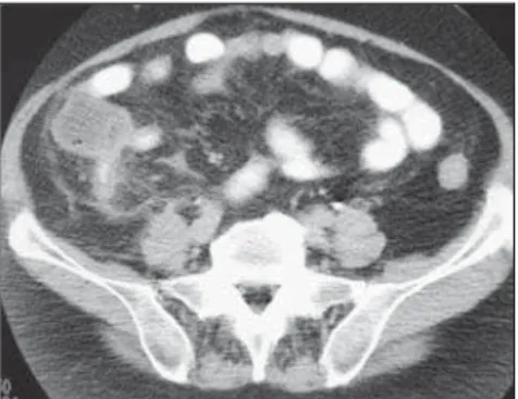
Inflammatory mass – In the case of an intense inflammatory process after appen-diceal perforation, large, ill-define inflam-matory masses may be observed in the right
Figure 6. Computed tomography. Female, 50-year-old patient. Acute appendicitis. Cecal thickening (ar-row), blurring of periappendicular fat and appendix thickening.
B A
➝
Figure 5. Computed tomography. Male, 46-year-old patient. Appendicitis demonstrated by marked thicken-ing of the appendix wall, and contrast-enhancthicken-ing..
Figure 4. Computed tomography. Female, 42-year-old patient. Marked liquid distention of the appendix in the right iliac fossa, with parietal thickening and blurring of adjacent fat, characterizing appendicitis.
iliac fossa, determined by the blockage of the adjacent intestinal loops and omentum (Figure 9). In some cases, the appendix may be totally destructed by infection, so its identification is unfeasible(2).
Free intraperitoneal fluid – The ap-pendix perforation results in spillage of pus into the abdominal cavity, with possible bacterial peritonitis(1) (Figure 10).
Extraluminal air – It may be found
within a collection or free inside the cav-ity (pneumoperitoneum) as a result of ap-pendiceal perforation (Figure 11). Pneumo-peritoneum pneumoPneumo-peritoneum is less fre-quent, and, if present, is small(1).
COMPLICATIONS
Complications occur as a result from delayed diagnosis and appendix perfora-tion(1,2), disseminating the infectious
pro-cess into the peritoneal cavity. Main com-plications are the following:
Abscess – A frequent complication, oc-curring in the appendix or in other sites inside the abdominal cavity, characterized by fluid collection, marginal enhancement after intravenous contrast administration, and many times blocked by adjacent intes-tinal loops (Figure 12).
Figure 8. Computed tomography. Appendicoliths. A: Normal appendix with appendicoliths in an asymp-tomatic patient (arrow). B,C,D: Early phase of appendicitis with appendicoliths. In this case, although the appendix thickness is only 6 mm, other positive signs such as liquid distention of the appendix, pres-ence of appendicolith, and compatible US results, have allowed the presurgical diagnosis. D: Photo of the surgical specimen. E,F,G: Appendicular abscess recognized by the presence of appendicolith in a three-year-old child using antibiotics for two weeks (double arrows).
E F
G D
➝
➝
➝
A
➝
B C
➝
➝
➝
Figure 7. Computed tomography. Male, 60-year-old patient. Acute appendicitis. Arrow head sign (ar-row).
A B
Figure 11. Computed tomography. Male, 59-year-old patient. Appendicitis complicated with wall abscess characterized by extraluminal air (arrows) surgically confirmed. The appendix has been com-pletely destructed by the infection, with unfeasible localization by means of CT.
Figure 9. Computed tomography. Female, 34-year-old patient. Inflammatory mass in the right iliac fossa. Appendicitis. Differential diagnosis with Crohn´s disease, ileocecal tuberculosis and neo-plastic process.
Figure 10. Computed tomography. Female, 71-year-old patient. Appendicitis with free intraperito-neal fluid suggesting peritonitis confirmed at the surgery (arrows).
A B
➝
➝
Figure 12. Computed tomography. Acute appen-dicitis complicated with abscess in the right iliac fossa. Two different cases, the second (B) with gaseous content (arrow).
A B
B A
A B
➝
➝
Venous thrombosis – Severe complica-tion resulting from infectious process dis-semination to the portal system. Also, he-patic abscesses may be observed.
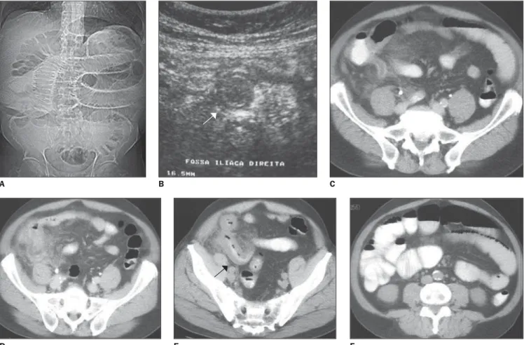
Intestinal obstruction – Initially, the blockage determined by the intestinal loops determines a regional ileum, however, as the disease progresses, obstruction may occur as a result from inflammatory process and ischemia (Figure 13).
Sepsis – Extremely severe complication detectable by clinical diagnosis, resulting from intracavitary abscesses or diffuse peri-tonitis, with consequent systemic dissemi-nation of the infectious process and high mortality.
Ureteral obstruction – The inflamma-tory process may determine ureteral ob-struction at right (Figure 14).
DIFFERENTIAL DIAGNOSIS
Main differential diagnoses are(2,4,6): di-verticulitis, epiploic appendagitis,
typhi-Figure 13. Male, 64-year-old patient. A: Digital radiogram B: Ultrasound (arrow). C–F: Classical findings of acute appendicitis besides signs of intestinal ob-struction characterized by dilatation of small bowel loops and mural thickening of the terminal ileum (arrow).
Figure 14. Computed tomography. Female, 27-year-old patient. Investigation for renal colic. Obstructive uropathy associated with signs of an inflammatory process in the retroperitoneum (arrows), with upper ureteral obstruction. Acute appendicitis confirmed at surgery.
D C
A B
➝
➝
A B C
D E F
litis, omental infarct, Crohn’s disease, coli-tis, acute cholelythiasis/cholecysticoli-tis, ure-teral calculus/pyelonephritis, pelvic in-flammatory disease/ovarian cyst, mesen-teric lymphadenopathy, neoplasm.
REFERENCES
1. Birnbaum BA, Wilson SR. Appendicitis at the millennium. Radiology 2000;215:337–348. 2. Gore RM, Miller FH, Pereles FS, Yaghmai V,
Berlin JW. Helical CT in the evaluation of the acute abdomen. AJR Am J Roentgenol 2000;174: 901–913.
3. Sivit CJ, Applegate KE, Berlin SC, et al. Evalua-tion of suspected appendicitis in children and
young adults: helical CT. Radiology 2000;216: 430–433.
4. Paulson EK, Harris JP, Jaffe TA, Haugan PA, Nelson RC. Acute appendicitis: added diagnos-tic value of coronal reformations from isotropic voxels at multi-detector row CT. Radiology 2005; 235:879–885.
5. Mullins ME, Kircher MF, Ryann DP, et al. Evalu-ation of suspected appendicitis in children using limited helical CT and colonic contrast material. AJR Am J Roentgenol 2001;176:37–41. 6. Lane MJ, Katz DS, Ross BA, Clautice-Engle TL,
Mindelzun RE, Jeffrey RB Jr. Unenhanced heli-cal CT for suspected acute appendicitis. AJR Am J Roentgenol 1997;168:405–409.
7. Freire Filho EO, Jesus PEM, D’Ippolito G, Szejn-feld J. Tomografia computadorizada sem contraste
intravenoso no abdome agudo: quando e por que usar. Radiol Bras 2006;39:51–62.
8. Menezes MR, Kay FU. Tomografia computado-rizada multidetectores não-contrastada na avalia-ção do abdome agudo: um novo paradigma no pronto-socorro? Radiol Bras 2006;39(2):IV–V. 9. Kamel IR, Goldberg SN, Keogan MT, Rosen MP,
Raptopoulos V. Right lower quadrant pain and suspected appendicitis: nonfocused appendiceal CT – review of 100 cases. Radiology 2000;217: 159–163.