O P E N A C C E S S
http://dx.doi.org/ 10.5339/jemtac.2012.7
Published: 10 May 2012 c
2012 Parambath et al., licensee Bloomsbury Qatar Foundation Journals. This is an open access article distributed under the terms of the Creative Commons Attribution License CC BY 3.0 which permits unrestricted use, distribution and reproduction in any medium, provided the original work is properly cited.
Case study
Spigelian hernia with Richter-type
herniation of the ileum: A rare cause
of right iliac fossa pain mimicking
acute appendicitis
Arif Nelliyulla Parambath1,*, Khairi Hajaji2, Shatha Ali Al Hilil1
1Emergency Radiology Department, Hamad Medical Corporation, Doha, Qatar
2General Surgery Department, Hamad Medical Corporation, Doha, Qatar *Corresponding author. Email: [email protected]
ABSTRACT
We report a case of a sixty-two-year-old man who presented to the emergency department with severe right iliac fossa pain. The patient was clinically diagnosed as having acute appendicitis and was referred for an abdominal CT scan. Abdominal CT revealed a Spigelian hernia with Richter-type herniation of the small bowel without any significant bowel obstruction. The patient underwent immediate surgical repair and the postoperative course was uneventful. This is a rare association of two rare conditions mimicking a common disease such as appendicitis.
Keywords:Spigelian hernia, Richter-type herniation, CT scan, Appendicitis
Page 2 of 4
Parambath et al. Journal of Emergency Medicine, Trauma and Acute Care 2012:7
INTRODUCTION
Richter’s hernia, named after August Gottlob Richter (1742–1812) is characterized by herniation of only a portion of the bowel wall circumference. The usual site is the terminal ileum, but it can occur anywhere in the gastrointestinal tract. Although there is an increased incidence of incarceration and strangulation for this type of hernia, intestinal obstruction is rare because of the patency of the lumen. Spigelian hernia, named after Adrian Van Deer Spiegel, a Belgian anatomist (1578–1625), is a rare type of hernia due to a defect in the Spigelian aponeurosis of the abdominal wall. This usually lies beneath the aponeurosis of the external oblique muscle, which is a relatively tough structure, making the clinical detection difficult. This type of hernia is more prone to complications such as incarceration, strangulation and obstruction. Complicated cases can mimic acute abdominal conditions such as appendicitis, diverticulitis and tubo-ovarian pathology. In this paper, we report a patient suffering with a Richter-type of herniation of the small bowel and Spigelian hernia, presenting with clinical signs and symptoms of acute appendicitis.
CASE REPORT
A sixty-two-year-old man presented to the emergency department with sudden onset of abdominal pain and vomiting. On physical examination he was hemodynamically stable and apyrexial. Palpation of the abdomen revealed an ill defined mass in the right iliac fossa with severe tenderness. Routine investigations including haemogram, urine analysis and serum chemistry were all within the normal limits. With a clinical diagnosis of complicated appendicitis, a CT scan was requested to exclude other possibilities. Multi-detector CT (MDCT) (64 slice) scan of the abdomen with the help of multiplanar reconstruction (MPR) identified a localized outpouching of the small bowel (Richter-type herniation) through a defect in the spigelian fascia—Spigelian hernia (Figs. 1,2(a)&2(b)). Apart from minimal distension of the adjacent small bowel, no significant diffuse dilatation of the small bowel suggestive of an intestinal obstruction was noticed. Emergency surgery was performed via a transverse incision in the right iliac fossa, confirming a rare combination of Spigelian hernia with Richter-type herniation of the terminal ileum (Fig. 3). The herniated bowel appeared edematous without any evidence of gangrene. After reducing the bowel, the defect in the internal oblique muscle was closed with sutures and mesh. The postoperative period was uneventful and the patient was discharged with advice.
DISCUSSION
Richter’s hernia is a rare type of bowel herniation commonly occurring in the terminal ileum, but can also occur anywhere in the gastrointestinal tract[1]. Pathophysiologically it is characterized by protrusion of part of the circumference of the bowel wall through the hernial orifice with retained patency of the lumen. With regard to location, common sites are the femoral ring (36–88%), inguinal canal (12–36%) and abdominal wall (4–25%)[2]. Rarer sites include umbilical, obturator,
supravesical, Spigelian, Morgagni, internal and diaphragmatic hernias. Recently more cases of this type of hernia are reported in patients that undergo laparaproscopic surgery due to herniation through the trocar site[3,4]. Even though the incidence of incarceration and strangulation is more
Page 3 of 4
Parambath et al. Journal of Emergency Medicine, Trauma and Acute Care 2012:7
Figure 2.(a) & (b) show the sagittal and coronal reformations of MDCT images. Spigelian hernia with Richter-type herniation of the ileum is shown. The herniated bowel is seen beneath the aponeurosis of the external oblique muscle (arrow).
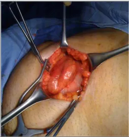
Figure 3.Pre-operative image showing the herniated ileum.
common for this type of hernia, the intestinal obstruction is rare because of patency of the lumen[5]. Spigelian hernias are a rare type of interparietal hernia anatomically located in the area known as the ‘spigelian belt’ in the anterior abdominal wall. ‘Spigelian belt’ is bordered medially by rectus muscle, laterally by the semilunar line, superiorly by the arcuate line of Douglas and inferiorly by the inferior epigastric vessel[6,7]. The herniated tissue can be bowel, preperitoneal fat or peritoneal sac through a defect in the spigelian aponeurosis (fascia formed by the aponeurosis of the internal oblique and transverse abdominal muscles). Compared to other types of hernias, the incidence of Spigelian hernias are low and etiologies considered are congenital or acquired, the latter being more
common[8,9]. Obesity, chronic obstructive pulmonary disease, prior surgery, and abdominal trauma are the suggested predisposing factors. It represents 1–2% of all hernias presenting to the
emergency department with a high incidence of incarceration and strangulation (14–21%)[10,11]. Unlike other forms of abdominal wall hernias, Spigelian hernias can frequently go unnoticed and present acutely with complications such as incarceration, strangulation and intestinal
Page 4 of 4
Parambath et al. Journal of Emergency Medicine, Trauma and Acute Care 2012:7
Spigelian hernias are either surgical or laparoscopic repair using non-absorbable sutures and the placement of mesh.
CONCLUSION
Richter-type herniation of the bowel in a Spigelian hernia is not a common association. This
occurrence rarely coexists to mimic a common condition like acute appendicitis. MDCT, by virtue of its high quality images and multiplanar reformation capability is a very useful diagnostic tool in the differentiation of this condition.
References
[1] Tito W.A. and Allen W.C. Richter and Littre Hernia. In Hernia. 3rd edition, Eds. Nyhus J.B. and Condon R.E. 1989; Lippincott, Philadelphia. 305–310.
[2] Frankau C. Strangulated hernias: a review of 1487 cases. Br J Surg. 1931;19:176–191.
[3] Bourke J.B. Small-intestinal obstruction from a Richter’s hernia at the site of insertion of a laparoscope. Br Med J. 1977;26:1393–1394.
[4] Stewart D. Richter’s hernia at site of insertion of laparoscope. Br Med J. 1977;2:1673.
[5] Ghahremani G.G. Abdominal and pelvic hernias. In Textbook of gastrointestinal radiology. 2nd ed., Eds. Gore R.M. and Levine M.S. 2000; Saunders, Philadelphia, Pa. 1993–2009.
[6] Larson D.W. and Farley D.R. Spigelian hernias: repair and outcome for 81 patients. World J Surg. 2002;26:1277–1281.
[7] Skandalakis P.N., Zoras O., Skandalakis J.E. et al. Spigelian hernia: surgical anatomy, embryology, and technique of repair. Am Surg. 2006;72:42–48.
[8] Al-Salem A.H. Congenital spigelian hernia and cryptorchidism: cause or coincidence?. Pediatr Surg Int. 2000;16:433–436.
[9] Walton J.M. and Bass J.A. Spigelian hernias in infants: report of two cases. Can J Surg. 1995;38:95–97. [10] Zacharakis E., Papadopoulos V., Ganidou M. et al. Incarcerated Spigelian hernia: a case report. Med Sci Monit.
2006;12:CS64–CS66.
[11] Spangen L. Spigelian hernia. Surg Clin North Am. 1984;64:351–366.
[12] Tsalis K., Zacharakis E., Lambrou I. et al. Incarcerated small bowel in a spigelian hernia. Hernia. 2004;8:384–386. [13] Spangen L. Spigelian hernia. World J Surg. 1989;13:573–580.