COMPUTED TOMOGRAPHY CONTRIBUTION TO THE STAGING
OF SUPRAGLOTTIC SQUAMOUS CELL CARCINOMA*
Antonio Gilson Monte Aragão Jr.1
, Ricardo Pires de Souza2
, Abrão Rapoport3
OBJECTIVE: The present study was aimed at evaluating the role of computed tomography in the local clini-cal staging of supraglottic tumors according to the TNM classification, as well as the interobserver agree-ment on the detection of the tumor extent. MATERIALS AND METHODS: Thirty-nine dossiers of inpatients of Hospital Heliópolis with supraglottic squamous cell carcinoma in the period between 1988 and 1998 were retrospectively evaluated. CT studies were individually analyzed by two radiologists. The kappa test was utilized for evaluating the interobserver agreement. RESULTS: Computed tomography has played a decisive role in the upstaging of 38.5% of cases, as a result of a deep tumor extent undetected at clinical examina-tion. CONCLUSION: Interobserver agreement was considered as excellent for vocal folds and subglottis; good for supraglottic, paraglottic and preepiglottic spaces, thyroid and cricoid cartilages and for extralaryngeal tumor extension; and regular for the base of the tongue.
Keywords: Laryngeal neoplasms; Squamous cell carcinoma; Computed tomography; Tumors staging.
Contribuição da tomografia computadorizada no estadiamento do carcinoma de células escamosas da su-praglote.
OBJETIVO: Este trabalho tem como objetivos avaliar a influência da tomografia computadorizada no estadia-mento local da classificação TNM dos tumores da supraglote e avaliar a concordância interobservadores na detecção da extensão tumoral. MATERIAIS E MÉTODOS: Foram avaliados, retrospectivamente, 39 pacien-tes com carcinoma de células escamosas da supraglote atendidos no Hospital Heliópolis entre 1988 e 1998. Os exames de tomografia computadorizada foram analisados por dois radiologistas individualmente. Para a avaliação da concordância interobservadores utilizou-se o índice kappa. RESULTADOS: A tomografia com-putadorizada foi determinante no estadiamento mais avançado em 38,5% dos casos, decorrente de exten-são tumoral profunda não-identificada no exame clínico. CONCLUSÃO: A concordância interobservadores foi considerada ótima para as pregas vocais e subglote; boa para a supraglote, espaços paraglótico e pré-epi-glótico, cartilagens cricóide e tireóide e para extensão tumoral extralaríngea; e regular para a base da língua. Unitermos: Neoplasias laríngeas; Carcinoma de células escamosas; Tomografia computadorizada; Estadia-mento de neoplasias.
Abstract
Resumo
* Study developed at Hospital Heliópolis, São Paulo, SP, Brazil. 1. Course of Post-Graduation, Department of Head & Neck at Hospital Heliópolis, São Paulo, SP, Brazil.
2. Coordinator for the Department of Radiology, Hospital Heliópolis, São Paulo, SP, Brazil.
3. Coordinator for the Course of Post-Graduation in Head & Neck Surgery, Hospital Heliópolis, São Paulo, SP, Brazil.
Mailing address: Dr. Antonio Gilson Monte Aragão Jr. Rua Henriqueta Galeno, 1080, ap. 1201, Bairro Dionísio Torres. Fortaleza, CE, Brazil, 60135-480. E-mail: [email protected] Received January 12, 2006. Accepted after revision Novem-ber 23, 2006.
INTRODUCTION
Laryngeal cancer is a potentially curable disease. About 30% of these tumors occur in the supraglottis, and 95% of them are squamous cell carcinomas, with a peak of incidence in the fifth decade of life and a 5/1 men/women ratio. Radiotherapy and conservative surgery, that can preserve functions such as deglutition and voice, have made the accurate staging and defini-tion of the tumor extent imperative for an appropriate therapeutic planning. For this
purpose, an accurate knowledge of the real tumor extent and condition of the laryngeal cartilaginous skeleton is indispensable(1–5).
Laryngoscopy is of help in the evaluation of the superficial tumor extent, while com-puted tomography (CT) plays a significant role in the evaluation of the deep tumor extension, for example, to the preepiglottic and paraglottic spaces)(6–9).
The present study is aimed at evaluat-ing the interobserver agreement in the de-tection of neoplastic involvement in several sites and extensions of the supraglottic lar-ynx, and the influence of CT utilization on the clinical staging as regards higher lev-els of the T parameter in the TNM classifi-cation of supraglottic tumors.
MATERIALS AND METHODS
The present study involved inpatients of the Department of Head & Neck at
Hospi-tal Heliópolis, with supraglottic squamous cell carcinoma, in the period between 1988 and 1998. The patients inclusion criteria adopted were the following: a) tumor clini-cally staged by means of direct/indirect laryngoscopy; b) squamous cell carcinoma diagnosed by biopsy; c) CT study per-formed before any treatment (chemo-therapy, radiation therapy or surgery).
Thirty-nine dossiers meeting the pre-es-tablished criteria were evaluated. Thirty-three patients (84.6%) were men and six (15.4%) women, corresponding to a men/ women ratio of 5.5/1. Patients’ ages ranged between 39–74 years among men, and 40– 67 years among women, with mean ages of 55 and 53 years, respectively, and 54.9 for both sexes. All of these patients were smok-ers, and 31 (79.5%) of them reported mod-erate or heavy use of alcohol.
axial slices with 5 mm thickness and incre-ment, after intravenous, iodinated contrast media injection at a dose of 1.0–2.0 ml/kg, and 60–76% concentration, with the pa-tient in supine position with neck exten-sion, during calm respiration and without deglutition.
The studies were individually evaluated by two radiologists with no previous knowledge of findings at physical exami-nations, laryngoscopic or histopathological studies. The aspects analyzed were con-cerned with the T parameter, according to the TNM classification defined by the In-ternational Union Against Cancer (UICC), reviewed in 1997.
The evaluation covered tumor extension towards the following supraglottic regions: epiglottis, aryepiglottic fold, ventricular folds and their eventual extensions towards the vocal folds, subglottis, base of the tongue, preepiglottic space, paraglottic space, thyroid cartilage, cricoid cartilage, and extralaryngeal soft tissues.
Deep structures were considered as af-fected when one or more of the following criteria were present: blurring of fatty tis-sues planes, local mass effect and direct involvement of the lesion, characterized by the presence of tumor with soft tissues den-sity in the region to be evaluated. The car-tilages invasion was considered in the pres-ence of lesions internal and externally to the cartilage, or when erosion was detected (these criteria were utilized for the car-tilages evaluated in the present study); scle-rosis was the invasion criterion in the evaluation of the cricoid cartilage involve-ment. The subglottic involvement was con-sidered in the presence of any tissue adja-cent to the inner surface of the cricoid car-tilage.
The staging was based on the T param-eters of tumor extent defined by the UICC (reviewed in 1997). The tomographic stag-ing was based on the analysis performed by the most experienced radiologist special-ized in head and neck.
The kappa test (κ) was utilized in the evaluation of interobserver agreement in the CT studies reading (Chart 1), consid-ering the significance level = 0.05 and a 95% confidence interval(10). Changes in the
clinical staging resulting from the CT evaluation were based on the T tumor
ex-Chart 1 Evaluation of interobserver agreement according to kappa index(10).
κ value
< 0.00 0.00–0.20 0.21–0.40 0.41–0.60 0.61–0.80 0.81–0.99 = 1.00 Agreement Bad Feeble Poor Regular Good Very good Excellent
CT studies was the following: seven pa-tients (17.9%) T2, 21 (53.8%) T3 and 11 (28.2%) T4. A comparison between clini-cal and CT staging showed a coincidence in 22 cases (56.4%) — five (12.8%) T2 stage, 12 (30.8%) T3 stage, and five (12.8%) T4 stage (Tables 1 and 2). The two patients with clinical staging T1 were up-staged as T2 and T3 at CT. No patient was staged as T1 by CT (Table 1, Graphic 1).
Of 13 patients (33.3%) with clinical staging T2, only five (38%) were coinci-dental at CT. The remaining eight patients (61%) were upstaged at CT as follows: seven T3 and one T4. Of 18 patients with clinical staging T3, 12 (67%) were coinci-dental at CT. The remaining six patients (33%) were not coincidental; five were upstaged as T4. One patient was substaged as T2 at CT (Table 1, Graphic 1).
Five of the six (83%) patients clinically staged as T4 had the same result at CT; in only one patient (17%) the CT staging (T3) was different from the clinical staging. This may be explained since there was a clini-cal suspicion of invasion of the cartilage by the tumor that had not been observed at CT (Table 1).
There was a coincidence between clini-cal and tomographic staging in 38.5%, 66.7%, and 83.3% of cases respectively classified as T2, T3 and T4.
The tomographic staging increased the clinical staging in 61.5% of T2-staged cases, and in 27.8% of T3-staged cases. As previously mentioned, two T1-staged cases were upstaged at CT. Two cases were substaged at CT; the first one because the TC failed to detect vocal fold fixation, de-creasing the clinical staging from T3 to T2; in the other case, the staging decreased from T3 to T2 as a result of the clinical suspicion of invasion of the cartilage by the tumor that had not been confirmed at CT.
Table 1 Distribution of patients according clinical and tomographic stagings.
Tomographic staging T1 T2 T3 T4 Total T1 0 1 1 0 2 (5.1%) T2 0 5 7 1 13 (33.3%) T3 0 1 12 5 18 (46.2%) T4 0 0 1 5 6 (15.4%) Total 0 7 (17.9%) 21 (53.8%) 11 (28.2%) 39 (100.0%) Clinical staging
tent criteria defined by the UICC, observ-ing the regions involved in the upstagobserv-ing.
RESULTS
1 – Interobserver agreement
Interobserver agreement was consid-ered as good for supraglottic sites — epig-lottis (κ = 0.655), aryepiglottic folds (κ = 0.754), and ventricular folds (κ = 0.794). Also, it was considered as good for evalu-ation of paraglottic (κ = 0.693) and preepiglottic (κ = 0.744) spaces. The evalu-ation of vocal folds reached a κ = 0.854, with interobserver agreement considered as very good. The agreement was good for evaluation of thyroid cartilage (κ = 0.674) and cricoid cartilage (κ = 0.687). A regu-lar interobserver agreement (κ = 0.480) was found in the evaluation of the base of the tongue; and for the subglottis, the agree-ment was very good (κ = 0.880), and good in the evaluation of the extralaryngeal tu-mor extension (κ = 0.747).
2 – Clinical and tomographic staging
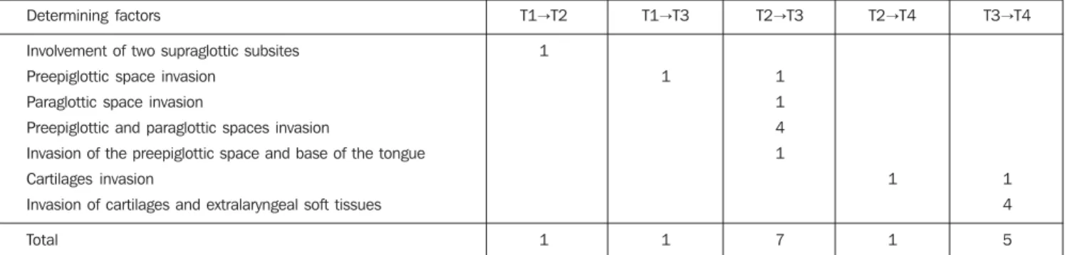
Table 2 Sites of neoplastic invasion determining upstaging at computed tomography.
Determining factors
Involvement of two supraglottic subsites Preepiglottic space invasion
Paraglottic space invasion
Preepiglottic and paraglottic spaces invasion
Invasion of the preepiglottic space and base of the tongue Cartilages invasion
Invasion of cartilages and extralaryngeal soft tissues
Total
T1→T2
1
1
T1→T3
1
1
T2→T3
1 1 4 1
7
T2→T4
1
1
T3→T4
1 4
5
Computed tomography has played a de-cisive role in the upstaging in 15 (38.5%) cases of the whole casuistic according to the factors listed on Table 2.
DISCUSSION
According to the literature, CT presents a higher staging accuracy (between 68% and 82.7%) than the clinical examination (between 52% and 74.3%) for supraglottic tumors. This staging accuracy potentialized with the association between CT and clini-cal evaluations, reaching 91.4% of effec-tiveness(4,7,11–17).
Also, according to data in the literature, CT determines an increase in clinical stag-ing rangstag-ing between 19.4% and 30.7%(3, 12,15,18). In the present study, this upstaging
Figure 1. Supraglottic squamous cell carcinoma with extension into the pre-epiglottic space. Clinical staging T1; tomographic staging T3. B: Aryepiglottic fold squamous cell carcinoma. Clinical staging T2; tomographic staging T3.
A B
Graphic 1. Tomographic staging as compared to clinical staging.
Tomographic staging
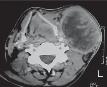
Figure 2. Axial tomographic view at the level of ventricular folds, where a volu-minous tumor-like lesion is observed, causing constriction of the larynx lumen. There is a tumor-like component invading the right paraglottic space. Sclerosis is observed on the right wing of the thyroid cartilage, as well as lymphadeno-megalies with hypodense nucleus at right.
Figure 3. CT axial view at the level of supraglottis showing tumor infiltration into the left paraglottic space and tumor on both sides of the thyroid carti-lage indicating invasion. Clinical staging T3, tomographic staging T4. Also, a voluminous necrotic lymph node block is observed at right.
Figure 4. Tumor-like lesion with an infiltrative aspect in the epig-lottis, invading the pre-epiglottic space, and tumor-like component infiltrating the extralaryngeal soft tissues at left. Clinical staging T2, tomographic staging T4.
Figure 5. Axial tomographic view of the thyroid cartilage region. In-filtrative process determining ero-sion of the left lamina of the thy-roid cartilage and infiltration to extralaryngeal soft tissues. Clini-cal and tomographic staging T4. Figure 5
Figure 4
was observed in 38.5% of cases, similarly to the findings of other authors; main rea-sons were: cartilages invasion, infiltration into the pre-epiglottic and paraglottic spaces, and extralaryngeal tumor extension that had not been detected by laryngoscopy (Table 2). Patients clinically staged as T2 were mostly upstaged at CT. Only two cases clinically staged as T1 met the initial inclusion criteria, and were upstaged. The low number of tumors initially staged as T1 may be explained by: low socio-economic level of the patients, delay in the search for medical assistance, late symptoms in supra-glottic tumors, tumors clinically staged as
T1 are not routinely submitted to prethera-peutic CT.
CT may determine a substaging in up to 10.6% of cases(3); in the present study, two
cases were substaged at CT in comparison with the clinical examination: one case where the clinical examination had de-tected fixation of vocal fold (not dede-tected at CT), and another where the CT substaged from T4 to T3, considering the clinical sus-picion of cartilage invasion not confirmed by CT.
It is important to note that false-positive cases in CT evaluation may occur as a re-sult of inflammatory alterations or
peritu-moral edema. On the other hand, false-negative cases result from tumors restricted to the mucosal surface or presenting with microscopic tumor infiltration(3,19–21).
The deep tumor extension into the preepiglottic and paraglottic spaces is ne-glected in the laryngoscopic evaluation, so CT becomes an essential method in the evaluation of these regions(22,23). The
infil-tration of these spaces has been implicated in the occurrence of lymph node metasta-sis(19) and tumor recurrence(24). An
Figure 6. Extensive tumor conditioning erosion of the thyroid cartilage and extension to extralaryngeal soft tissues. Almost complete aerial column obliteration is observed. Clinical and tomographic staging T4.
Figure 7. Asymmetrical thickening and heterogeneous attenuation of the right vocal fold. Histopathology: chronic inflammatory alterations. False-positive re-sult.
The interobserver agreement was con-sidered as good for all of the supraglottic sites, as well as for the pre-epiglottic and paraglottic spaces. An excellent interob-server agreement was found in the evalua-tion of vocal folds and subglottic space. A previous study has also demonstrated a good interobserver agreement for supra-glottic and subsupra-glottic spaces, and very good for vocal folds(26).
In one case where both observers had considered that the left vocal fold was in-filtrated by the tumor, the histopathologi-cal analysis demonstrated that its thicken-ing was a result from a chronic inflamma-tory reaction, characterizing a false-posi-tive case; this finding being indistinguish-able from tumor infiltration. In another case of disagreement, where clinical staging was T3 — because of vocal fold fixation —, the CT substaged as T2, since this method had not detected this finding; the histopatho-logical analysis detected the involvement of the vocal fold by the tumor. The rel-evance of the correlation between clinical/ endoscopic and tomographic evaluations becomes clear, as the actual tumor extent is established with higher accuracy.
The CT evaluation of laryngeal carti-lages is highly specific, however, its sen-sitivity is low, with a high rate of false-negative results, and may affect the thera-peutic indication for partial laryngectomy or radical radiation therapy(7). Gross
carti-lage destruction may be easily identified, while mild macroscopic or microscopic in-vasions are unlikely to be detected by CT(4,27–29). The irregular calcification
pat-tern is the factor that represents one of the major difficulties in the evaluation of cartilages, especially thyroid cartilage(8,28– 30). Good interobserver agreement was
found for evaluation of tumor invasion of the thyroid cartilage and cricoid cartilage. Hermans et al.(30) have found an
interob-server agreement considered as just regu-lar. This may be explained by the fact that infiltration criteria were adopted for each of the cartilages. Sclerosis was considered as a sign of infiltration just for the cricoid cartilage, since infiltration into the thyroid cartilage frequently is just an indication of inflammatory reaction(5).
The CT evaluation of the base of the tongue is not effective principally because of secretions deposition that may simulate tumor involvement, as well as the presence of redundant lymphatic tissue(3,17). CT
failed to identify one third of cases of in-filtration into the base of the tongue. Inter-observer agreement was considered as just regular in the evaluation of tumor infiltra-tion into the base of the tongue. The histo-pathological analysis demonstrated two patients with this finding, one of them iden-tified by both observers, and the other, by only one observer. These two cases pre-sented invasion of the preepiglottic space
confirmed by anatomopathological analy-sis.
Subglottic invasion may occur both by superficial mucosal dissemination and lat-eral infiltration of the elastic cone(3). On the
other hand, tumor extension to other extra-laryngeal soft tissues may occur through the larynx or thyrohyoid or cricoid mem-branes(17). Laryngoscopy is useful in the
evaluation of subglottis, but its accuracy may be impaired in case of exophytic tu-mors, and extralaryngeal extension may not be detected; this is one of the main reasons for clinical substaging(7). CT may represent
a useful tool in this evaluation, presenting a very good interobserver agreement for subglottis, and good for extralaryngeal ex-tension.
substaging. However, the good CT repro-ducibility becomes more significant in face of the tendency towards more conservative, and frequently non-surgical treatments for laryngeal tumors. As a result CT becomes an important tool in the pretherapeutic evaluation of these patients.
CONCLUSIONS
In the present study, computed tomog-raphy presented a very good interobserver agreement in the evaluation of tumor in-volvement in the vocal folds and tumor extension into the subglottis. Interobserver agreement was considered as good for analysis of epiglottis, aryepiglottic folds, ventricular folds, preepiglottic space, paraglottic space, thyroid and cricoid cartilages, and for evaluation of extralaryn-geal tumor extension. For evaluation of tumor involvement in the base of the tongue, the interobserver agreement was considered as regular.
CT upstaged 38.5% of patients with supraglottic cancer. The greatest difference between clinical and tomographic stagings was found in the group of patients with clinically staged T2 tumors, and CT up-staged 61.5% of these patients.
REFERENCES
1. Sagel SS, AufderHeide JF, Aronberg DJ, Stanley RJ, Archer CR. High resolution computed tomog-raphy in the staging of carcinoma of the larynx. Laryngoscope 1981;91:292–300.
2. Sexton CC, Anderson CG. Computed tomography in carcinoma of the larynx. Australas Radiol 1984;28:330–334.
3. Charlin B, Brazeau-Lamontagne L, Guerrier B, Leduc C. Assessment of laryngeal cancer: CT scan versus endoscopy. J Otolaryngol 1989;18: 283–288.
4. Sulfaro S, Barzan L, Querin F, et al. T staging of the laryngohypopharyngeal carcinoma. A 7-year multidisciplinary experience. Arch Otolaryngol Head Neck Surg 1989;115:613–620.
5. Becker M, Zbären P, Delavelle J, et al. Neoplas-tic invasion of the laryngeal cartilage: reassess-ment of criteria for diagnosis at CT. Radiology 1997;203:521–532.
6. Zbären P, Becker M, Lang H. Pretherapeutic stag-ing of laryngeal carcinoma. Clinical findstag-ings, computed tomography, and magnetic resonance imaging compared with histopathology. Cancer 1996;77:1263–1273.
7. Zbären P, Becker M, Lang H. Staging of laryngeal cancer: endoscopy, computed tomography and magnetic resonance versus histopathology. Eur Arch Otorhinolaryngol 1997;254 Suppl 1:S117– S122.
8. Kazkayasi M, Önder T, Özkaptan Y, Can C, Pa-busçu Y. Comparison of preoperative computed tomographic findings with postoperative histo-pathological findings in laryngeal cancers. Eur Arch Otorhinolaryngol 1995;252:325–331. 9. Mukherji SK, Pillsbury HR, Castillo M. Imaging
squamous cell carcinomas of the upper aerodiges-tive tract: what clinicians need to know. Radiol-ogy 1997;205:629–646.
10. Pereira MG. Epidemiologia: teórica e prática. 1ª ed. Rio de Janeiro, RJ: Guanabara Koogan, 1995. 11. Katsantonis GP, Archer CR, Rosenblum BN, Yeager VL, Friedman WH. The degree to which accuracy of preoperative staging of laryngeal car-cinoma has been enhanced by computed tomog-raphy. Otolaryngol Head Neck Surg 1986;95:52– 62.
12. Deschepper C, Casselman J, Van de Voorde W, Lemahieu S, Van Damme B, Baert AL. The con-tribution of CT to the T-staging of laryngeal car-cinoma. J Belge Radiol 1989;72:191–197. 13. Kolbenstvedt A, Charania B, Natvig K, Tausjo J.
Computed tomography in T1 carcinoma of the lar-ynx. Acta Radiol 1989;30:467–469.
14. Werber JL, Lucente FE. Computed tomography in patients with laryngeal carcinoma: a clinical perspective. Ann Otol Rhinol Laryngol 1989;98(1 Pt 1):55–58.
15. Dullerud R, Johansen JG, Dahl T, Faye-Lund H. Influence of CT on tumor classification of laryn-geal carcinomas. Acta Radiol 1992;33:314–318. 16. Thabet HM, Sessions DG, Gado MH, Gnepp DA, Harvey JE, Talaat M. Comparison of clinical evalu-ation and computed tomographic diagnostic ac-curacy for tumors of the larynx and hypopharynx. Laryngoscope 1996;106(5 Pt 1):589–594. 17. Williams DW 3rd. Imaging of laryngeal cancer.
Otolaryngol Clin North Am 1997;30:35–58. 18. Barbera L, Groome PA, Mackillop WJ, et al. The
role of computed tomography in the T classifica-tion of laryngeal carcinoma. Cancer 2001;91:394– 407.
19. Larsson S, Mancuso A, Hoover L, Hanafee W. Differentiation of pyriform sinus cancer from su-praglottic laryngeal cancer by computed tomog-raphy. Radiology 1981;141:427–432. 20. Reid MH. Laryngeal carcinoma: high-resolution
computed tomography and thick anatomic sec-tions. Radiology 1984;151:689–696. 21. Silverman PM, Bossen EH, Fisher SR, Cole TB,
Korobkin M, Halvorsen RA. Carcinoma of the lar-ynx and hypopharlar-ynx: computed tomographic-histopathologic correlations. Radiology 1984; 151:697–702.
22. Weinstein GS, Laccourreye O, Brasnu D, Yousem DM. The role of computed tomography and mag-netic resonance imaging in planning for conser-vation laryngeal surgery. Neuroimaging Clin N Am 1996;6:497–504.
23. Dursun G, Keser R, Aktürk T, Akiner MN, Demireller A, Sak SD. The significance of pre-epiglottic space invasion in supraglottic laryngeal carcinomas. Eur Arch Otorhinolaryngol 1997;254 Suppl. 1:S110–112.
24. Hermans R, Van den Bogaert W, Rijnders A, Baert AL. Value of computed tomography as outcome predictor of supraglottic squamous cell carcinoma treated by definitive radation therapy. Int J Radiat Oncol Biol Phys 1999;44:755–765.
25. Gregor RT. The preepiglottic space revisited: is it significant? Am J Otolaryngol 1990;11:161– 164.
26. Paiva RGS, Souza RS, Rapoport A, Soares AH. Avaliação por tomografia computadorizada do envolvimento loco-regional do carcinoma espino-celular de corda vocal. Radiol Bras 2001;34:193– 200.
27. Mafee MF, Schild JA, Michael AS, Choi KH, Capek V. Cartilage involvement in laryngeal car-cinoma: correlation of CT and pathologic macro-section studies. J Comput Assist Tomogr 1984;8: 969–973.
28. Castelijns JA, Gerritsen GJ, Kaiser MC, et al. In-vasion of laryngeal cartilage by cancer: compari-son of CT and MR imaging. Radiology 1988;167: 199–206.
29. Castelijns JA, Golding RP, Van Schalk C, Valk J, Snow GB. MR findings of cartilage invasion by laryngeal cancer: value in predicting outcome of radiation therapy. Radiology 1990;174(3 Pt 1): 669–673.