279
External laryngocele: sonographic appearance – a case report
Radiol Bras 2007;40(4):279–282 Case Report
EXTERNAL LARYNGOCELE: SONOGRAPHIC APPEARANCE – A CASE
REPORT*
Marco da Cunha Pinho1
, Publio César Viana1
, Mauricio Omokawa2
, Cezar Simões3 , Eloisa M.M. Santiago Gebrim4
, Giovanni Guido Cerri5
, Maria Cristina Chammas6
Laryngoceles are fairly unusual diseases defined as anomalous saccular dilatation of the laryngeal ventricles. The usual classification divides laryngoceles into internal, external and mixed types. Internal laryngoceles are those located medially to the thyrohyoid membrane and usually compress the false vocal cords causing hoarseness or airway obstructive symptoms. External laryngoceles extend through the thyrohyoid membrane, presenting as cervical masses; and mixed laryngoceles present both the internal and external components with their respective symptoms. Diagnosis is usually defined by computed tomography and/or laryngoscopy. This is a report of a case of mixed laryngocele diagnosed by ultrasonography in a patient referred for inves-tigation with a history of palpable cervical mass.
Keywords: Laryngocele; Cervical mass; Ultrasonography; Laryngoscopy.
Laringocele: aspecto ultra-sonográfico – relato de caso.
Laringoceles são lesões relativamente raras definidas como dilatações anômalas dos sáculos dos ventrículos laríngeos. A classificação usual divide a laringocele em interna, externa e combinada ou mista. Laringoceles internas são as que se localizam medialmente à cartilagem tireóidea e geralmente causam compressão nas bandas ventriculares levando a rouquidão e sintomas compressivos na via aérea. As externas se estendem através da membrana tireóidea, apresentando-se como massas cervicais, e as mistas são as que ocupam as duas regiões, podendo causar ambos os sintomas. O diagnóstico é geralmente feito por tomografia compu-tadorizada e/ou laringoscopia. Apresentamos um caso de laringocele mista em que o diagnóstico foi sugerido no exame de ultra-sonografia, num paciente encaminhado com história de massa cervical.
Unitermos: Laringocele; Massa cervical; Ultra-sonografia; Laringoscopia. Abstract
Resumo
* Study developed in the Division of Ultrasonography at Insti-tuto de Radiologia do Hospital das Clínicas da Faculdade de Medicina da Universidade de São Paulo (InRad/HC-FMUSP), São Paulo, SP, Brazil.
1. MDs, Ex-Residents, Department of Radiology, Hospital das Clínicas da Faculdade de Medicina da Universidade de São Paulo (HC-FMUSP), São Paulo, SP, Brazil.
2. MD, Ex-Resident in Head & Neck Surgery at Hospital das Clínicas da Faculdade de Medicina da Universidade de São Paulo (HC-FMUSP), São Paulo, SP, Brazil.
3. MD, Collaborator, Department of Laryngoscopy, Division of Head & Neck Surgery, Hospital das Clínicas da Faculdade de Medicina da Universidade de São Paulo (HC-FMUSP), São Pau-lo, SP, Brazil.
4. PhD, Director for the Department of Computed Tomogra-phy at Instituto de Radiologia do Hospital das Clínicas da Facul-dade de Medicina da UniversiFacul-dade de São Paulo (InRad/HC-FMUSP), São Paulo, SP, Brazil.
5. Full Professor, Department of Radiology, Hospital das Clíni-cas da Faculdade de Medicina da Universidade de São Paulo (HC-FMUSP), São Paulo, SP, Brazil
6. PhD, Director for the Department of Ultrasonography at Instituto de Radiologia do Hospital das Clínicas da Faculdade de Medicina da Universidade de São Paulo (InRad/HC-FMUSP), São Paulo, SP, Brazil.
Mailing address: Dra. Maria Cristina Chammas. Avenida Ma-noel dos Reis Araújo, 453, Jardim Marajoara. São Paulo, SP, Brazil, 04664-000. E-mail: [email protected]
Received April 20, 2005. Accepted after revision June 10, 2005.
INTRODUCTION
The laryngeal ventricles constitute a recess located between the false vocal cords above and the true vocal cords below.
The anterosuperior aspect of this recess ends in a blind pouch called laryngeal sac-cule that extends upward through the paralaryngeal space, laterally limited by the thyrohyoid cartilage, and medially by the laryngeal wall(1–3). They vary in size (5–15
mm in length), and typically may be ob-served in up to 30% of the adult population on routine computed tomography (CT) studies(1).
Laryngocele can be defined as an abnor-mal dilatation or herniation of the laryngeal saccule forming an air sac. When this cav-ity is filled with mucus or pus, it is called respectively laryngomucocele and laryngo-pyocele.
Laryngoceles are classified into inter-nal, external and mixed or combined. Inter-nal laryngocele is laterally limited by the thyrohyoid cartilage, and medially by the laryngeal wall(4). When the hernial sac
ex-tends through the thyrohyoid membrane, proximal to the entry of the superior laryn-geal vessels and nerves, it is classified as external laryngocele. In the mixed or com-bined laryngocele both the internal and
external components are present(4). Some
authors classify laryngoceles only into ex-ternal and inex-ternal, considering the term “mixed” as redundant because external laryngoceles always present an associated internal component(5,6). Etiopathogenesis
of laryngoceles is considered as multifac-torial, and is related to the increase in the transglottic pressure, a factor usually present in glassblowers or musicians who play wind instruments(4,7). Typically, the
incidence is higher in white, male individu-als, and most frequently is unilateral and combined(4).
Laryngoceles appearance at CT and magnetic resonance imaging (MRI) has been already well characterized in the lit-erature(1–3,8), however few reports have
described their features at ultrasonogra-phy(9,10). The present paper reports the case
280
Pinho MC et al.
Radiol Bras 2007;40(4):279–282 CASE REPORT
A male, 53-year-old patient has been referred to the Department of Ultrasonog-raphy of Instituto de Radiologia do Hospi-tal das Clínicas da Faculdade de Medicina da Universidade de São Paulo, with a his-tory of a painless, slow-growth cervical mass in the right submandibular region for one year. Additionally, the patient reported a progressive hoarseness developed during the same period. Symptoms like dyspnea or high dysphagia were absent. The mass was submitted to fine needle aspiration biopsy whose specimen was considered as unsat-isfactory.
Clinically, the patient presented with a soft, painless, compressible mass in the right submandibular region (Figure 1). Also, hoarseness and low vocal intensity were observed.
The ultrasound study demonstrated a superficial mass, just below the subcutane-ous plane, in the right submandibular para-median region, medial to the carotid bulb (Figure 2).
The mentioned mass presented a sur-face with an intense sound reflection, and a large posterior reverberation artifact, so its measurement as well as characterization of its deep portion was difficult. The
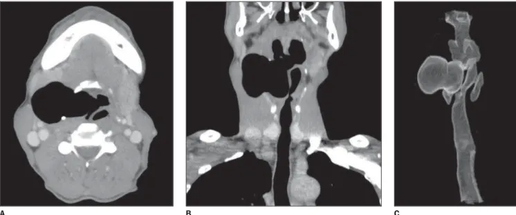
de-sions in the larynx, and a large laryngocele was clearly characterized, with no evidence of an obstructive factor (Figure 5).
The patient was submitted to surgical intervention that confirmed the imaging findings (Figure 6).
DISCUSSION
Laryngoceles etiopathogenesis is still to be determined. However, several theories try to explain the development of the dis-ease, from congenital predisposition to a multifactorial nature. The congenital theory suggests that in some cases, during the normal development of the larynx, between the second and third gestational months, there is an abnormal growth of the saccule (long saccule), and this is a predisposing factor for the future development of laryn-gocele. Other authors suggest that the stress caused by a continuous increased intra-laryngeal pressure leads to the saccule di-latation and herniation. This theory is supported by the fact that laryngoceles are most frequently found in musicians who play wind instruments and in patients with chronic respiratory diseases (“chronic coughers”). Although this is the most ac-cepted theory, some authors postulate that there is an association between congenital
Figure 2. Sonographic images. A: Axial view of a palpable mass showing hypoechogenic formation with an intense sound reverberation on its surface. B: As the transducer is medially moved, it is possible to observe that the mass (M, at left) communicates with the larynx by means of an isthmus (at right). C: Axial image with extended vision showing more clearly the mass topogra-phy, laterally to larynx and medially to the great cervical vessels (right internal jugular vein – VJID; right common carotid artery – ACCD).
C
A B
scribed appearance suggested the presence of air within the lesion. Later in the study, an equally air-filled connection was found between the mass and the larynx at the same level, suggesting the hypothesis of laryngocele.
Plain posteroanterior and lateral radio-graphs have confirmed the hypothesis of external laryngocele at right (Figure 3).
Subsequently, the patient was submitted to laryngoscopy that demonstrated a bulg-ing of the right ventricular recess, without identification of the isthmus communicat-ing with the laryngocele (Figure 4).
A CT study was requested for surgical planning and investigation of mucous
281
External laryngocele: sonographic appearance – a case report
Radiol Bras 2007;40(4):279–282
Figure 3. Plain posteroanterior (A) and lateral (B) radiographs demonstrating an air-filled mass (white arrows) in the right, anterior, paramedian region laterally displacing and compressing the airway (black arrowheads).
B A
Figure 4. Laryngoscopy during inspiration. A little bulging of the vestibular region (white arrows) is noted. The isthmus communicating with the laryn-gocele could not be identified. The analysis of the laryngeal mucosa has not demonstrated neoplas-tic lesions; only an edema in the postcricoid region (arrowheads) is observed, constituting an indirect sign of the presence of gastroesophageal reflux dis-ease in this patient.
predisposition and prolonged exposure to high-pressure within the larynx.
The clinical features of this entity are highly variable and non-specific. Most fre-quently, it may be asymptomatic, and the diagnosis occurs incidentally during imag-ing studies for other reasons or clinical suspicion, for example, staging of laryngeal carcinomas(8,11–13).
In summary, symptoms may be divided into compressive, caused by internal la-ryngoceles, and those symptoms related to
the mass effect, like in cases of external laryngoceles. Main complaints among pa-tients are hoarseness and cough. On the other hand, some patients with external laryngoceles report a palpable cervical mass, and, less frequently, dysphagia and dyspnea, associated or not with a peculiar professional or pathological history. In patients who present with a palpable cer-vical mass associated with inflammation, laryngoceles constitute an important differ-ential diagnosis.
Imaging studies play a significant role in the diagnosis of this lesion, and many authors suggest that CT is the golden stan-dard, with MRI playing an adjuvant role in the diagnosis of laryngoceles.
Ultrasonography is generally utilized for initial evaluation of cervical masses, mainly for differentiating the nature of the lesion as well as defining its contents and location(9,10).
In the present case, the location, the presence of sound reverberation in the
le-A B C
282
Pinho MC et al.
Radiol Bras 2007;40(4):279–282
sion, and the ventricular appendage extend-ing toward the laryngeal wall have sug-gested the diagnosis.
CT, not only suggests the diagnosis, but also may classify the lesion, and directly affect the therapeutic choice. This imaging method allows the visualization of the di-lated air-filled saccule, as well as its limits and anatomical relations. In external laryn-goceles, it may be observed that the hernial sac passes through the thyrohyoid mem-brane, extending superiorly into the para-laryngeal space. Sometimes, a causal fac-tor, for example, a squamous-cell carci-noma located in the opening of the laryn-geal ventricle, may be found (secondary laryngocele). In these cases, the tumor par-tially obstructs the communication between the saccule and the larynx, creating a valve-like mechanism. Also, a cystic lesion with soft-tissue attenuation may be observed, suggesting a diagnosis of laryngomucocele or laryngopyocele, depending on the clini-cal context.
MRI is important, particularly in cases where laryngocele is associated with a lar-ynx squamous-cell carcinoma. This imag-ing method is useful to corroborate the di-agnosis, in staging the tumor, for evaluat-ing the disease extent in soft-tissues, as well as to provide, by means of multiplanar ac-quisition, a better surgical timing planning by otolaryngologists and head and neck surgeons(8,12,14,15).
Few reports describe possible appear-ance of laryngoceles at ultrasonography(8,9).
The commonest presentation is a mass with an intense sound reflection, determining a posterior reverberation artifact, suggesting the presence of air in the paramedian line. Sometimes, it is possible to demonstrate the isthmus between the mass and the lar-ynx so as to raise the diagnostic hypothesis of laryngocele. Main differential diagnoses in these cases, would be Zenker’s diverticu-lum or an air-filled abscess. Additionally, a mass presenting hypo- or non-echogenic contents and posterior acoustic shadowing may be characterized, defining the cystic nature of the lesion. Differential diagnoses of this mass range from thyroglossal duct cyst (median line) to dermoid cyst, cystic hygroma and abscess or lymphadeno-megaly with liquefaction. Both the pa-tient’s clinical condition and age constitute significant factors when these hypotheses are considered.
REFERENCES
1. Swartz JD, D’Angelo AJ, Harnsberger HR, Zwillenberg S, Marlowe FI. The laryngeal muco-cele. Imaging analysis of a rare lesion. Clin Im-aging 1990;14:110–115.
2. Nazaroglu H, Ozates M, Uyar A, Deger E, Simsek M. Laryngopyocele: signs on computed tomog-raphy. Eur J Radiol 2000;33:63–65.
3. Kumar G, Bradley PJ, Wastie ML. Case of the month. What a blow! Laryngocele. Br J Radiol 1998;71:799–800.
4. Abrahão M, Santos RO, Cervantes O. Tratamento dos tumores benignos da laringe. In: Carvalho MB,
organizador. Tratado de cirurgia de cabeça e pes-coço e otorrinolaringologia. São Paulo, SP: Ate-neu, 2001;905.
5. Curtin HD. The larynx. In: Som PM, Curtin HD, editors. Head and neck imaging. 4th ed. St. Louis, MO: Mosby, 2003;1595–1699.
6. Curé J. Laryngocele. In: Hansberger R, Wiggins R, Hudgins P, et al., editors. Diagnostic imaging: head and neck. 1st ed. Salt Lake City, UT: Amyr-sys, 2004;III, 3–6.
7. Sobol SM, Bailey SB. Evaluation and surgical management of tumors of the neck: benign tu-mors. In: Thawley SE, Panje WR, Batsakis JG, Lindberg RD, editors. Comprehensive manage-ment of head and neck tumors. Philadelphia, PA: WB Saunders, 1999;1435–1439.
8. Harvey RT, Ibrahim H, Yousem DM, Weinstein GS. Radiologic findings in a carcinoma-associ-ated laryngocele. Ann Otol Rhinol Laryngol 1996; 105:405–408.
9. Youssefzadeh S, Steiner E, Turetschek K, Gritz-mann N, Kursten R, Franz P. The sonography of laryngeal cysts. Rofo 1993;159:38–42. 10. Heppt W, Born IA, Maier H. Use of B-mode
sono-graphy in the diagnosis of laryngocele. Laryngo-rhinootologie 1990;69:378–380.
11. Helmberger RC, Croker BP, Mancuso AA. Lei-omyosarcoma of the larynx presenting as a laryngopyocele. AJNR Am J Neuroradiol 1996; 17:1112–1114.
12. Glazer HS, Mauro MA, Aronberg DJ, Lee JK, Johnston DE, Sagel SS. Computed tomography of laryngoceles. AJR Am J Roentgenol 1983;140: 549–552.
13. Celin SE, Johnson J, Curtin H, Barnes L. The as-sociation of laryngoceles with squamous cell car-cinoma of the larynx. Laryngoscope 1991;101: 529–536.
14. Ettema SL, Carothers DG, Hoffman HT. Laryn-gocele resection by combined external and endo-scopic laser approach. Ann Otol Rhinol Laryngol 2003;112:361–364.
15. Martinez Devesa P, Ghufoor K, Lloyd S, Howard D. Endoscopic CO2 laser management of laryngo-cele. Laryngoscope 2002;112(8 Pt 1):1426–1430.
Figure 6. A: Intraoperative photo of the patient with Valsalva maneuver to show laryngocele. B: Laryngocele exposed after cervical incision. Note the isthmus communicating with the airway (arrows). C: The laryngocele is open for evaluation of the mucosa; signs of neoplastic lesion are absent.