Radiol Bras. 2014 Jan/Fev;47(1):43–48 43
Scrotal collections: pictorial essay correlating sonographic
with magnetic resonance imaging findings
*
Coleções na bolsa testicular: ensaio iconográfico correlacionando achados ultrassonográficos com a ressonância magnética
Resende DAQP, Souza LRMF, Monteiro IO, Caldas MHS. Scrotal collections: pictorial essay correlating sonographic with magnetic resonance imaging findings. Radiol Bras. 2014 Jan/Fev;47(1):43–48.
Abstract
R e s u m o
The present study is aimed at describing scrotal collections observed at ultrasonography and magnetic resonance imaging. The authors describe the main features of hydrocele, hematocele and pyocele, as well as the most common causes, clinical manifestations and associated diseases, with a brief review of the embryology and anatomy of the scrotum. Collections are frequently found in the evaluation of the scrotum, which is often performed on an emergency basis, and in most cases can be differentiated by means of imaging studies. With the consolidation of magnetic resonance imaging as the method of choice complementary with ultrasonography, the authors also describe magnetic resonance imaging findings of scrotal collections as well as the situations where such method is indicated.
Keywords: Testis; Testicular hydrocele; Hematocele; Abscess; Ultrasonography: Magnetic resonance imaging.
O objetivo deste trabalho é descrever coleções na bolsa testicular vistas na ultrassonografia e na ressonância magnética. São descritas as principais características da hidrocele, hematocele e piocele, assim como as causas mais comuns, manifestações clínicas e doenças associadas, com uma breve revisão da embriologia e anatomia da bolsa testicular. Coleções são achados frequentes na avaliação da bolsa testicular, muitas vezes realizada em caráter de urgência, e podem ser diferenciadas por meio de exames de imagem. Com a consolidação da ressonância magnética como exame de escolha em complemento à ultrassonografia, são também descritas as carac-terísticas das coleções escrotais na ressonância magnética, além das indicações para a sua realização.
Unitermos: Testículo; Hidrocele testicular; Hematocele; Abscesso; Ultrassonografia; Ressonância magnética.
* Study developed at Department of Imaging Diagnosis, Universidade Federal do Triângulo Mineiro (UFTM), Uberaba, MG, and at Central de Diagnóstico Ribeirão Preto (Cedirp), Ribeirão Preto, SP, Brazil.
1. MD, Resident of Radiology and Imaging Diagnosis, Universidade Federal do Triângulo Mineiro (UFTM), Uberaba, MG, Brazil.
2. PhD, Associate Professor, Unit of Radiology and Imaging Diagnosis at Univer-sidade Federal do Triângulo Mineiro (UFTM), Uberaba, MG, Brazil.
3. MD, Trainee Radiologist (Computed Tomography and Magnetic Resonance Im-aging) at Central de Diagnóstico Ribeirão Preto (Cedirp), Ribeirão Preto, SP, Brazil.
4. MD, Trainee in Radiology and Imaging Diagnosis at Central de Diagnóstico Ribeirão Preto (Cedirp), Ribeirão Preto, SP, Brazil.
Mailing Address: Dr. Daniel de Almeida Queiroz Prata Resende. Rua 12, nº 399, ap. 401, Setor Oeste. Goiânia, GO, Brazil, 74140-040. E-mail: dresende72@yahoo. com.br.
Received February 28, 2013. Accepted after revision July 19, 2013.
the scrotum, with excellent spatial resolution, providing al-most 100% sensitivity in the detection and differentiation of intra- and extratesticular diseases. Additionally, it has the ad-vantage of being rapidly performed (which is imprescindible in cases of testicular emergency), is a low-cost method and does not emit ionizing radiation(3).
The technique of scrotal ultrasonography includes lon-gitudinal and cross-sectional B-mode scans and utilization of color Doppler. The latter is essential in the differentia-tion between ischemia (absence of flow) and inflammadifferentia-tion (increased flow)(1), situations which are frequently found in testicular emergencies. Additionally, there are resources such as trapezoidal scanning and extended field of view. Thus, the lesions can be better delimited and the symmetry of the scro-tal contents can be evaluated. Such a technique is useful in the assessment of testicular diseases.
However, in some situations, the sonographic evalua-tion is not conclusive. Magnetic resonance imaging (MRI) has shown to be the complementary method of choice, avoid-ing unnecessary interventions and reducavoid-ing general costs re-lated to the testicular disease(4,5). The basic protocol includes the acquisition of axial, sagittal and coronal, T1- and T2-weighted images, and may include supplementary T1-weighted, gradient-echo images which are useful in the de-tection of hemorrhages(5,6). The utilization of paramagnetic
Daniel de Almeida Queiroz Prata Resende1, Luís Ronan Marquez Ferreira de Souza2, Isabela de Oliveira Monteiro3, Marcel Henrique de Souza Caldas4
INTRODUCTION
Collections represent frequent findings in the assessment of the scrotum, not only in investigations performed in emer-gency situations, but also on an outpatient basis. Such col-lections are caused by different factors ranging from physi-ological events to varied diseases. Such situations should be differentiated and the nature of the collections should be determined when possible. Hydrocele is the most frequently found type of collection, followed by hematocele and, most rarely, pyocele(1).
The scrotum originates from genital protuberances which, under the influence of testosterone, dilate and fuse to form two testicular cavities, also called scrotum. The vagi-nal process starts developing at about the eighth week of fetal development, corresponding to an evagination of the pari-etal peritoneum, which extends caudally through the abdomi-nal wall into the testicular cavities(3). Through the vaginal process, the testis moves down from the abdomen into the scrotum between the 7th and 9th months of the fetal life. Once the testes have descended, the vaginal process is oblit-erated, and its scrotal portion remains as a cavity, the tunica vaginalis. Failure of the testes to descend and patency or incomplete closure of the vaginal process may result in cryp-torchidism, inguinoscrotal hernia and hydrocele(1).
The tunica vaginalis involves the testis, with exception of its posterior aspect, and is constituted by a visceral
por-Hydrocele
It is an abnormal collection of serous fluid located be-tween the visceral and parietal layers of the tunica vaginalis, but also may be adjacent to the spermatic cord. At US, the hydrocele fluid is most commonly anechoic (Figure 1), and may present subtle echoes or fibrin septa(2) (Figure 2). In cases of chronic collections, findings of thickened walls and development of calculi may be added(7).
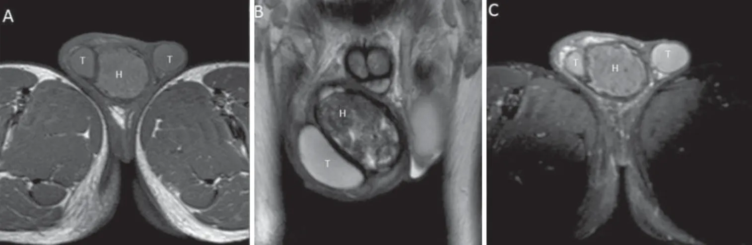
At MRI, hydroceles present the typical behavior of liq-uids, with homogeneously low signal on T1-and high signal on T2-weighted sequences (Figure 3). Septations and cal-culi may also be identified.
In children, hydrocele is the most common cause of painless scrotal edema(1), but it may be associated with pain or diffuse discomfort(7). Theoretically, in neonates, all hydroceles are congenital and associated with a patent
vagi-Figure 1. B-mode US image with ex-tended field of view in a neonate: homo-geneous hydrocele at right, and usual aspect of the left scrotum. Observe that the collections does not extend toward the posterior region of the testis.
Figure 2. B-mode US image. A and B
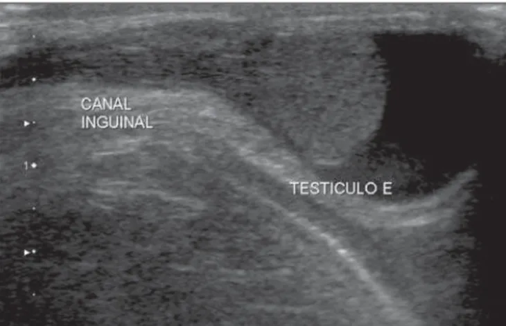
nal process that allows the passage of peritoneal fluid into the scrotum (Figure 4). About 80% of congenital hydroceles resolve in up to two years. The vaginal process closure above the testis and below the inner inguinal ring leads to a less common type of hydrocele, that is also known as spermatic cord cyst, which is seen as a fluid collection along the cord (Figure 5).
Abdominoscrotal hydrocele is extremely uncommon, with about 80 reported cases. It is described as a large collection protruding through the inner inguinal ring by means of an unknown mechanism and is clinically manifested as an abdominoscrotal communicating mass. Both abdomino-scrotal hydroceles and spermatic cord cysts require surgical treatment.
In older children, adolescents and adults, hydrocele is usually acquired and related to inflammatory processes, tes-ticular torsion, trauma, tumor(1), or may be idiopathic. The idiopathic hydrocele mechanism is still unknown, but it is believed that it results either from an imbalance between the fluid production and reabsorption, or from the absence of efferent lymphatics(7).
In cases of testicular torsion, the presence of hydrocele may demonstrate a failure in the fixation of the testis to the scrotal wall – bell-clapper deformity (Figure 6) –, which allows free movement of the testis within the scrotum, thus increasing the chance of torsion. Such a condition is rather associated with testicular torsion in the peripuberal period (the other peak of testicular torsion incidence occurs in the perinatal period)(2).
Some differential diagnoses must be taken into consid-eration, namely, indirect inguinal hernia, where the presence of fluid and gas involved by the bowel loop wall is observed Figure 3. Hydrocele secondary to extratesticular leiomyoma. B-mode US image of right scrotum (A): hydrocele (asterisk) adjacent to the leiomyoma (L). B,C,D: Magnetic resonance imaging demonstrating the hydrocele behavior at different weightings, with low signal intensity on T1-weighted (B) as compared with high signal on T2-weighted image (C) and absence of enhancement on T1-weighted image after contrast agent injection (D). Observe the physiological amount of fluid within the left scrotum.
Figure 4. Congenital hydrocele. B-mode US image of an one-month-old infant: incomplete left testis descent (the testis is near the distal orifice of the inguinal canal) in association with hydrocele.
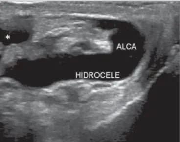
Figure 5. B-mode US with extended Field of view: spermatic cord cyst in asso-ciation with rete testis ectasia (arrow) and adjacent physiological amount of fluid.
Figure 7. Inguinal hernia associated with hydrocele. B-mode US showing an ir-regular mass with thickened walls, containing a small amount of fluid, and sur-rounded by free fluid with anechoic appearance.
Figure 8. Spermatocele. B-mode US image: cyst with slightly lobulated margins, anechoic contents, with small fluctuating echoes (spermatozoids), located in the epidydimis. Observe the location and delimitation of the lesion.
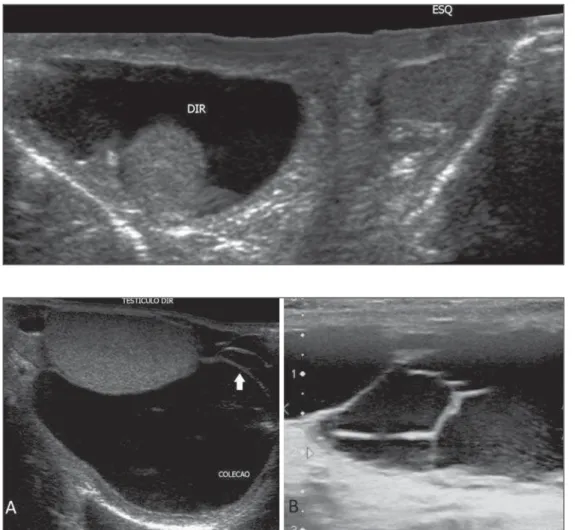
ance(5) with complex septations(7), fluid-fluid level and subtle echoes(8) (Figure 10). US had 87% specificity and 89% sen-sitivity in the diagnosis of hematocele as compared with in-traoperative findings(2).
MRI findings follow the pattern of methemoglobin deg-radation in other tissues (Table 1). The T2-weighted images may have a variable appearance, and chronic hematomas may present with a hyposignal halo secondary to hemosiderin deposition(5) (Figure 11). Magnetic resonance imaging is also important in the detection of tunica albuginea rupture, which indicates surgical intervention in cases of testicular trauma(4).
Figure 9. B-mode US image. Acute hematocele secondary to trauma, with an echogenic fluid collection within the left scrotum.
The main causes of hematocele include trauma, tumor, torsion and surgery(3). In neonates, such condition may be secondary to intra- and extraperitoneal abdominal bleedings. In such age range, the finding of non-traumatic hematocele should lead to the investigation of abdominal foci, since there are reports in the literature about hematocele secondary to adrenal hemorrhage(10,11).
Testicular trauma is the most common cause of hema-tocele and the third most common cause of acute scrotal pain. The right testis is most frequently affected, probably because of its location slightly above the left testis in most men, which facilitates its compression against the pelvis(8). It may be caused by intra- or extratesticular bleeding and not rarely hematocele and intratesticular hematoma are found on a single study; and for this reason it is nonspecific for testicu-lar rupture(9).
In the pediatric age range, hematocele may also be sec-ondary to abdominal traumas(8). A subtle trauma may go un-noticed and, in such cases, the bleeding is frequently associ-ated with varicoceles, with rupture of a dilassoci-ated vessel(3).
The acute onset of a voluminous hematocele may reduce the testicular blood flow due extrinsic vessels compression, mimicking a partial or even complete torsion, requiring drainage of the collection(8). Additionally, large collections make the tunica albuginea identification more difficult at US and may lead to false-negative diagnoses of intratesticular hematoma where, in fact, hematocele is present(2). The iden-tification of an intact tunica albuginea allows for ruling out the presence of testicular rupture(8).
Table 1—Phases of hemoglobin degradation and corresponding signal at MRI.
Phase
Oxyhemoglobin Deoxyhemoglobin Intracellular methemoglobin Extracellular methemoglobin
T1-weighted signal
Low Low High High
T2-weighted signal
High Very low
LOw High
Figure 12. B-mode US image. Collection with multiple, incomplete septa inside, in a patient with hyperemia and pain in the scrotum.
Pyocele
Most frequently, pyocele occurs as a complication of orchiepidydimitis, particularly in cases where the latter crosses the mesothelial layer of the tunica vaginalis(3), or secondarily to a reactive infected hydrocele(6). The patients present with acutely swollen and painful scrotum, frequently in association with fever and leukocytosis.
tion of the transducer – a finding that is described as “fall-ing snow” sign(12).
Considering that in most cases pyocele is secondary to testicular inflammatory processes, the diagnosis depends on the recognition of such processes. Typically, at MRI, abscesses present hyposignal on T1- and hypersignal on T2-weighted sequences, which is characteristic of fluid contents, with a halo of hypersignal on T2-weighted sequences. On contrast-enhanced images, only the perilesional parenchyma presents with intense enhancement (Figure 13).
Possible complications include: Fournier’s gangrene, ne-crotizing and potentially fatal perineal infection. Frequently, patients with scrotal abscess have a history of diabetes, HIV infections or other immunosuppressive conditions. Anaerobic and gas-producing bacteria, including those of the Clostridium genus are the main etiological agents of such a condition(3).
CONCLUSION
US is the first method of choice in the assessment of the scrotum, frequently demonstrating collections. The present essay was aimed at demonstrating imaging findings which allows for the differentiation of hydrocele, hemato-cele and pyohemato-cele, besides describing common causes and clinical characteristics which may be associated with such findings. It is important to highlight that, in some situations, sonographic findings alone do not allow the distinction of the material contained in the collection, so the clinical his-tory and physical examination of the patient play a relevant role in the final diagnosis. History and time span from the trauma, previous surgeries and comorbidities must not be neglected. As the wide utilization of US is added to the high incidence of scrotal collections, it is important to be famil-iar with the characteristics of each collection.
With the MRI consolidation as a method to solve doubts in the diagnosis of testicular diseases, it is important that radiologists are aware of the signal characteristics which allow the differentiation of each disease.
REFERENCES
1. Aso C, Enríquez G, Fité M, et al. Gray-scale and color Doppler sonography of scrotal disorders in children: an update. Radiographics. 2005;25:1197–214.
2. Guichard G, El Ammari J, Del Coro C, et al. Accuracy of ultra-sonography in diagnosis of testicular rupture after blunt scrotal trauma. Urology. 2008;71:52–6.
3. Woodward PJ, Schwab CM, Sesterhenn IA. From the archives of the AFIP: Extratesticular scrotal masses: radiologic-pathologic cor-relation. Radiographics. 2003;23:215–40.
4. Parenti GC, Feletti F, Brandini F, et al. Imaging of the scrotum: role of MRI. Radiol Med. 2009;114:414–24.
5. Kim W, Rosen MA, Langer JE, et al. US MR imaging correlation in pathologic conditions of the scrotum. Radiographics. 2007;27:1239– 53.
6. Cassidy FH, Ishioka KM, McMahon CJ, et al. MR imaging of scro-tal tumors and pseudotumors. Radiographics. 2010;30:665–83. 7. Schul MW, Keating MA. The acute pediatric scrotum. J Emerg Med.
1993;11:565–77.
8. Bhatt S, Dogra VS. Role of US in testicular and scrotal trauma. Radiographics. 2008;28:1617–29.
9. Vital RJ, Mattos LA, Souza LRMF, et al. Aspectos ultra-sonográfi-cos das alterações não-neoplásicas do testículo. Radiol Bras. 2007;40: 61–7.
10. Gonçalves R, Abuabara A, Abuabara RF, et al. Scrotal hematoma as a sign of adrenal hemorrhage in newborns. Sao Paulo Med J. 2011; 129:113–5.
11. Ibáñez Godoy I, Mora Navarro D, Delgado Rioja MA, et al. He-matoma escrotal unilateral. An Pediatr (Barc). 2004;60:477–8. 12. Mahmood NS, Suresh HB. Role of Doppler sonography in
uncov-ering the testis within a pyocele: the “falling snow” sign. J Ultra-sound Med. 2009;28:557.