LETTER TO THE EDITOR
Anomalous origin of left coronary artery diagnosed
by magnetic resonance imaging
Ricardo Oliveira Falca˜o,IMarcelo Souto Nacif,II,III,IVSongtao Liu,III David A. Bluemke,III Carlos Eduardo Rochitte,V Edson MarchioriII
IPlani Diagno´sticos Me´dicos, Sa˜o Jose´ dos Campos, SP, Brazil.IIRadiology and Imaging Diagnosis, Federal Fluminense University, Federal University of Rio
de Janeiro, Rio de Janeiro, Brazil.IIIDepartment of Radiology and Imaging Sciences, National Institutes of Health Clinical Center, Bethesda, MD, USA. IVCardiology Department, Johns Hopkins School of Medicine, Baltimore, MD, USA.VDepartment of Cardiovascular MRI and CT, Instituto do Corac¸a˜o
(InCor), Hospital das Clinicas, Faculdade de Medicina, Universidade de Sa˜o Paulo, Sa˜o Paulo, Brasil.
Email: [email protected] Tel.: 1 301-4351481
Coronary arteries normally arise from the sinuses of Valsalva on the ascending aorta. The incidence of anom-alous origin of the left coronary artery from the trunk of the pulmonary artery is about 1 in 300,000 live births.1
The clinical course of patients with this anomaly, which includes heart failure early in life, depends on either the development of coronary collaterals after birth2or invasive correction.3–4 Here, we report a case of a five-year-old female with exertional dyspnea and changes in her electro-cardiographic examination who was referred for magnetic resonance imaging (MRI).
CASE PRESENTATION
The patient was a five-year-old Caucasian, female, born at term with no history of complications during labor. In the fourth month of life, she developed prolonged crying during feedings. A constitutional growth delay was dis-covered at her first consultation with her pediatrician. In her seventh month of life, the child exhibited mild dyspnea and cyanosis during feeding. The patient presented clinically with delayed physical development and was always below the growth curve at her regular consultations. Nevertheless, there were no suspected abnormalities based on her examinations until the age of five. Symptoms of heart failure were detected when the patient began school; she suffered from constant fatigue, and her dyspnea was increased and occurred earlier than her colleagues during physical exercise. The child underwent a clinical examina-tion by a cardiologist, and the results of the electrocardio-gram (EKG) suggested a change in heart vascularization.
The patient was referred for an MRI using a 1.5-Tesla Achieva system (Philips Medical Systems, Best, The Netherlands) with a five-element cardiac phase-array coil. The study was performed under anesthesia with gadoli-nium contrast (0.2 mmol/kg). The multiplanar images (Figure 1) obtained from the cardiac MRI synchronized to the ECG during full expiration and the magnetic resonance angiography of the coronary arteries (Figure 2) were
analyzed and processed in using an Extended MR Work-Space 5.2 workstation (Philips Healthcare, Best, The Netherlands).
DISCUSSION
The rare anomaly of the origin of the left coronary artery1 from the trunk of the pulmonary artery (ALCAPA -Anomalous Left Coronary Artery from Pulmonary Artery) is often referred to as Bland-White-Garland syndrome. The estimated incidence of Bland-White-Garland syndrome is 1/300,000 live births and represents 0.5% of congenital heart disease cases. During fetal life, this anomaly is tolerated because there is no difference in blood pressure between systemic and pulmonary circulation, nor is there a differ-ence in the oxygen gradient between the aorta and the pulmonary artery6. The presence of symptoms depends on the type of collateral circulation between the right and left
Copyrightß2010CLINICS– This is an Open Access article distributed under the terms of the Creative Commons Attribution Non-Commercial License (http:// creativecommons.org/licenses/by-nc/3.0/) which permits unrestricted non-commercial use, distribution, and reproduction in any medium, provided the original work is properly cited.
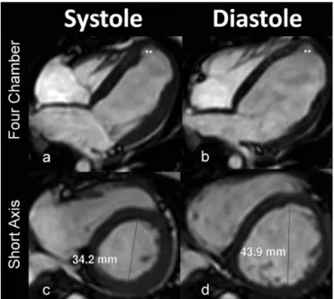
Figure 1 - Magnetic resonance images taken during cardiac systole and end diastole. The images above the plane show four chambers with aneurysmal formation of the left ventricle, and the images below demonstrate ventricular dilation.
CLINICS 2010;65(11):1215-1216 DOI:10.1590/S1807-59322010001100027
coronary arteries. Development of these collaterals may be crucial for the onset of myocardial ischemia.
The anomaly can be classified according to collateral pattern. When collaterals are present it is called the ‘‘adult type’’, and the ‘‘infantile type’’ occurs when there are no collaterals.7 Patients usually show signs and symptoms of congestive heart failure due to ischemic heart disease and myocardial infarcted segments.1,8,9
The infantile form has a potential mortality of about 80– 90%, even with medical treatment, and exhibits clinical symptoms of myocardial ischemia, such as sweating, irritability and fatigue.10 The onset of symptoms often occurs around the eighth week of life. An important differ-ential diagnosis in adults is dilated cardiomyopathy.5,10
In the adult form, patients usually have significant collateral circulation between the right and left coronary arteries; however, the amount of blood perfusion through the collaterals is not adequate, especially in the subendo-cardial region. Therefore, the disease manifests as chronic ischemia, which can result in a malignant ventricular arrhythmia and a risk of sudden death in 80–90% of cases. Patients with this form of the anomaly can also be asymptomatic.2
Coronary artery angiography can characterize changes in the origin, course and morphology of the coronary tree. It is invasive and has a low, but present, risk of complications. MRI is a noninvasive method that does not require radiation and can demonstrate the origin of the coronary arteries and assess the degree of valvular insufficiency, ventricular function, regional contractility and myocardial viability, all
of which are important considerations during the preopera-tive evaluation and postoperapreopera-tive follow up.7,9
Surgery is the proposed treatment for both forms of ALCAPA.3,4The Takeuchi procedure is a technique used to correct the infantile form of this syndrome, which consists of creating a communication between the aorta and the left coronary ostium, through the pulmonary artery, using tubular material (graft).7 Usually, this technique is
per-formed when direct implantation of the anomalous artery into the aorta is difficult due to unfavorable conditions. In the adult form, ligation of the origin of the coronary artery at the pulmonary artery is performed in a combined manner so that flow is either restored or persists through a con-nection with either the internal thoracic artery or a saphe-nous vein graft from the ascending aorta.11
Final considerations: The patient described in this case report underwent surgical ligation of the origin of the left coronary artery, and flow was restored by a graft to the left internal mammary artery. The patient had a good recovery in the early postoperative period and was discharged with her cardiac function under control.
All authors were involved in the conception and design of this manuscript, the drafting the manuscript, critical revisions of the manuscript for intellectual content, and final approval. Marcelo Souto Nacif was involved in the data acquisition.
REFERENCES
1. Keith JD. The anomalous origin of the left coronary artery from the pulmonary artery. Br Heart J. 1959;21:149-61, doi: 10.1136/hrt.21.2.149. 2. Wesselhoft H, Fawcet JS, Johnson AL. Anomalous origin of the left
coronary from the pulmonary trunk: its clinical spectrum, patology and pathophysiology, based on a review of 140 cases with seven further cases. Circulation. 1968;38:403-25.
3. Saeed BT, Rosin MD, Murray RG. Successful operation in an old survivor of anomalous origin of the left coronary artery from the pulmonary trunk (Bland-White-Garland syndrome). Br Heart J. 1994;71:193-5, doi: 10. 1136/hrt.71.2.193.
4. Moodie DS, Fyfe D, Gill CC, Cook SA, Lytle BW, Taylor PC, et al. Anomalous origin of the left coronary artery from the pulmonary artery (Bland-White-Garland syndrome) in adult patients: long term follow-up after surgery. Am Heart J. 1983;106:381-8, doi: 10.1016/0002-8703(83)90207-7.
5. Perloff JK. Anomalous origin f the left coronary artery from the pulmonary trunk. In the clinical recognition of congenital heart disease. 4th edition. Edited by: Perloff JK. Philadelphia: WB Saunders Co. 1984:546-561.
6. Werzmann M, Salehian O, Elliot T, Merchant N, Siu SC, Webb GD. Anomalous origin of the left coronary artery from the main pulmonary artery in adults: coronary collateralization at its best. Circulation 2004;110;e511-e513.
7. Barbetakis N, Efstathiou A, Efstathiou N, Papagiannopoulou P, Soulountsi V, Fessatidis I. A long-term survivor of Bland-White-Garland syndrome with systemic collateral supply: case report and review of the literature. BMC Surg. 2005;5:23, doi: 10.1186/1471-2482-5-23.
8. Askenazi J, Nadas AS. Anomalous left coronary artery originating from the pulmonary artery. Circulation. 1975;51:976-87.
9. Arciniegas E, Farooki ZQ, Hakimi M, Green EW. Management of anomalous left coronary artery from the pulmonary artery. Circulation. 1980;62(Suppl 1):180-9.
10. Takimura CH, Nakamoto A, Hotta VT, Campos MF, Ma´lamo M, Otsubo R. Origem anoˆmala da corona´ria esquerda no tronco pulmonar. Relato de um caso em adulto. Arq Bras Cardiol 2002;78:309-11.
11. Dodge-Khatami A, Mavroudis C, Backer CL. Anomalous origin of the left coronary artery from the pulmonary artery: collective review of surgical therapy. Ann Thorac Surg. 2002;74:946–55, doi: 10.1016/S0003-4975(02)03633-0.
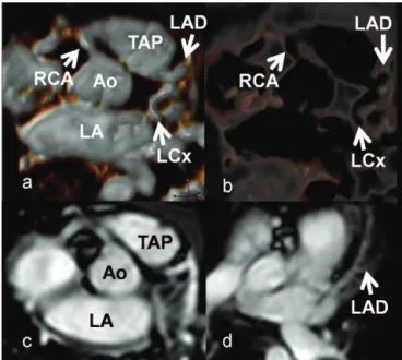
Figure 2 - Magnetic resonance angiography of the coronary arteries with 3D reconstructions showing the ostia of the coronary arteries. Note that the left coronary artery has its origin in the pulmonary trunk (TAP – trunk of pulmonary artery, Ao - ascending aorta, LA - left atrium, Cx - circumflex coronary artery, DA - left anterior descending coronary artery, CD - right coronary artery).
ALCAPA by cardiac MRI
Falca˜o RO et al. CLINICS 2010;65(11):1215-1216