Department of Orthopedics and Traumatology, Clinics Hospital, Faculty of Medicine, University of Sao Paulo/ SP, Brazil.
Email:zumiotti@hcnet.usp.br
Received for publication on February 26, 2006. Accepted for publication on May 10, 2006.
ORIGINAL RESEARCH
COMPARATIVE MULTICENTER STUDY OF
TREATMENT OF MULTI-FRAGMENTED TIBIAL
DIAPHYSEAL FRACTURES WITH NONREAMED
INTERLOCKING NAILS AND WITH BRIDGING PLATES
Hélio Jorge Alvachian Fernandes, Marcos Hideyo Sakaki Jorge dos Santos Silva, Fernando Baldy dos Reis, Arnaldo Valdir Zumiotti
Fernandes HJA, Sakaki MH, Silva JS, Reis FB, Zumiotti AV. Comparative multicenter study of treatment of multi-fragmented tibial diaphyseal fractures with nonreamed interlocking nails and with bridging plates. Clinics. 2006;61(4):333-38.
OBJECTIVE: A prospective, randomized study to compare patients with closed, multi-fragmented tibial diaphyseal fractures treated using one of two fixation methods undertaken during minimally invasive surgery: nonreamed interlocking intramedullary nails or bridging plates.
MATERIALS AND METHODS: Forty-five patients were studied; 22 patients were treated with bridging plates, 23 with interlocking nails without reaming. All fractures were Type B and C (according to the AO classification).
RESULTS: Clinical and radiographic healing occurred in all cases. No cases of infection occurred. The healing time for patients who received nails was longer (4.32 weeks on average) than the healing time for those who received plates (P = 0.026). No
significant differences were observed between the two methods regarding ankle mobility for patients in the two groups.
CONCLUSIONS: The healing time was shorter with the bridging plate technique, although no significant functional differences were found.
KEY WORDS: Intramedullary fixation of fractures. Tibial fractures. Bridging plates. Diaphysis. Comminuted fractures.
INTRODUCTION
Most tibial fractures occur in the long bones of adults. A review of the literature did not provide papers that were appropriate to reach a decision about the best management method.1 Usually, low-energy fractures are not treated
sur-gically, but rather by bloodless reduction and plastered im-mobilization, while for fractures secondary to high-energy traumas, a trend exists towards using the surgical approach. With the purpose of comparing surgical methods of treatment, a prospective, randomized, multicenter study on multi-fragmented tibial diaphyseal fractures was conducted
in 2 institutions, namely the Departments of Orthopedics of the University of São Paulo and of the Federal Univer-sity of São Paulo.
MATERIALS AND METHODS
The inclusion criteria for this study were patients with closed, multi-fragmented tibial diaphyseal fractures. Threshold cases of very proximal or distal fractures were excluded using the square rule2. A protocol was developed
Forty-five patients were studied, with 22 patients receiv-ing bridgreceiv-ing plates and 23 receivreceiv-ing interlockreceiv-ing nails, and with the location of treatment as follows: 26 patients (13 plates, 13 nails) at the Federal University of São Paulo, and 19 patients (9 plates, 10 nails) at the Institute of Ortho-pedics and Traumatology, Faculty of Medicine, University of Sao Paulo, SP (Brazil). The study period was from Janu-ary 2002 to June 2003. The follow-up period varied be-tween 6 months to 1 year. This protocol was approved by the Ethics Committees of the participant institutions and signed informed consent obtained from each included pa-tient.
The surgical technique consisted of indirect reduction for both forms of osteosynthesis without violation of the fracture focus. The blocked intramedullary nails used were AO universal steel nails introduced with no medullary chan-nel reaming. We used a narrow plate for large fragments (4.5 mm AO).
The average values for age, time from accident to sur-gery, and follow-up period were, respectively, 34 years, 23 days, and 51 weeks for the group treated with intramedul-lary nails, and 34 years, 34 days, and 32 weeks for the group treated with bridging plates.
We used Student’s t test for independent samples3 to
compare the numerical measurements of patients for these preliminary conditions
In the group treated with intramedullary nails, 4 patients were women and 19 were men, while in the group treated with a bridging plate, 3 were women and 19 were men.
Eleven individuals were smokers and 12 were nonsmok-ers in the group treated with nails, while 13 were smoknonsmok-ers and 7 were nonsmokers in the group treated with plates.
Tibial fracture was accompanied by fibular fracture in 18 patients of the group treated with nails and in 10 pa-tients of the group treated with plates; 5 and 12 papa-tients, respectively, presented with intact fibulae.
We used the chi-square test or Fisher’s exact test (as required for each case2) to compare groups according to
categorical variables.
RESULTS
The results obtained regarding healing time as well as complications such as angulation, shortening, infection, healing delay, pseudoarthrosis, and ankle mobility are pre-sented in Tables 4 and 5 for patients who received bridg-ing plates and blocked intramedullary nails, respectively.
The results of the statistical analysis of cases are shown in Table 6.
The groups were homogeneous concerning age, and on average, they had the same time from accident to surgery, while patients who received nails had significantly longer follow-up periods.
The groups were not different regarding sex or smoking. More isolated fractures were found among the individuals who received nails as compared with those who received plates.
The healing time for patients who received nails was sig-nificantly longer, by an average of 4.30 weeks, than the heal-ing time for those who received bridgheal-ing plates (P = 0.019,
Student’s t test applied for independent samples) (Table 7).
There were no significant differences when comparing the two groups for the following parameters: infection, healing delay (P = 0.109, Fisher’s test), pseudoarthrosis,
angulation > 10 degrees, shortening > 1 cm, and ankle mo-bility (P = 0.243, Fisher’s test).
DISCUSSION
According to the AO classification, multi-fragmented
Table 3 - Distribution of patients by group and fracture site
Group
Site Nail Plate Total
Distal 9 4 13
Medial 13 13 26
Medial/Distal 1 3 4
Proximal 0 1 1
Proximal/Medial/Distal 0 1 1
Total 23 22 45
Table 2 - Distribution of patients per group and according to the AO classification
Group
AO Nail Plate Total
B1 6 1 7
B2 7 10 17
B3 5 5 10
C1 1 0 1
C2 3 4 7
C3 1 2 3
Total 23 22 45
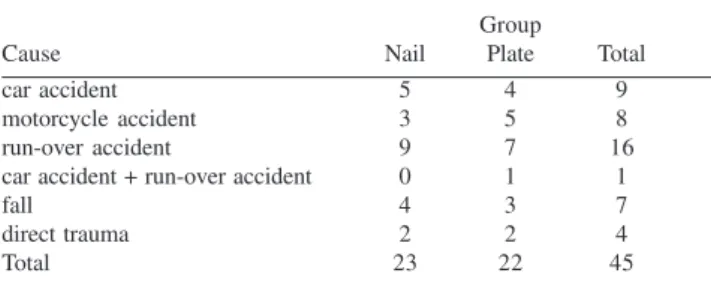
Table 1 - Distribution of patients per group and cause of fracture
Group
Cause Nail Plate Total
car accident 5 4 9
motorcycle accident 3 5 8
run-over accident 9 7 16
car accident + run-over accident 0 1 1
fall 4 3 7
direct trauma 2 2 4
tibial diaphyseal fractures are named 42 (4 for tibia; 2 for diaphysis) and subdivided into B and C. Type B fractures present contact between the proximal and distal fragments after reduction, while Type C fractures are more fragmented
and do not show this contact. The statistical analysis showed that both groups (nails and plates) were homoge-neous concerning age, time from accident to surgery, sex, and smoking; the only difference was the increased
inci-Table 5 - Individual results for patients undergoing surgical treatment with intramedullary nails: ordinal number, healing time, infection, healing delay, pseudoarthrosis, angulation, shortening, and ankle mobility
Ordinal number Healing time Infection Healing Pseudo- Angulation Shortening Ankle
(weeks) delay arthrosis > 10º > 1 cm mobility
1 40 no yes no no no complete
2 20 no no no no no complete
3 36 no yes no no no complete
4 14 no no no no no complete
5 14 no no no no no complete
6 16 no no no no no complete
7 26 no yes no no no incomplete
8 32 no yes no no no complete
9 22 no no no no no complete
10 16 no no no no no complete
11 16 no no no no no incomplete
12 14 no no no no no complete
13 18 no no no no no complete
14 18 no no no no no complete
15 20 no no no no no complete
16 20 no no no no no complete
17 16 no no no no no complete
18 13 no no no no no complete
19 14 no no no no no complete
20 15 no no no no no complete
21 15 no no no no no complete
22 32 no no no no no complete
23 20 no no no no no complete
Sources: Sao Paulo Hospital (EPM/UNIFESP) and Institute of Orthopedics and Traumatology (USP)
Table 4 - Individual results for patients undergoing a surgical treatment with bridging plates: ordinal number, healing time, infection, healing delay, pseudoarthrosis, angulation, shortening, and ankle mobility
Ordinal Healing time Infection Healing Pseudo- Angulation Shortening Ankle
number (weeks) delay arthrosis > 10º > 1 cm mobility
1 20 no no no no no incomplete
2 16 no no no no no complete
3 15 no no no no no complete
4 22 no no no no no incomplete
5 13 no no no no no complete
6 22 no no no no no complete
7 16 no no no no no incomplete
8 18 no no no no no complete
9 13 no no no no no complete
10 13 no no no no no complete
11 13 no no no no no complete
12 13 no no no no no complete
13 14 no no no no no complete
14 13 no no no no no complete
15 16 no no no no no incomplete
16 14 no no no no no complete
17 14 no no no no no complete
18 15 no no no no no complete
19 13 no no no no no complete
20 18 no no no no no incomplete
21 20 no no no no no complete
22 21 no no no no no complete
dence of the presence of an associated fibular fracture in the nails group.
This study was designed to compare the efficacy of treatment of closed, multi-fragmented tibial diaphyseal fractures with nonreamed interlocking intramedullary nails and with bridging plates. In both types of osteosynthesis, the aim was to apply the principle of fixation with relative stability. By using this principle for the fractures, the formation ratio is better tolerated and leads to a lower de-gree of implant loading. In both cases, healing is favored by the nonviolation of the fracture focus, the formation of a secondary bone callus being expected.
For multi-fragmented fractures, surgical treatment us-ing the open reduction technique may compromise the blood supply and lead to a healing disorder. Wide surgical exposure is required to achieve anatomical reduction and fixation with the absolute stability principle; however, in
multi-fragmented fractures, this strategy can lead to a se-vere disturbance of the blood supply.2,4 For this reason, open
reduction surgery is reserved only for single-trace diaphy-seal fractures, where direct or primary healing is expected. It should be emphasized that the deformation ratio in such cases is less tolerated, and small technical inaccuracies greatly increase the loading of the implant and can lead to nonhealing and, therefore, to a defective osteosynthesis.
Many authors consider blocked intramedullary nails the implant of choice in tibial diaphyseal fractures.5-10 This was
the main argument and also the main difficulty in this study, since most staff at the various Orthopedics Services who were contacted were not willing to participate in this study, alleging that intramedullary nails was the standard treat-ment for these fractures. Thus, only 3 institutions partici-pated in this study, although one of them did not present enough data for inclusion in the protocol.
Bridging plates are used more often in diaphyseal frac-tures that compromise the proximal and distal ends of the tibia11-15; however, in this study this aspect was not
consid-ered, since patients were randomly allocated. The place-ment of the plate on the anterior-medial tibial face is tech-nically easier and leads to less compromise of its vascu-larization.16
We compared similar groups and observed that the clini-cal and radiologiclini-cal parameters analyzed, such as articu-lar function, deformities, infection, and pseudarthrosis, were similar in both groups (Tables 4 and 5). The healing time was the only significant difference found. On aver-age, bone healing in patients receiving bridging plates oc-curred 4 weeks earlier compared with patients who received nonreamed interlocking intramedullary nails (Table 7).
We can conclude that the healing times were signifi-cantly shorter in patients undergoing surgery with the bridg-ing plate technique, and the functional results were not dif-ferent among patients of both groups.
Table 7 - Descriptive measurements of patients’ healing time per group
Group
Measurement Nail Plate p*
Mean 20.30 16.00 0.019
Std. deviation 7.68 3.19
Minimum 13.00 13.00
Maximum 40.00 22.00
Asymmetry 1.41 0.78
Curtosis 1.04 -0.81
*student's t test for healing time
Table 6 - Results of comparison between groups
Variable P Test used
Age 0.949 Student’s t test
Time from accident to surgery 0.230 Student’s t test
Follow-up period 0.003 Student’s t test
Sex 0.999 Fisher’s exact test
Smoking 0.258 Chi-square test
Single fracture 0.023 Chi-square test
RESUMO
Fernandes HJA, Sakaki MH, Silva JS, Reis FB, Zumiotti AV. Estudo multicêntrico comparativo do tratamento de fraturas diafisárias multifragmentárias de tíbia com hastes bloqueadas não-fresadas e placas em ponte. Clinics. 2006;61(4):333-38.
OBJETIVOS: Estudo prospectivo e randomizado
comparou pacientes com fraturas diafisárias multifrag-mentárias fechadas de tíbia, tratados com dois métodos de fixação: hastes intramedulares bloqueadas não-fresadas e placas em ponte.
RESULTADOS: A consolidação clínica e radiográfica ocorreu em todos os casos. Não houve caso de infecção. Verificou-se que o tempo de consolidação dos pacientes que receberam haste foi maior (em média 4,32 semanas) do que o tempo de consolidação daqueles que receberam placa (p = 0,026). Não foram observadas diferenças estatisticamente significantes entre os dois métodos no tocante à mobilidade
do tornozelo nos pacientes dos dois grupos.
CONCLUSÕES:O tempo de consolidação foi menor com uso de placas em ponte, porém sem diferenças funcionais significantes.
UNITERMOS: Fixação intramedular de fraturas. Fraturas da tíbia. Placas Ósseas. Diáfises. Fraturas cominutivas.
REFERENCES
1. Littenberg B, Weinstein LP, McCarren M, Mead T, Swiontkowski MF, Rudicel SA, et al. Closed fractures of the tibial shaft. A meta-analysis of three methods of treatment. J Bone Joint Surg Am. 1998;80:174-83. 2. Müller ME, Allgöwer M, Schneider R, Willenegger H. Manual de
osteossíntese. 3rd ed. Sao Paulo: Manole; 1993.
3. Altman D. Practical statistics for medical research. Chapman and Hall: London; 1997.
4. Perren SM, Claes L. Biology and biomechanics in fracture management. In: Rüedi TP, Murphy WM, editors. AO principles of fracture management. New York: Thieme; 2000. p 7-31.
5. Alho A, Ekeland A, Strömsoe K, Folleras G, Thöresen BO. Locked intramedullary nailing for displaced tibial shaft fractures. J Bone Joint Surg Am. 1990;72:805-9.
6. Court-Brown C, Christie J, McQueen M. Closed intramedullary tibial nailing. Its use in closed and type I open fractures. J Bone Joint Surg Br. 1990;72:605-11.
7. Court-Brown CM, Gustilo T, Shaw AD. Knee pain after intramedullary tibial nailing: its incidence, etiology, and outcome. J Orthop Trauma. 1997;11:103-15.
9. Tornetta P 3rd, Bergman M, Watnik N, Berkowitz G, Steuer J. Treatment of grade-IIIB open tibial fractures. A prospective randomised comparison of external fixation and non-reamed locked nailing. J Bone Joint Surg Br. 1994;76:13-9.
10. Angliss R, Tran A, Edwards E, Doig SG. Unreamed nailing of tibial shaft fractures in the multiply injured patients. Injury. 1996;27:255-60. 11. Redfern D, Syed S, Davies S. Fractures of the distal tibia: minimally
invasive plate osteosynthesis. Injury. 2004;35:615-20.
12. Helfet D, Suk M. Minimally invasive percutaneous plate osteosynthesis of fractures of the distal tibia. Instr Course Lect. 2004;53:471-5.
13. Oh C, Kyung H, Park IH, Kim PT, Ihn JC. Distal tibia metaphyseal fractures treated by percutaneous plate osteosynthesis. Clin Orthop. 2003;408:286-9.
14. Cole P, Zlowodzki M, Kregor P. Less Invasive Stabilization System (LISS) for fractures of the proximal tibia: indications, surgical technique and preliminary results of the UMC Clinical Trial. Injury. 2003;34:16-29.
15. Stannard J, Wilson T, Volgas D, Alonso JE. Fracture stabilization of proximal tibial fractures with the proximal tibial LISS: early experience in Bimingham, Alabama (USA). Injury. 2003;34(Suppl 1):A36-42. 16. Borrelli J Jr, Prickett W, Song E, Becker D, Ricci W. Extraosseous blood


