Intermittent claudication and severe renal artery stenosis
are independently associated in hypertensive patients
referred for renal arteriography
Thiago Andrade Macedo,I,*Luciano Ferreira Drager,IRodrigo Pinto Pedrosa,IHenrique Cotchi Simbo Muela,I Valeria Costa-Hong,ILuiz Junia Kajita,IILuiz Aparecido BortolottoI
IUnidade de Hipertensao, Divisao de Cardiologia, Instituto do Coracao (InCor), Hospital das Clinicas HCFMUSP, Faculdade de Medicina, Universidade de
Sao Paulo, Sao Paulo, SP, BR. IILaboratorio de Hemodinamica, Divisao de Cardiologia, Instituto do Coracao (InCor), Hospital das Clinicas HCFMUSP,
Faculdade de Medicina, Universidade de Sao Paulo, Sao Paulo, SP, BR.
OBJECTIVE:The purpose of this study was to evaluate the association between the presence of clinical symptoms of peripheral artery disease and severe renal artery stenosis in patients referred for renal angiography.
METHOD: We included 82 patients with clinical suspicion of renovascular hypertension and performed an imaging investigation (renal Doppler ultrasound and/or renal scintigraphy) for possible renal artery stenosis. All patients underwent renal arteriography and were examined for peripheral artery disease based on the presence of intermittent claudication and ankle-brachial index test results. Severe renal artery stenosis was defined as a lesion causing 70% obstruction.
RESULTS:Severe renal artery stenosis was present in 32 of 82 (39%) patients. Patients with severe renal artery stenosis were older (63±12vs56±12 years,p=0.006), had more intermittent claudication (55vs45%,p=0.027),
and had a greater prevalence of an ankle-brachial indexo0.9 (44%vs20%,p=0.021) than patients without
severe renal artery stenosis. Multivariate logistic regression analysis showed that the presence of intermittent claudication was independently associated with renal artery stenosis X70% (OR: 3.33; 95% CI 1.03–10.82, p=0.04), unlike the ankle-brachial index, which showed no association (OR: 1.44; 95% CI 0.37–5.66,p=0.60).
CONCLUSION:Intermittent claudication is independently associated with severe renal artery stenosis (X70%) in
patients clinically suspected of having renovascular hypertension.
KEYWORDS: Intermittent Claudication; Renal Artery Stenosis; Renal Angiography.
Macedo TA, Drager LF, Pedrosa RP, Muela HC, Costa-Hong V, Kajita LJ, et al. Intermittent claudication and severe renal artery stenosis are independently associated in hypertensive patients referred for renal arteriography. Clinics. 2017;72(7):411-414
Received for publication onJuly 24, 2016;First review completed onAugust 24, 2016;Accepted for publication onMarch 9, 2017
*Corresponding author. E-mail: [email protected]
’ INTRODUCTION
Atherosclerotic renal stenosis is a significant cause of sec-ondary arterial hypertension and is strongly associated with cardiovascular events (1,2), independent of the degree of hyper-tension (3,4). Previous studies have shown the coexistence of renal artery stenosis (RAS) with atherosclerosis at other vascular sites (5-7) and high blood pressure is linked to an increased incidence of peripheral artery disease (PAD) (8). Most patients with PAD have no symptoms but do expe-rience intermittent claudication (IC), the main symptom of PAD, which is frequently underdiagnosed (2) in patients
with hypertension. During the clinical investigation, the presence of symptoms suggestive of PAD should be further investigated using the ankle-brachial index (ABI) test. This test, besides being an important diagnostic tool, provides significant information about the presence of subclinical atherosclerosis (2) and the increased risk of cardiovascular morbidity and mortality (3). Furthermore, in the presence of vascular risk factors such as hypertension, the detection of asymptomatic PAD may represent the need for a change from primary to secondary prevention, resulting in more rigorous treatment strategies.
Evidence has shown that RAS is a predictor of coronary artery disease (CAD) and a marker of diffuse atherosclerosis (9-11). In a previous study, the authors identified predictors of serious CAD (X70%) in patients with clinical suspicion of severe RAS (X70%). Severe RAS is a strong predictor of serious CAD independent of angina, and dual investigation should be considered in patients referred for renal angio-graphy (9). The purpose of our study was to determine whether IC could be a clinical predictor of severe RAS in
DOI:10.6061/clinics/2017(07)04
Copyright&2017CLINICS–This is an Open Access article distributed under the terms of the Creative Commons License (http://creativecommons.org/licenses/by/ 4.0/) which permits unrestricted use, distribution, and reproduction in any medium or format, provided the original work is properly cited.
No potential conflict of interest was reported.
411
hypertensive patients referred for renal angiography because the relationship between severe RAS and the presence of clinical symptoms of PAD is not clear.
’ MATERIALS AND METHODS
All subjects included in the study had established hyper-tension and suspicion of RAS based on clinical data. One or more of the following conditions were considered: resistant hypertension (sustained hypertension despite the use of 3 different classes of antihypertensive agents), previous hyper-tensive pulmonary edema, congestive heart failure, malig-nant hypertension or progressive renal failure. Subjects were referred to the Hypertension Unit of the Heart Institute (InCor) - Hospital das Clínicas da Faculdade de Medicina da Universidade de São Paulo over a 2-year period. The procedures were conducted in accordance with institutional guidelines, and the institutional review committee approved the protocol (1125/07 – Ethics Committee for Analysis of Research Projects HC-FMUSP). All patients gave written informed consent.
Patients were selected whose angiography would be use-ful for the diagnosis of significant RAS and according to the risk of RAS based on clinical data and additional diagnostic noninvasive imaging that suggested the presence of RAS as follows: decreased renal perfusion by renal scintigraphy, increased renal blood flow velocity (X180 cm/s) observed on Doppler ultrasound of the renal arteries, and/or magnetic resonance angiography of renal arteries suggesting stenosis. Patients with known PAD (prior ABIo0.9, peripheral lower
extremity vascular procedures, or major lower extremity amputation) or RAS detected by prior renal angiography were excluded.
Risk factors were measured at each clinical examination and included age, resting blood pressure, smoking status, presence of diabetes, and measurement of serum cholesterol levels. Blood tests were performed to measure total choles-terol, low-density lipoprotein cholescholes-terol, high-density lipo-protein cholesterol, and serum creatinine. The diagnosis of hypercholesterolemia was based on whether the patients had been prescribed cholesterol-lowering agents or had a fasting total cholesterol level 4200 mg/dL. Patients were consid-ered to have diabetes mellitus if dietary or pharmacological interventions were required to maintain normal blood glucose levels (o126 mg/dL) or if they were using specific
medications. Blood pressure (BP) was measured using an automatic digital sphygmomanometer (OMRON-705CP, Japan) after the patient had been in a resting position for 5 minutes. The classification of BP was based on the average of 2 readings taken by the examining physician and according to the recom-mendations of the Eighth Joint National Committee on High Blood Pressure (4).
Evaluation for the presence of IC was based on a struc-tured history and questions from the Edinburgh Claudica-tion QuesClaudica-tionnaire (5). Patients with a positive quesClaudica-tionnaire (6 answers) were diagnosed as having IC. The clinical evaluation also included the ABI measured with the patient in the supine position after a 10-minute rest. A portable vascular Doppler device (DV 610, Medmega, SP, Brazil) and a mercury sphygmomanometer with an appropriate cuff size for the patient’s arm circumference were used for measure-ments. Systolic blood pressure measurements were performed in the following order: right brachial artery, right dorsalis pedis artery, right posterior tibial artery, left dorsalis pedis
artery, left posterior tibial artery, and right brachial artery. The ABI was calculated for each lower limb to determine the ratio between the highest ankle pressure (dorsalis pedis or posterior tibial) and highest blood pressure (right or left arm) (6). The lowest ABI value obtained was used for the analysis of the results (right or left ABI).
Evaluation of renal arteries
All patients underwent renal angiography after a detailed medical history and complete clinical examination were com-pleted. For a statistical comparison of clinical and laboratory variables, the study population was divided into 2 groups, according to the presence (X70%) or absence (o70%) of
severe renal obstruction verified by renal angiography. Serum creatinine levels were determined twice during the evalua-tion, 1 day before the procedure and 3 days afterwards. The glomerular filtration rate (GFR) was estimated with the Modification of Diet in Renal Disease (MDRD) formula (7). To prevent nephropathy induced by iodinated contrast, patients who had an estimated GFR (eGFR) below 60 mL/min/m2 received a normal saline infusion (1 mL/kg/h) before and 24 h after the procedure plus 600 mg of oralN-acetyl-cysteine twice daily and low-osmolar contrast for the procedure, as previously described (8).
Statistical analysis
Data were analyzed using SPSS 20.0 statistical software. Descriptive analysis was used to define the study population. Parametric data are expressed as the mean±standard
devia-tion, and nonparametric data are expressed as the median followed by the interquartile range or as percentages, when appropriate. Student’sttest for independent samples and the Mann-Whitney U test were used to compare quantitative variables of groups with and without significant RAS. A Chi-squared test was used to analyze qualitative variables, and the Fisher correction was used when necessary. Univariate and multiple logistic regression analyses were used to deter-mine the factors associated with significant RAS in the entire population. Variables with a value ofpo0.1 in the univariate
analysis were included in multivariate models. Apvalueo0.05
was considered significant.
’ RESULTS
We evaluated 82 patients with clinical suspicion of RAS, consisting of 62% females, with a mean age of 59±13 years.
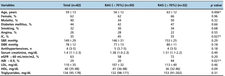
The main characteristics of the total sample and groups categorized by the presence or absence of severe RAS are shown in Table 1. A high frequency of diabetes, IC, and smoking were observed. Severe RAS diagnosed by renal arteriography was present in 32 of 82 (39%) patients. The prevalence of atherosclerosis risk factors was compared between groups (RASo70% and RASX70%). Patients with severe RAS were older (63±12vs56±12,p=0.006) and had a
greater prevalence of IC (55%vs45%;p=0.027) and ABIo0.9
(44%vs20%,p=0.021). No significant differences were observed between the 2 groups concerning diabetes, smoking status, angina, blood pressure, renal function, and dyslipidemia. The average contrast volume used in renal arteriography was 110 mL. Only 7 (8.5%) patients had a transient and mild/moderate increase in creatinine levels suggestive of contrast-induced nephropathy. Dialysis was not required, and no patient developed serious complications during renal arteriography. In the univariate analysis, the variables that
412 IC and RAS in Hypertensive Patients
were significantly correlated with the presence of severe RAS included age, ABI, and IC (Table 2). In the multivariate logistic regression analysis, the presence of IC was an inde-pendent variable associated with severe RAS (OR: 3.33, 95% CI: 1.03–10.82,p=0.04).
’ DISCUSSION
The present study produced the following important find-ings: a) in patients with hypertension who were referred for renal angiography under clinical suspicion of renovascular hypertension, the frequencies of IC and an ABI o0.9 were
35% and 28%, respectively; and b) a validated clinical param-eter (presence of IC defined by a standardized questionnaire) was independently associated with severe RAS in patients with hypertension. Atherosclerotic RAS is a clinical condition frequently observed in patients with multiple cardiovascular risk factors and concomitant atherosclerosis in other arterial beds (10,11). Because atherosclerosis is a systemic disease, it is conceivable that a significant proportion of patients with hypertension who have confirmed RAS also have PAD; however, current evidence is scant, even though PAD has a high prevalence (12) and is associated with increased cardio-vascular morbidity and mortality (13). Although IC is the symptomatic expression of PAD, approximately 50% of patients with IC remain asymptomatic (14). Conversely, we found a high prevalence (35%) of IC among patients with suspicion of RAS, and IC was more prevalent in the group with RAS X70% than in the group without severe RAS (55%vs45%,p=0.027). IC has a significant effect on quality of
life and is strongly associated with impaired functional capacity (15). In addition, IC is related to a twofold to fourfold increased risk of mortality, predominantly from cardiovascular disease (16), and its presence is a predictor of higher rates of myocardial infarction, stroke, and disability in older adults (17). Similar to the elevated prevalence of the clinical presentation of IC, we found an elevated prevalence of an ABIo0.9 (29%). This parameter was observed more
frequently in patients with severe RAS (44%vs20%,p=0.021) and is equally associated with an unfavorable prognosis as described in previous studies (6,13,18).
The clinical expression of severity in the group with RASX70% can be verified by uncontrolled blood pressure (SBP median=149 mmHg) despite patients’use of 4 different classes of antihypertensive medications (Table 1). The pre-valence of diabetes mellitus in our study was 44%, which is higher than that reported in other studies of patients with PAD (between 20 and 29%) (19). The group with severe RAS was older and possibly had more diffuse and serious atherosclerosis, which could explain the higher prevalence of PAD (20). We did not find studies that investigated the prediction of RAS based on the presence of PAD defined by clinical parameters or the ABI.
The main strength of the present investigation was the systematic evaluation of IC symptoms and PAD in con-secutive patients diagnosed according to the gold standard method used for identifying RAS. We found a potential role for IC symptoms, which can be easily identified when the clinical history is obtained, for predicting severe RAS in high-risk patients. This finding may reinforce the indication of RAS investigation as important in patients with resistant hypertension. Some limitations should also be addressed. First, the sample size was small, which is primarily explained by our single-center study design, and we only investigated patients at high risk, suggesting the presence of RAS. Second, the selection of patients with hypertension at high risk for atherosclerotic RAS could overestimate the number of individuals affected by atherosclerosis in other vessels, including the peripheral arteries. However, both limitations are related to the standard indications for investigating RAS
per seand could partially explain the high rate (35%) of IC in
selected patients despite of prevalence of severe RAS in the total group.
Table 2 -Unadjusted and adjusted odds ratio variables in patients according to severe renal artery stenosis (X70%).
Univariate Multivariate
Variables OR (CI) pvalue OR (CI) pvalue
Age 1.05 (1.01-1.10) 0.009 1.26 (0.97-1.08) 0.70 ABI 3.11 (1.16-1.01) 0.009 1.44 (0.37-5.66) 0.60 IC 2.85 (1.11-7.27) 0.029 3.33 (1.03-10.82) 0.04*
ABI: Ankle-brachial index; OR: odds ratio; CI: confidence interval; IC: intermittent claudication.
* Adjusted for age, ABI, and IC.
Table 1 -Clinical characteristics of the 82 patients according to the presence of severe renal artery stenosis.
Variables Total (n=82) RAS (o70%) (n=50) RAS (X70%) (n=32) pvalue
Age, years 59±13 56±12 63±12 0.006*
Female, % 62 62 66 0.96
Mulatto, % 40 34 50 0.32
Diabetes mellitus, % 44 42 47 0.66
Smoking, % 32 30 34 0.68
Angina, % 26 28 22 0.55
IC, % 35 45 55 0.027*
SBP, mmHg 149±29 146±31 153±25 0.29
DBP, mmHg 78±12 77±13 80±11 0.18
Antihypertensives, n 4 (3-5) 5 (3.7-5) 4 (3-5) 0.18
Serum creatinine, mg/dL 1.4 (1.1-2.3) 1.38 (1.0-2.3) 1.51 (1.1-2.2) 0.49
eGFRo60 mL/min/m2, % 63 58 72 0.20
ABIo0.9, % 29 20 44 0.021*
LDL, mg/dL 110±35 107±32 113±40 0.46
HDL, mg/dL 40 (35-48) 41 (36-48) 36 (32-46) 0.09
Triglycerides, mg/dL 134 (95-178) 132 (98-171) 153 (91-202) 0.31
RAS: renal artery stenosis; IC: intermittent claudication; SBP: systolic blood pressure; DBP: diastolic blood pressure; eGFR: estimated glomerular filtration rate; ABI: Ankle-brachial index; LDL: low-density lipoprotein cholesterol; HDL: high-density lipoprotein cholesterol.
413
CLINICS 2017;72(7):411-414 IC and RAS in Hypertensive Patients
The presence of IC was independently associated with severe RAS in patients suspected of having renovascular hypertension based on clinical evidence.
’ AUTHOR CONTRIBUTIONS
Macedo TA conceived and designed the work, wrote the draft and con-tributed to the correction of the manuscript, was responsible for the acquisition, analysis and interpretation of data for the work, managed the literature searches, conducted research procedures, supervised the work, agreed to be accountable for all aspects of the work and to answer questions related to the accuracy or integrity of any part of the work and approved thefinal version of the manuscript to be published. Drager LF conceived and designed the work, wrote the manuscript draft and contributed to the correction of the manuscript, supervised the work and approved the final version of the manuscript to be published. Muela HC, Pedrosa RP, Costa-Hong V and Kajita LJ were responsible for the acquisition, analysis and interpretation of data for the work, managed the literature searches, conducted research procedures and approved thefinal version of the manu-script to be published. Bortolotto LA conceived and designed the work, wrote the draft and contributed to the correction of the manuscript, was responsible for the acquisition, analysis and interpretation of data for the work, managed the literature searches, supervised the work, agreed to be accountable for all aspects of the work and to answer questions related to the accuracy or integrity of any part of the work, approved thefinal version of the manuscript to be published. All authors read and approved thefinal manuscript and have participated sufficiently in the work to take public responsibility for appropriate portions of the content.
’ REFERENCES
1. Colyer WR Jr, Cooper CJ. Cardiovascular morbidity and mortality and renal artery stenosis. Prog Cardiovasc Dis. 2009;52(3):238-42, http://dx. doi.org/10.1016/j.pcad.2009.09.004.
2. Bendermacher BL, Teijink JA, Willigendael EM, Bartelink ML, Büller HR, Peters RJ, et al. Symptomatic peripheral arterial disease: the value of a validated questionnaire and a clinical decision rule. Br J Gen Pract. 2006;56(533):932-7.
3. Fowkes FG, Price JF, Stewart MC, Butcher I, Leng GC, Pell AC, et al. Aspirin for prevention of cardiovascular events in a general population screened for a low ankle brachial index: a randomized controlled trial. JAMA. 2010;303(9):841-8, http://dx.doi.org/10.1001/jama.2010.221. 4. James PA, Oparil S, Carter BL, Cushman WC, Dennison-Himmelfarb C,
Handler J, et al. 2014 evidence-based guideline for the management of high blood pressure in adults: report from the panel members appointed to the Eighth Joint National Committee (JNC 8). JAMA. 2014;311(5): 507-20, http://dx.doi.org/10.1001/jama.2013.284427.
5. Leng GC, Fowkes FG. The Edinburgh Claudication Questionnaire: an improved version of the WHO/Rose Questionnaire for use in epidemio-logical surveys. J Clin Epidemiol. 1992;45(10):1101-9, http://dx.doi.org/ 10.1016/0895-4356(92)90150-L.
6. Hirsch AT, Haskal ZJ, Hertzer NR, Bakal CW, Creager MA, Halperin JL, et al. ACC/AHA 2005 guidelines for the management of patients with peripheral arterial disease (lower extremity, renal, mesenteric, and abdo-minal aortic): executive summary a collaborative report from the American Association for Vascular Surgery/Society for Vascular Surgery, Society for Cardiovascular Angiography and Interventions, Society for Vascular Medi-cine and Biology, Society of Interventional Radiology, and the ACC/AHA
Task Force on Practice Guidelines (Writing Committee to Develop Guide-lines for the Management of Patients With Peripheral Arterial Disease) endorsed by the American Association of Cardiovascular and Pulmonary Rehabilitation; National Heart, Lung, and Blood Institute; Society for Vas-cular Nursing; TransAtlantic Inter-Society Consensus; and VasVas-cular Disease Foundation. J Am Coll Cardiol. 2006;47(6):1239-312, http://dx.doi.org/ 10.1016/j.jacc.2005.10.009.
7. Ruilope LM, Zanchetti A, Julius S, McInnes GT, Segura J, Stolt P, et al. Prediction of cardiovascular outcome by estimated glomerular filtration rate and estimated creatinine clearance in the high-risk hypertension population of the VALUE trial. J Hypertens. 2007;25(7):1473-9, http://dx. doi.org/10.1097/HJH.0b013e328133246c.
8. Drager LF, Andrade L, Barros de Toledo JF, Laurindo FR, Machado César LA, Seguro AC. Renal effects of N-acetylcysteine in patients at risk for contrast nephropathy: decrease in oxidant stress-mediated renal tubular injury. Nephrol Dial Transplant. 2004;19(7):1803-7, http://dx.doi.org/ 10.1093/ndt/gfh261.
9. Macedo TA, Pedrosa RP, Costa-Hong V, Kajita LJ, Morais GR, De Lima JJ, et al. Renal artery stenosis predicts coronary artery disease in patients with hypertension. PLoS One. 2013;8(3):e58635, http://dx.doi.org/10.1371/ journal.pone.0058635.
10. Kuroda S, Nishida N, Uzu T, Takeji M, Nishimura M, Fujii T, et al. Pre-valence of renal artery stenosis in autopsy patients with stroke. Stroke. 2000;31(1):61-5, http://dx.doi.org/10.1161/01.STR.31.1.61.
11. Iglesias JI, Hamburger RJ, Feldman L, Kaufman JS. The natural history of incidental renal artery stenosis in patients with aortoiliac vascular disease. Am J Med. 2000;109(8):642-7, http://dx.doi.org/10.1016/S0002-9343(00) 00605-7.
12. Ogren M, Hedblad B, Isacsson SO, Janzon L, Jungquist G, Lindell SE. Non-invasively detected carotid stenosis and ischaemic heart disease in men with leg arteriosclerosis. Lancet. 1993;342(8880):1138-41, http://dx. doi.org/10.1016/0140-6736(93)92123-B.
13. Criqui MH, Langer RD, Fronek A, Feigelson HS, Klauber MR, McCann TJ, et al. Mortality over a period of 10 years in patients with peripheral arterial disease. N Engl J Med. 1992;326(6):381-6, http://dx.doi.org/ 10.1056/NEJM199202063260605.
14. Ramos R, Quesada M, Solanas P, Subirana I, Sala J, Vila J, et al. Prevalence of symptomatic and asymptomatic peripheral arterial disease and the value of the ankle-brachial index to stratify cardiovascular risk. Eur J Vasc Endovasc Surg. 2009;38(3):305-11, http://dx.doi.org/10.1016/j.ejvs.2009. 04.013.
15. Breek JC, Hamming JF, De Vries J, Aquarius AE, van Berge Henegouwen DP. Quality of life in patients with intermittent claudication using the World Health Organisation (WHO) questionnaire. Eur J Vasc Endovasc Surg. 2001;21(2):118-22, http://dx.doi.org/10.1053/ejvs.2001.1305. 16. Kannel WB, McGee DL. Update on some epidemiologic features of
intermittent claudication: the Framingham Study. J Am Geriatr Soc. 1985; 33(1):13-8, http://dx.doi.org/10.1111/j.1532-5415.1985.tb02853.x. 17. Simonsick EM, Guralnik JM, Hennekens CH, Wallace RB, Ostfeld AM.
Intermittent claudication and subsequent cardiovascular disease in the elderly. J Gerontol A Biol Sci Med Sci.1995;50A(1):M17-22.
18. Murabito JM, Evans JC, Larson MG, Nieto K, Levy D, Wilson PW, et al. The ankle-brachial index in the elderly and risk of stroke, coronary dis-ease, and death: the Framingham Study. Arch Intern Med. 2003;163(16): 1939-42, http://dx.doi.org/10.1001/archinte.163.16.1939.
19. Hirsch AT, Criqui MH, Treat-Jacobson D, Regensteiner JG, Creager MA, Olin JW, et al. Peripheral arterial disease detection, awareness, and treat-ment in primary care. JAMA. 2001;286(11):1317-24, http://dx.doi.org/ 10.1001/jama.286.11.1317.
20. Kröger K, Stang A, Kondratieva J, Moebus S, Beck E, Schmermud A, et al. Prevalence of peripheral arterial disease - results of the Heinz Nixdorf recall study. Eur J Epidemiol. 2006;21(4):279-85, http://dx.doi.org/10.1007/ s10654-006-0015-9.
414 IC and RAS in Hypertensive Patients