Detection value of free cancer cells in peritoneal washing
in gastric cancer: a systematic review and meta-analysis
Francisco Tustumi,
*Wanderley Marques Bernardo, Andre Roncon Dias, Marcus Fernando Kodama Pertille Ramos,
Ivan Cecconello, Bruno Zilberstein, Ulysses Ribeiro-Ju´nior
Hospital das Clı´nicas da Faculdade de Medicina da Universidade de Sa˜o Paulo, Sa˜o Paulo/SP, Brazil.
Intraperitoneal free cancer cells in gastric adenocarcinoma are associated with a poor outcome. However, the
true prognostic value of intraperitoneal free cancer cells is still unclear, leading to a lack of consensus in the
management of gastric cancer. The aim of the present study is to perform a systematic review and meta-analysis
to analyze intraperitoneal free cancer cells-positive patients with regard to tumor oncologic stage, recurrence,
grade of cellular differentiation, and survival rates and to analyze the clinical significance of intraperitoneal
free cancer cells with regard to prognosis. Databases were searched up to January 2016 for prognostic factors
associated with intraperitoneal free cancer cells, including oncologic stage, depth of neoplasm invasion, lymph
nodal spread, differentiation grade of the tumor, and recurrence and survival rates. A total of 100 studies were
identified. Meta-analysis revealed a clear association between intraperitoneal free cancer cells and a poor
prognosis. intraperitoneal free cancer cells -positive patients had higher rates of nodal spread (risk difference:
0.29;
p
o
0.01), serosal invasion (risk difference: 0.43;
p
o
0.01), recurrence (after 60 months of follow-up, risk
difference: 0.44;
p
o
0.01), and mortality (after 60 months of follow-up, risk difference: 0.34;
p
o
0.01).
Intra-peritoneal free cancer cells are associated with a poor outcome in gastric cancer. This surrogate biomarker
should be used to guide therapy both prior to and after surgery.
KEYWORDS:
Gastric Carcinoma; Peritoneal Washing; Peritoneal Lavage; Cytology; Carcinoembryonic Antigen; RT-PCR.
Tustumi F, Bernardo WM, Dias AR, Ramos MF, Cecconello I, Zilberstein B, et al. Detection value of free cancer cells in peritoneal washing in gastric cancer: a systematic review and meta-analysis. Clinics. 2016;71(12):733-745
Received for publication onJuly 23, 2016;First review completed onAugust 29, 2016;Accepted for publication onSeptember 9, 2016 *Corresponding author. E-mail: [email protected]
’
INTRODUCTION
Peritoneal dissemination is the most common pattern of
recurrence in gastric cancer, even after a potentially
cura-tive resection. This characteristic may be attributable to
pos-sible intraperitoneal dissemination of malignant cells already
present at the time of surgery or to surgical manipulations.
Current knowledge on intraperitoneal free cancer cell (IFCC)
positivity in gastric cancer demonstrates that these cells are
associated with a poor prognosis and advanced oncologic
stages. Additionally, high recurrence rates, mainly due to
peritoneal dissemination, and poor median survival are
associated with cytology detection (1-3).
Based on these data, the Japanese Classification of Gastric
Carcinoma: 3
rdEnglish Edition (4) and the 7
thEdition of the
AJCC Cancer Staging Manual: Stomach (5) consider
conven-tional cytology positivity in peritoneal fluid to be an
indi-cator of stage IV disease.
Several institutional protocols are used to manage
IFCC-positive patients, including chemotherapy, prompt
gastrect-omy, neoadjuvant treatment, peritoneal infusion, hyperthermic
peritoneal chemotherapy, or palliation alone. However, none
of these techniques are accepted worldwide as a gold
stan-dard therapy.
The investigation of peritoneal washing for IFCCs in gastric
cancer patients remains controversial. Little is known about the
actual burden of IFCC positivity and its accuracy for predicting
an outcome. Moreover, a lack of consensus exists in its routine
practice, methods of detection (6), and association with clinical
pathological variables.
Thus, the aim of this study was to perform a systematic
review and meta-analysis, investigating patients positive for
IFCCs detected via different methods, regarding the
neo-plasm oncologic stage, recurrence rates, grade of cellular
differentiation, and survival rates and to analyze the clinical
significance of IFCCs with regard to prognosis.
’
METHODS
The construction and modeling of the present study
were guided by the Preferred Reporting Items for Systematic
Reviews and Meta-Analyses (PRISMA) statement (7).
Database search
A literature search was performed in MEDLINE using the
following search terms: (((
‘‘
Stomach Neoplasms/cytology
’’
[Mesh]) AND ((Peritoneum OR Peritoneal OR abdominal
cavity OR ascitic fluid OR washing OR lavage)))) OR
(((cytology AND gastric cancer)) AND ((Peritoneum OR
DOI:10.6061/clinics/2016(12)10
Peritoneal OR abdominal cavity OR ascitic fluid))). Other
databases searched included LILACS, CENTRAL, Cochrane,
CINAHL, and Scopus as well as grey literature.
No attempts were made to locate unpublished material.
Inclusion criteria
Patients with confirmed gastric adenocarcinoma submitted
to preoperative peritoneal washing/lavage evaluation (open,
laparoscopic, or by paracentesis) for IFCCs (conventional
cytology with Papanicolaou, Giemsa, or Hematoxylin-eosin
staining); molecular methods, such as RT-PCR for
carcino-embryonic antigen (CEA), cytokeratin (CK20), and
melanoma-associated gene (MAGE); and immunohistochemistry.
Studies that evaluated the prognosis (i.e., oncologic stage,
survival, recurrence rate, or grade of cellular differentiation).
Prospective or retrospective studies.
Studies selected by both of two reviewers.
Exclusion criteria
Data could not be extracted from pooled results.
Patients submitted to a neoadjuvant approach prior to the
peritoneal washing/lavage procedure.
Presence of other primary malignancy.
Case series, case reports, animal models, conference
pro-ceedings, editorials, and letters.
Review articles and meta-analyses were excluded from
meta-analysis.
Studies with no full-text.
Idiom
No restriction.
Search period
No restriction. The search was performed up to January
2016.
Outcomes
Recurrence rate
Recurrence site: lymph node, peritoneal, or other organs
(local recurrence or hematogenous spread)
Mortality
Oncologic stage
Serosal invasion
Lymph node spread
Grade of cellular differentiation
Statistical analysis
Absolute numbers for the outcome parameters were
extracted and analyzed with Review Manager Version 5.3
software (Copenhagen: The Nordic Cochrane Centre; The
Cochrane Collaboration, 2014).
We performed subgroup analysis and sensitivity tests
to explore the causes of statistical heterogeneity in which
the effect of single studies on the heterogeneity value was
tested. Forest plots were used for graphical exploration of
heterogeneity. A funnel plot was used to identify
publica-tion bias.
Studies characteristics
Of the selected articles, 20 were excluded because they
lacked the information necessary for meta-analysis, such as
serosal invasion, oncologic stage, neoplasm dissemination,
and grade of cellular differentiation. In total, 100 (1-3, 8-104)
eligible trials were identified and reviewed, and 91 were
included in the meta-analysis. Cumulatively, 16,913 gastric
cancer patients were evaluated. In 41 studies analyzed, all
patients were submitted to curative intention surgery.
We assessed the quality of the studies using the
Newcastle-Ottawa Scale (NOS). In terms of study quality, the cohort
studies were considered to be of fair (scores of 4–6) to good
(scores of 7–9) quality, but two articles were considered low
quality (scores of 1-3).
Of the 100 eligible trials, data describing conventional
cytology were available for 68 papers; data regarding
PCR-CEA were available for 27; and data regarding PCR-CK20
were available for 5. Other studies also evaluated Ber-Ep4,
MAGE, RT-LAMP, or a combination of techniques used to
detect IFCCs.
Most studies performed peritoneal washing/lavage
simi-larly to the method described by Nakajima et al. (69). The
peritoneal cavity was washed with 50 to 200 ml of normal
saline. After stirring, the fluid was collected. Thirty-three
studies collected fluid from the Douglas space, 16 collected
fluids from the Douglas and left subphrenic spaces, and
5 collected fluid from the perigastric surroundings. The
remaining studies collected fluid from different combinations of
recesses. Peritoneal washing/lavage was performed by
lapa-rotomy in 81%, by laparoscopy in 15.2%, and by drainage tube
in 3.8% of the studies.
Data were collected from 16 countries. The median
follow-up across all studies was 36 months (range 12-108 months).
The prevalence of IFCCs ranged from 2 to 72%, with a
median of 27%. Considering only conventional cytology
studies, the median prevalence was 19.3% (range 2-61%).
Considering only PCR-CEA, the median prevalence was
27.8% (range 15-63%). Considering only PCR-CK20, the
median prevalence was 27.9% (range 15-39%).
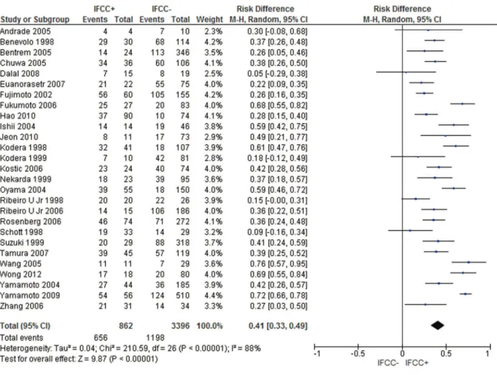
IFCC and oncologic stage
The present study analyzed the oncologic stage according
to the UICC/AJCC system 6
thedition (105). For this purpose,
IFCC detection alone was not considered stage IV.
The pooled data of the network meta-analysis showed
that IFCC detection was associated with a significantly
higher risk of stage III or IV compared with stage I or II (risk
difference: 0.41; 95% CI: 0.33–0.49; n=4,258 patients; I
2=88%,
p
o
0.00001) (see Figure 1). The sensitivity analysis failed to
identify outliers. A random-effects analysis method was used
to adjust for inter-study heterogeneity.
For the subgroup analysis, conventional cytology studies
(1-3,12,16-19,24,49,54,79,94,96) (risk difference: 0.34; 95% CI:
0.2–0.48; n=2,373 patients; I
2=93%;
p
o
0.00001) and PCR-CEA
(31,36,38,49,77,93,94,97,104) (risk difference: 0.5; 95% CI: 0.36–
0.63; n=1,073 patients; I
2=83%;
p
o
0.00001) were reviewed by
comparing stage III or IV patients with stage I or II patients.
Comparable results were identified (risk difference: 0.32;
95% CI: 0.19–0.44; n=600 patients; I
2=55%;
p
o
0.00001) when
analyzing studies that evaluated oncologic stages according
to the UICC/AJCC system 7
thedition (26,31,38,59,97).
IFCC and serosal invasion
The pooled data of the network meta-analysis showed that
IFCC detection was associated with a significantly higher
risk of serosal invasion than tumors that did not invade
the serosa (risk difference: 0.43; 95% CI: 0.38–0.48; n=11,511
patients; I
2=89%,
p
o
0.00001) (see Figure 2). The sensitivity
analysis failed to identify outliers. A random-effects analysis
method was used to adjust for inter-study heterogeneity.
For the subgroup analysis, conventional cytology studies
(2,3,10,11,15,16,18,19,22,25,29,34,40,41,43,46,48-57,60,63,69,70,
78,81,83,87,92,94,96,99,101) (risk difference: 0.39; 95% CI: 0.35–
0.43; n=2,374 patients; I
2=93%;
p
o
0.00001) and PCR-CEA
(28,36-38,48-51,57,58,64,70,76,77,83,89,93,94,97,101,104) (risk
dif-ference: 0.51; 95% CI: 0.45–0.57; n=2,612 patients; I
2=66%;
p
o
0.00001) were reviewed.
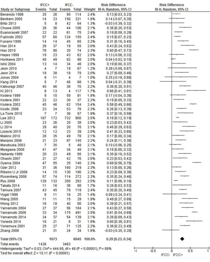
IFCC and lymph node spread
The pooled data of the network meta-analysis showed that
IFCC detection was associated with a significantly increased
risk of lymph node spread compared to cancer with no lymph
node involvement (risk difference: 0.29; 95% CI: 0.23–0.34;
n=7,718 patients; I
2=89%,
p
o
0.00001) (see Figure 3). The
sensi-tivity analysis failed to identify outliers. A random-effects
anal-ysis method was used to adjust for inter-study heterogeneity.
For the subgroup analysis, conventional cytology studies
(1-3,12,14,16,18,19,25,29,41,43,46,51,52,54-57,61,63,78,81,92,94,
96,101) (risk difference: 0.25; 95% CI: 0.18–0.31; n=5,008 patients;
I
2=87%;
p
o
0.00001) and PCR-CEA (31,36,38,48,57,58,64,76,
77,93,94,97,104) (risk difference: 0.3; 95% CI: 0.15–0.45; n=
1,464 patients; I
2=93%;
p
o
0.00001) were reviewed.
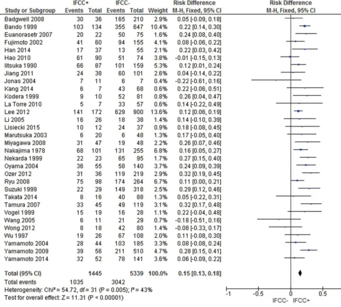
IFCC and grade of cellular differentiation
The pooled data of the network meta-analysis showed that
IFCC detection was associated with a significantly increased
probability of having poorly differentiated tumors compared
to well or moderately differentiated tumors (risk difference:
0.15; 95% CI: 0.12–0.17; n=7,232; I
2=65%,
p
o
0.00001).
A sensitivity analysis was performed by repeating the
net-work analysis after omitting 3 studies with a high risk of
bias (31,102,103). The final result revealed a risk difference
of 0.15 (95% CI: 0.13–0.18; n=6,784; I
2=43%,
p
o
0.00001) (see
Figure 4).
For the subgroup analysis, conventional cytology studies
(2,10,11,18,19,34,40,41,43,55-57,69,78,81,87,89,92,94-96,
102,103) (risk difference after excluding 2 outliers (102,103):
0.17; 95% CI: 0.14–0.2; n=5,437 patients; I
2=39%;
p
o
0.00001)
and PCR-CEA (31,57,64,77,93,94,97) (risk difference: 0.08;
95% CI: 0.01–0.15; n=805 patients; I
2=55%;
p
o
0.04) were
reviewed.
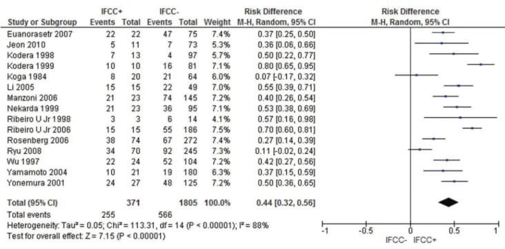
IFCC and recurrence
The recurrence rate was assessed for gastric cancers treated
with curative intention surgery.
The pooled data of the network meta-analysis showed that
IFCC detection was associated with a significantly increased
risk of recurrence. For recurrence after 24 months of
follow-up (15,16,28,72), the risk difference was 0.38 (95% CI: 0.25–
0.51; n=360 patients; I
2=57%,
p
o
0.00001). For recurrence
after 60 months, the risk difference was 0.44 (95% CI: 0.32–
0.56; n=2,176 patients; I
2=88%,
p
o
0.00001) (see Figure 5).
For IFCC-positive patients, the mean recurrence rate was
55.35% after 24 months and 68.73% after 60 months. For
IFCC-negative patients, the mean recurrence rate was 16.77%
after 24 months and 31.36% after 60 months.
IFCC and sites of recurrence
For gastric cancers treated with curative intent surgery,
studies were assessed regarding peritoneal recurrence, lymph
nodal recurrence, or recurrence in other organs.
For peritoneal recurrence, the presence of IFCCs predicted
a risk difference of 0.48 (95% CI: 0.38–0.59; n=2,683 patients;
I
2=86%,
p
o
0.00001) (see Figure 6). The sensitivity analysis
failed to identify outliers. A random-effects analysis method
was used to adjust for inter-study heterogeneity.
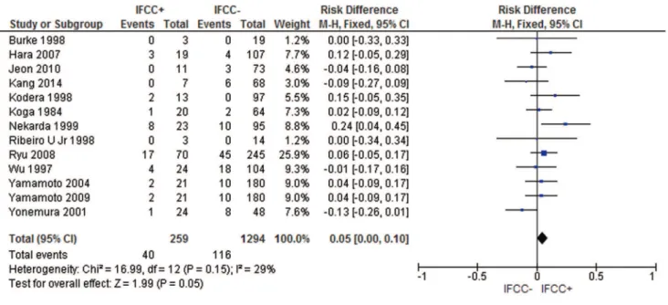
For lymph nodal recurrence, the presence of IFCCs
pre-dicted a risk difference of 0.05 (95% CI: 0.00–0.1; n=1,553
patients; I
2=29%,
p
=0.05) (see Figure 7).
For local or hematogenous recurrence, the presence of
IFCCs did not predict a poor prognosis (risk difference: 0.02;
95% CI: -0.03, 0.07; n=1,355 patients; I
2=23%,
p
=0.22) (see
Figure 8).
IFCC and mortality
The pooled data of the network meta-analysis showed that
IFCC detection was associated with a significantly increased
risk of mortality.
For mortality after 12 months of follow-up (9,13,15,20,56,
57,63,85,92), the risk difference was 0.26 (95% CI: 0.19–0.33;
n=1,765 patients; I
2=48%,
p
o
0.00001). One study was
omit-ted after sensitivity analysis (92).
For mortality after 24 months (9,13,20,29,61,63,84,92), the
risk difference was 0.4 (95% CI: 0.33–0.48; n=934 patients;
I
2=35%,
p
o
0.00001). One study was omitted after sensitivity
analysis (9).
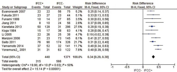
For mortality after 60 months, the risk difference was 0.34
(95% CI: 0.29–0.38; n=1,811 patients; I
2=50%,
p
o
0.00001).
Two studies were omitted after sensitivity analysis (69,72)
(see Figure 9).
For IFCC-positive patients, the mean mortality rate was
43.5% after 12 months, 75% after 24 months, and 72.3% for
studies that analyzed mortality after 60 months. For
IFCC-negative patients, the mean mortality rate was 16.6% after
12 months, 43.2% after 24 months, and 41.2% after 60 months.
For the subgroup analysis, studies that exclusively
eval-uated patients who submitted to curative intention surgery
were assessed.
For mortality after 12 months, the risk difference was 0.35
(95% CI: 0.24–0.45; n=799 patients; I
2=13%,
p
o
0.00001). One
study was omitted after sensitivity analysis (92).
For mortality after 24 months, the risk difference was 0.34
(95% CI: 0.24–0.44; n=717 patients; I
2=35%,
p
o
0.00001). One
For mortality after 60 months, the risk difference was
0.42 (95% CI: 0.37–0.47; n=995 patients; I
2=58%,
p
o
0.00001).
One study was omitted after sensitivity analysis (18).
’
DISCUSSION
The present study evaluated the burden of IFCC positivity
in gastric cancer by analyzing the individual data of each
included study. The strengths of our study include the
following: the study strategy was designed to be
comprehen-sive; the inclusion and exclusion criteria and data extraction
were determined to reduce bias; no idiom restrictions allowed
the avoidance of cultural and racial bias; and this is the one of
the first studies to analyze the true burden effect of IFCC
positivity on recurrence rates (and sites of recurrence) and
mortality rates. A limitation of this study was that some of the
comparisons had a high level of heterogeneity.
Pecqueux et al. (106) also analyzed the relationship between
IFCCs and survival and recurrence rates. However, they
compared studies that evaluated recurrence and survival rates
at different times, which compromised the validity of the
findings.
Therefore, the present study assessed survival and
recur-rence rates at 1-, 2-, and 5-year follow-up evaluations. IFCC
was associated with higher early and late mortality.
Additi-onally, our study evaluated sites of recurrence and showed
that recurrence was mainly due to peritoneal dissemination.
Similar to Pecqueux et al. (106), our study also revealed a
high level of heterogeneity for recurrence rates. This finding
could be explained by the different follow-up programs of
each oncologic center, including different adjuvant therapies
and methodologies for diagnosing recurrence.
To explore the causes of statistical heterogeneity, we
per-formed subgroup and sensitivity analyses in which the
effects of single studies on the heterogeneity value were
tested. A funnel plot was used to identify publication bias.
If publication bias was identified, the study was excluded
from the analysis.
Figure 5 -Recurrence rate. IFCCs were associated with a higher probability of recurrence after 60 months of follow-up.
IFCC was associated with lymphatic spread (risk
dif-ference: 0.29; 95% CI: 0.23–0.34,
p
o
0.00001) and lymph
nodal recurrence (risk difference 0.05; 95% CI: 0.00–0.1,
p
o
0.05). This result may be explained by
confound-ing variables (IFCC actually could be associated with
neoplasm depth, which would subsequently be
asso-ciated with lymph nodal spread and recurrence). None
of the trials explored these data, and future studies could
investigate a possible link between lymphatic and
peri-toneal dissemination, which has been suggested by some
authors (107).
IFCCs are also associated with locally advanced tumors,
especially those with serosal invasion, but can also be found
in earlier clinical stages of gastric cancer. Part of the
mech-anism by which advanced tumors disseminate into the
peri-toneum is likely associated with the area of serosal invasion
(53,54,79), which could contribute to tumoral cell exfoliation
and seeding into the peritoneal surface (108).
When assessing for subgroup analysis, different methods
of detecting IFCC presented similar results for the prognosis.
Both conventional cytology and PCR-CEA were associated
with advanced stage cases, serosal invasion, nodal spread,
and poorly differentiated neoplasms.
Most IFCC-positive patients who were treated with
cura-tive intent surgery likely experienced minimal to no benefit
from the surgery, with most experiencing early recurrence
(55.35% in 24 months). The survival rate of IFCC-positive
patients was 25% after 2 years.
Figure 7 -IFCC detection can also predict a higher probability of lymph nodal recurrence.
Accordingly, preoperative peritoneal washing/lavage in
gastric cancer should be strongly advised for high
sur-gical risk patients (e.g., the elderly, low status performance
patients, and patients with incapacitating comorbidities).
If IFCC positivity is determined, palliative therapy may be
considered.
In low surgical risk and oncologic low risk (no serosa
inva-sion, no lymph nodal spread, moderate or well differentiated
neoplasm) patients, immediate surgery should be performed,
and intraoperative peritoneal washing/lavage should be
added. If IFCC positivity is determined, postoperative
chemo-therapy could be indicated. Clinical trials of hyperthermic
intra-peritoneal chemotherapy may be proposed.
The pooled data demonstrate that IFCC findings are an
independent prognostic factor in gastric cancer. From this
work, it can be concluded that the prognosis in surgically
treated patients with gastric carcinoma is significantly affected
by the presence of IFCCs at the time of gastrectomy and should
guide gastric cancer management.
’
AUTHOR CONTRIBUTIONS
Tustumi F was responsible for the elaboration of the project and manu-script writing. Bernardo WM was responsible for the statistical analysis. Dias AR and Ramos MF helped with manuscript revision. Cecconello I and Zilberstein B selected the articles. Ribeiro-Junior U was responsible for the elaboration of the project.
’
REFERENCES
1. Bentrem D, Wilton A, Mazumdar M, Brennan M, Coit D. The value of peritoneal cytology as a preoperative predictor in patients with gastric carcinoma undergoing a curative resection. Ann Surg Oncol. 2005; 12(5):347-53, http://dx.doi.org/10.1245/ASO.2005.03.065.
2. Kodera Y, Yamamura Y, Shimizu Y, Torii A, Hirai T, Yasui K, et al. Peritoneal washing cytology: prognostic value of positive findings in patients with gastric carcinoma undergoing a potentially curative resection. J Surg Oncol. 1999;72(2):60-4, http://dx.doi.org/10.1002/ (SICI)1096-9098(199910)72:2o60::AID-JSO343.0.CO;2-1.
3. Ribeiro U Jr, Safatle-Ribeiro AV, Zilberstein B, Mucerino D, Yagi OK, Bresciani CC, et al. Does the intraoperative peritoneal lavage cyto-logy add prognostic information in patients with potentially curative gastric resection? J Gastrointest Surg. 2006;10(2):170-6, http://dx.doi. org/10.1016/j.gassur.2005.11.001.
4. Ajani JA, Bentrem DJ, Besh S, D’Amico TA, Das P, Denlinger C, et al. 2013 NCCN clinical practice guidelines in oncology: gastric cancer. 2013; Version 2. www.nccn.org.
5. Washington K. 7th edition of the AJCC cancer staging manual: stomach. Ann Surg Oncol. 2010;17(12):3077-9, http://dx.doi.org/10.1245/s10434-010-1362-z.
6. Brar SS, Mahar AL, Helyer LK, Swallow C, Law C, Paszat L, et al. Processes of care in the multidisciplinary treatment of gastric cancer: results of a RAND/UCLA expert panel. JAMA Surg. 2014;149(1):18-25, http://dx.doi.org/10.1001/jamasurg.2013.3959.
7. Moher D, Shamseer L, Clarke M, Ghersi D, Liberati A, Petticrew M, et al. Preferred reporting items for systematic review and meta-analysis pro-tocols (PRISMA-P) 2015 statement. Syst Rev. 2015;4:1, http://dx.doi. org/10.1186/2046-4053-4-1.
8. Andrade RJ, Iturriza JF, Loyo EL, Martinez LE. Adenocarcinoma del Sistema digestivo: utilidad diagnóstica de la citología peritoneal. Rev Venez Oncol. 2005;17(2):79-88.
9. Asao T, Fukuda T, Yazawa S, Nagamachi Y. Carcinoembryonic antigen levels in peritoneal washings can predict peritoneal recurrence after curative resection of gastric cancer. Cancer. 1991;68(1):44-7, http://dx. doi.org/10.1002/1097-0142(19910701)68:1o44::AID-CNCR28206801094
3.0.CO;2-J.
10. Badgwell B, Cormier JN, Krishnan S, Yao J, Staerkel GA, Lupo PJ, et al. Does neoadjuvant treatment for gastric cancer patients with positive peritoneal cytology at staging laparoscopy improve survival? Ann Surg Oncol. 2008;15(10):2684-91, http://dx.doi.org/10.1245/s10434-008-0055-3. 11. Bando E, Yonemura Y, Takeshita Y, Taniguchi K, Yasui T, Yoshimitsu Y, et al. Intraoperative lavage for cytological examination in 1,297 patients with gastric carcinoma. Am J Surg. 1999;178(3):256-62, http://dx.doi. org/10.1016/S0002-9610(99)00162-2.
12. Benevolo M, Mottolese M, Cosimelli M, Tedesco M, Giannarelli D, Vasselli S, et al. Diagnostic and prognostic value of peritoneal immuno-cytology in gastric cancer. J Clin Oncol. 1998;16(10):3406-11.
13. Bonenkamp JJ, Songun I, Hermans J, van de Velde CJ. Prognostic value of positive cytology findings from abdominal washings in patients with gastric cancer. Br J Surg. 1996;83(5):672-4, http://dx.doi.org/10.1002/ bjs.1800830526.
14. Brito AM, Sarmento BJ, Mota ED, Fraga AC Jr, Campoli PM, Milhomem LM, et al. Prognostic role of positive peritoneal cytology in patients with resectable gastric cancer. Rev Col Bras Cir. 2013;40(2):121-6, http://dx. doi.org/10.1590/S0100-69912013000200007.
15. Burke EC, Karpeh MS Jr, Conlon KC, Brennan MF. Peritoneal lavage cytology in gastric cancer: an independent predictor of outcome. Ann Surg Oncol. 1998;5(5):411-5, http://dx.doi.org/10.1007/BF02303859. 16. Chuwa EW, Khin LW, Chan WH, Ong HS, Wong WK. Prognostic
signi-ficance of peritoneal lavage cytology in gastric cancer in Singapore. Gastric Cancer. 2005;8(4):228-37, http://dx.doi.org/10.1007/s10120-005-0343-6.
17. Dalal KM, Woo Y, Kelly K, Galanis C, Gonen M, Fong Y, et al. Detection of micrometastases in peritoneal washings of gastric cancer patients by the reverse transcriptase polymerase chain reaction. Gastric Cancer. 2008;11(4):206-13, http://dx.doi.org/10.1007/s10120-008-0483-6.
18. Euanorasetr C, Lertsithichai P. Prognostic significance of peritoneal washing cytology in Thai patients with gastric adenocarcinoma under-going curative D2 gastrectomy. Gastric Cancer. 2007;10(1):18-23, http:// dx.doi.org/10.1007/s10120-006-0402-7.
19. Fujimoto T, Zhang B, Minami S, Wang X, Takahashi Y, Mai M. Evaluation of intraoperative intraperitoneal cytology for advanced gastric carcinoma. Oncology. 2002;62(3):201-8, http://dx.doi.org/10.1159/ 000059566.
20. Fujimura T, Kinami S, Ninomiya I, Kitagawa H, Fushida S, Nishimura G, et al. Diagnostic laparoscopy, serum CA125, and peritoneal metastasis in gastric cancer. Endoscopy. 2002;34(7):569-74, http://dx.doi.org/10.1055/ s-2002-33228.
21. Fujiwara Y, Okada K, Hanada H, Tamura S, Kimura Y, Fujita J, et al. The clinical importance of a transcription reverse-transcription concerted (TRC) diagnosis using peritoneal lavage fluids in gastric cancer with clinical serosal invasion: A prospective, multicenter study. Surgery. 2014;155(3):417-23, http://dx.doi.org/10.1016/j.surg.2013.10.004. 22. Fukagawa T, Katai H, Saka M, Morita S, Sasajima Y, Taniguchi H, et al.
Significance of lavage cytology in advanced gastric cancer patients. World J Surg. 2010;34(3):563-8, http://dx.doi.org/10.1007/s00268-009-0355-1.
23. Fukuda N, Sugiyama Y, Wada J. Prognostic factors of T4 gastric cancer patients undergoing potentially curative resection. World J Gastro-enterol. 2011;17(9):1180-4, http://dx.doi.org/10.3748/wjg.v17.i9.1180. 24. Fukumoto Y, Ikeguchi M, Matsumoto S, Inoue M, Osaki T, Fukuda K,
et al. Detection of cancer cells and gene expression of cytokines in the peritoneal cavity in patients with gastric cancer. Gastric Cancer. 2006; 9(4):271-6, http://dx.doi.org/10.1007/s10120-006-0390-7.
25. Funami Y, Tokumoto N, Miyauchi H, Ochiai T, Kuga K. Prognostic value of peritoneal lavage cytology and chemotherapy during surgery for advanced gastric cancer. Int Surg. 1999;84(3):220-4.
26. Han J, Lv P, Yu JL, Wu YC, Zhu X, Hong LL, et al. Circulating methy-lated MINT2 promoter DNA is a potential poor prognostic factor in gastric cancer. Dig Dis Sci. 2014;59(6):1160-8, http://dx.doi.org/10.1007/ s10620-013-3007-0.
27. Hao YX, Zhong H, Yu PW, Qian F, Zhao YL, Shi Y, et al. Influence of laparoscopic gastrectomy on the detection rate of free gastric cancer cells in the peritoneal cavity. Ann Surg Oncol. 2010;17(1):65-72, http://dx.doi. org/10.1245/s10434-009-0703-2.
28. Hara M, Nakanishi H, Jun Q, Kanemitsu Y, Ito S, Mochizuki Y, et al. Comparative analysis of intraperitoneal minimal free cancer cells between colorectal and gastric cancer patients using quantitative RT-PCR: possible reason for rare peritoneal recurrence in colorectal cancer. Clin Exp Metastasis. 2007;24(3):179-89, http://dx.doi.org/10.1007/s10585-007-9067-9.
29. Hayes N, Wayman J, Wadehra V, Scott DJ, Raimes SA, Griffin SM. Peritoneal cytology in the surgical evaluation of gastric carcinoma. Br J Cancer. 1999;79(3-4):520-4, http://dx.doi.org/10.1038/sj.bjc.6690081. 30. Homma Y, Ushida S, Yamada M, Kobayashi H, Suzuki K. Positive
peritoneal washing cytology in multiple cavities can predict poor pro-gnosis of advanced gastric cancer patients. Ann Surg Oncol. 2010;17(2): 455-60, http://dx.doi.org/10.1245/s10434-009-0764-2.
31. Horikawa M, Iinuma H, Inoue T, Ogawa E, Fukushima R. Clinical significance of intraperitoneal CD44 mRNA levels of magnetically separated CD45-negative EpCAM-positive cells for peritoneal recurrence and prognosis in stage II and III gastric cancer patients. Oncol Rep. 2011;25(5):1413-20.
32. Iida T, Iwahashi M, Katsuda M, Ishida K, Nakamori M, Nakamura M, et al. Prognostic significance of IL-17 mRNA expression in peritoneal lavage in gastric cancer patients who underwent curative resection. Oncol Rep. 2014;31(2):605-12.
33. Iitsuka Y, Kaneshima S, Tanida O, Takeuchi T, Koga S. Intraperitoneal free cancer cells and their viability in gastric cancer. Cancer. 1979; 44(4):1476-80, http://dx.doi.org/10.1002/1097-0142(197910)44:4o1476::
AID-CNCR282044044243.0.CO;2-R.
34. Iitsuka Y, Shiota S, Matsui T, Murata Y, Kimura A, Koga S. Relationship between the cytologic characteristics of intraperitoneal free cancer cells and the prognosis in patients with gastric cancer. Acta Cytol. 1990; 34(3):437-42.
35. Ishigami S, Uenosono Y, Arigami T, Yanagita S, Okumura H, Uchikado Y, et al. Clinical utility of perioperative staging laparoscopy for advanced gastric cancer. World J Surg Oncol. 2014;12:350, http://dx.doi.org/ 10.1186/1477-7819-12-350.
36. Ishii T, Fujiwara Y, Ohnaka S, Hayashi T, Taniguchi H, Takiguchi S, et al. Rapid genetic diagnosis with the transcription-reverse transcription concerted reaction system for cancer micrometastasis. Ann Surg Oncol. 2004;11(8):778-85, http://dx.doi.org/10.1245/ASO.2004.12.043. 37. Ito S, Nakanishi H, Kodera Y, Mochizuki Y, Tatematsu M, Yamamura Y.
Prospective validation of quantitative CEA mRNA detection in perito-neal washes in gastric carcinoma patients. Br J Cancer. 2005;93(9):986-92, http://dx.doi.org/10.1038/sj.bjc.6602802.
38. Jeon CH, Kim IH, Chae HD. Prognostic value of genetic detection using CEA and MAGE in peritoneal washes with gastric carcinoma after
curative resection: result of a 3-year follow-up. Medicine (Baltimore). 2014;93(11):e83, http://dx.doi.org/10.1097/MD.0000000000000083. 39. Jeon CH, Shin IH, Park JB, Chae HD. Prognostic significance of
MAGE in peritoneal washes in gastric carcinoma patients without peritoneal metastasis: results of a 5-year follow-up study. J Clin Gas-troenterol. 2010;44(10):682-6, http://dx.doi.org/10.1097/MCG.0b013e 3181d6bb0b.
40. Jiang CG, Xu Y, Wang ZN, Sun Z, Liu FN, Yu M, et al. Clinico-pathological analysis and prognostic significance of peritoneal cytology in Chinese patients with advanced gastric cancer. ANZ J Surg. 2011; 81(9):608-13, http://dx.doi.org/10.1111/j.1445-2197.2010.05536.x. 41. Jonas S, Weinrich M, Tullius SG, Al-Abadi H, Steinbrich R, Radke C,
et al. Microscopic tumor cell dissemination in gastric cancer. Surg Today. 2004;34(2):101-6, http://dx.doi.org/10.1007/s00595-003-2666-4. 42. Kanetaka K, Ito S, Susumu S, Yoneda A, Fujita F, Takatsuki M, et al.
Clinical significance of carcinoembryonic antigen in peritoneal lavage from patients with gastric cancer. Surgery. 2013;154(3):563-72, http://dx. doi.org/10.1016/j.surg.2013.03.005.
43. Kang KK, Hur H, Byun CS, Kim YB, Han SU, Cho YK. Conventional cytology is not beneficial for predicting peritoneal recurrence after cura-tive surgery for gastric cancer: results of a prospeccura-tive clinical study. J Gastric Cancer. 2014;14(1):23-31, http://dx.doi.org/10.5230/jgc.2014. 14.1.23.
44. Kano Y, Kosugi S, Ishikawa T, Otani T, Muneoka Y, Sato Y, et al. Prog-nostic significance of peritoneal lavage cytology at three cavities in patients with gastric cancer. Surgery. 2015;158(6):1581-9, http://dx.doi. org/10.1016/j.surg.2015.04.004.
45. Katsuragi K, Yashiro M, Sawada T, Osaka H, Ohira M, Hirakawa K. Prognostic impact of PCR-based identification of isolated tumour cells in the peritoneal lavage fluid of gastric cancer patients who underwent a curative R0 resection. Br J Cancer. 2007;97(4):550-6, http://dx.doi.org/ 10.1038/sj.bjc.6603909.
46. Ki YJ, Ji SH, Min JS, Jin SH, Park S, Yu HJ, et al. Test execution variation in peritoneal lavage cytology could be related to poor diagnostic accu-racy and stage migration in patients with gastric cancer. J Gastric Cancer. 2013;13(4):214-25, http://dx.doi.org/10.5230/jgc.2013.13.4.214. 47. Kodera Y, Nakanishi H, Ito S, Mochizuki Y, Ohashi N, Yamamura Y, et al.
Prognostic significance of intraperitoneal cancer cells in gastric carci-noma: analysis of real time reverse transcriptase-polymerase chain reaction after 5 years of followup. J Am Coll Surg. 2006;202(2):231-6, http://dx.doi.org/10.1016/j.jamcollsurg.2005.09.008.
48. Kodera Y, Nakanishi H, Ito S, Yamamura Y, Kanemitsu Y, Shimizu Y, et al. Quantitative detection of disseminated free cancer cells in perito-neal washes with real-time reverse transcriptase-polymerase chain reaction: a sensitive predictor of outcome for patients with gastric carcinoma. Ann Surg. 2002;235(4):499-506, http://dx.doi.org/10.1097/00000658-200204000-00007.
49. Kodera Y, Nakanishi H, Yamamura Y, Shimizu Y, Torii A, Hirai T, et al. Prognostic value and clinical implications of disseminated cancer cells in the peritoneal cavity detected by reverse transcriptase-polymerase chain reaction and cytology. Int J Cancer. 1998;79(4):429-33, http://dx. doi.org/10.1002/(SICI)1097-0215(19980821)79:4o429::AID-IJC2043.0.
CO;2-Z.
50. Kodera Y, Nakanishi H, Ito S, Yamamura Y, Fujiwara M, Koike M, et al. Prognostic significance of intraperitoneal cancer cells in gastric carcinoma: detection of cytokeratin 20 mRNA in peritoneal washes, in addition to detection of carcinoembryonic antigen. Gastric Cancer. 2005; 8(3):142-8, http://dx.doi.org/10.1007/s10120-005-0318-7.
51. Kodera Y, Nakanishi H, Ito S, Yamamura Y, Kanemitsu Y, Shimizu Y, et al. Quantitative detection of disseminated cancer cells in the greater omentum of gastric carcinoma patients with real-time RT-PCR: a com-parison with peritoneal lavage cytology. Gastric Cancer. 2002;5(2):69-76, http://dx.doi.org/10.1007/s101200200012.
52. Kodera Y, Yamamura Y, Ito S, Kanemitsu Y, Shimizu Y, Hirai T, et al. Is Borrmann type IV gastric carcinoma a surgical disease? An old problem revisited with reference to the result of peritoneal washing cytology. J Surg Oncol. 2001;78(3):175-81, http://dx.doi.org/10.1002/jso.1144. 53. Koga S, Kaibara N, Iitsuka Y, Kudo H, Kimura A, Hiraoka H. Prognostic
significance of intraperitoneal free cancer cells in gastric cancer patients. J Cancer Res Clin Oncol. 1984;108(2):236-8, http://dx.doi.org/10.1007/ BF00402474.
54. KostićZ, Cuk V, Bokun R, IgnjatovićD, Usaj-KnezevićS, IgnjatovićM. Detection of free cancer cells in peritoneal cavity in patients surgically treated for gastric adenocarcinoma. Vojnosanit Pregl. 2006;63(4):349-56, http://dx.doi.org/10.2298/VSP0604349K.
55. La Torre M, Ferri M, Giovagnoli MR, Sforza N, Cosenza G, Giarnieri E, et al. Peritoneal wash cytology in gastric carcinoma. Prognostic sig-nificance and therapeutic consequences. Eur J Surg Oncol. 2010;36(10): 982-6, http://dx.doi.org/10.1016/j.ejso.2010.06.007.
57. Li JK, Zheng M, Miao CW, Zhang JH, Ding GH, Wu WS. Peritoneal lavage cytology and carcinoembryonic antigen determination in pre-dicting peritoneal metastasis and prognosis of gastric cancer. World J Gastroenterol. 2005;11(46):7374-7, http://dx.doi.org/10.3748/wjg.v11. i46.7374.
58. Li Z, Zhang D, Zhang H, Miao Z, Tang Y, Sun G, et al. Prediction of peritoneal recurrence by the mRNA level of CEA and MMP-7 in peri-toneal lavage of gastric cancer patients. Tumour Biol. 2014;35(4):3463-70, http://dx.doi.org/10.1007/s13277-013-1458-8.
59. Lisiecki R, Spycha"a A, Pater K, Murawa D. Analysis of risk factors of positive peritoneal cytology in patients treated for gastric cancer -preliminary report. Pol Przegl Chir. 2015;87(10):506-12.
60. Majima T, Ichikura T, Mochizuki H. Prognostic significance of the cytologic features of free cancer cells in the peritoneal cavity of patients with gastric cancer. Surg Today. 2002;32(1):35-9, http://dx.doi.org/ 10.1007/s595-002-8110-6.
61. Makino T, Fujiwara Y, Takiguchi S, Miyata H, Yamasaki M, Nakajima K, et al. The utility of pre-operative peritoneal lavage examination in serosa-invading gastric cancer patients. Surgery. 2010;148(1):96-102, http://dx.doi.org/10.1016/j.surg.2009.11.025.
62. Mandorwski S, Lourenco LG, Forones NM. CA72-4 e CEA no soro e no lavado peritonial de doentes com câncer gástrico. Arq. Gastro-enterol. 2002;39(1):17-21, http://dx.doi.org/10.1590/S0004-2803200200 0100004.
63. de Manzoni G, Verlato G, Di Leo A, Tomezzoli A, Pedrazzani C, Pasini F, et al. Peritoneal cytology does not increase the prognostic information provided by TNM in gastric cancer. World J Surg. 2006;30(4):579-84, http://dx.doi.org/10.1007/s00268-005-7901-2.
64. Marutsuka T, Shimada S, Shiomori K, Hayashi N, Yagi Y, Yamane T, et al. Mechanisms of peritoneal metastasis after operation for non-Serosa-invasive gastric carcinoma: an ultrarapid detection system for intraperitoneal Free cancer cells and a prophylactic strategy for perito-neal metastasis. Clin Cancer Res. 2003;9(2):678-85.
65. Miyagawa K, Sakakura C, Nakashima S, Yoshikawa T, Fukuda K, Kin S, et al. Overexpression of RegIV in peritoneal dissemination of gastric cancer and its potential as A novel marker for the detection of peritoneal micrometastasis. Anticancer Res. 2008;28(2B):1169-79.
66. Miyashiro I, Takachi K, Doki Y, Ishikawa O, Ohigashi H, Murata K, et al. When is curative gastrectomy justified for gastric cancer with positive peritoneal lavage cytology but negative macroscopic peritoneal implant? World J Surg. 2005;29(9):1131-4, http://dx.doi.org/10.1007/s00268-005-7703-6.
67. Nakagawa S, Nashimoto A, Yabusaki H. Role of staging laparoscopy with peritoneal lavage cytology in the treatment of locally advanced gastric cancer. Gastric Cancer. 2007;10(1):29-34, http://dx.doi.org/10.1007/ s10120-006-0406-3.
68. Nakagohri T, Yoneyama Y, Kinoshita T, Konishi M, Inoue K, Takahashi S. Prognostic significance of peritoneal washing cytology in patients with potentially resectable gastric cancer. Hepato Gastroenterol. 2008; 55(86-87):1913-5.
69. Nakajima T, Harashima S, Hirata M, Kajitani T. Prognostic and ther-apeutic values of peritoneal cytology in gastric cancer. Acta Cytol. 1978;22(4):225-9.
70. Nakanishi H, Kodera Y, Yamamura Y, Ito S, Kato T, Ezaki T, et al. Rapid quantitative detection of carcinoembryonic antigen-expressing free tumor cells in the peritoneal cavity of gastric-cancer patients with real-time RT-PCR on the LightCycler. Int J Cancer. 2000;89(5):411-7, http://dx.doi. org/10.1002/1097-0215(20000920)89:5o411::AID-IJC343.0.CO;2-5.
71. Nakanishi H, Kodera Y, Yamamura Y, Kuzuya K, Nakanishi T, Ezaki T, et al. Molecular diagnostic detection of Free cancer cells in the peritoneal cavity of patients with gastrointestinal and gynecologic malignancies. Cancer Chemother Pharmacol. 1999;43 Suppl:S32-6, http://dx.doi.org/ 10.1007/s002800050869.
72. Nekarda H, Gess C, Stark M, Mueller JD, Fink U, Schenck U, et al. Immunocytochemically detected free peritoneal tumour cells (FPTC) are a strong prognostic factor in gastric carcinoma. Br J Cancer. 1999; 79(3-4):611-9, http://dx.doi.org/10.1038/sj.bjc.6690096.
73. Nishiyama M, Takashima I, Tanaka T, Yoshida K, Toge T, Nagata N, et al. Carcinoembryonic antigen levels in the peritoneal cavity: useful guide to peritoneal recurrence and prognosis for gastric cancer. World J Surg. 1995;19(1):133-7, http://dx.doi.org/10.1007/BF00316997.
74. Nishizawa M, Seshimo A, Miyake K, Amano K, Kameoka S. Usefulness of the TRC method in the peritoneal washing cytology for gastric cancer. Hepatogastroenterology. 2014;61(129):240-4.
75. Oh CA, Bae JM, Oh SJ, Choi MG, Noh JH, Sohn TS, et al. Long-term results and prognostic factors of gastric cancer patients with only posi-tive peritoneal lavage cytology. J Surg Oncol. 2012;105(4):393-9, http:// dx.doi.org/10.1002/jso.22091.
76. Ohashi N, Nakanishi H, Kodera Y, Ito S, Mochizuki Y, Koike M, et al. Intraoperative quantitative detection of CEA mRNA in the peritoneal lavage of gastric cancer patients with transcription reverse-transcription concerted (TRC) method. A comparative study with real-time quantita-tive RT-PCR. Anticancer Res. 2007;27(4C):2769-77.
77. Oyama K, Terashima M, Takagane A, Maesawa C. Prognostic sig-nificance of peritoneal minimal residual disease in gastric cancer detec-ted by reverse transcription-polymerase chain reaction. Br J Surg. 2004; 91(4):435-43, http://dx.doi.org/10.1002/bjs.4455.
78. Ozer I, Bostanci EB, Dalgic T, Karaman K, Ulas M, Ozogul YB, et al. Presence of free cancer cells in the peritoneal cavity of patients who underwent curative gastrectomy with lymph node dissection. Hepa-togastroenterology. 2012;59(117):1657-60, http://dx.doi.org/10.5754/ hge11562.
79. Ribeiro U Jr, Gama-Rodrigues JJ, Safatle-Ribeiro AV, Bitelman B, Ibrahim RE, Ferreira MB, et al. Prognostic significance of intraperitoneal free cancer cells obtained by laparoscopic peritoneal lavage in patients with gastric cancer. J Gastrointest Surg. 1998;2(3):244-9, http://dx.doi.org/ 10.1016/S1091-255X(98)80019-X.
80. Rosenberg R, Nekarda H, Bauer P, Schenck U, Hoefler H, Siewert JR. Free peritoneal tumour cells are an independent prognostic factor in curatively resected stage IB gastric carcinoma. Br J Surg. 2006;93(3): 325-31, http://dx.doi.org/10.1002/bjs.5196.
81. Ryu CK, Park JI, Min JS, Jin SH, Park SH, Bang HY, et al. The Clinical Significance and Detection of Intraperitoneal Micrometastases by ThinPrep(R) Cytology with Peritoneal Lavage Fluid in Patients with Advanced Gastric Cancer. J Korean Gastric Cancer Assoc. 2008; 8(4):189-97.
82. Saito H, Kihara K, Kuroda H, Matsunaga T, Tatebe S, Ikeguchi M. Surgical outcomes for gastric cancer patients with intraperitoneal free cancer cell, but no macroscopic peritoneal metastasis. J Surg Oncol. 2011;104(5):534-7, http://dx.doi.org/10.1002/jso.21983.
83. Sakakura C, Hagiwara A, Shirasu M, Yasuoka R, Fujita Y, Nakanishi M, et al. Polymerase chain reaction for detection of carcinoembryonic anti-gen-expressing tumor cells on milky spots of the greater omentum in gastric cancer patients: a pilot study. Int J Cancer. 2001;95(5):286-9, http://dx.doi.org/10.1002/1097-0215(20010920)95:5o286::AID-IJC10494
3.0.CO;2-Q.
84. Schott A, Vogel I, Krueger U, Kalthoff H, Schreiber HW, Schmiegel W, et al. Isolated tumor cells are frequently detectable in the peritoneal cavity of gastric and colorectal cancer patients and serve as a new pro-gnostic marker. Ann Surg. 1998;227(3):372-9, http://dx.doi.org/10.1097/ 00000658-199803000-00009.
85. Song KY, Kim JJ, Kim SN, Park CH. Staging laparoscopy for advanced gastric cancer: is it also useful for the group which has an aggressive surgical strategy? World J Surg. 2007;31(6):1228-3, http://dx.doi.org/ 10.1007/s00268-007-9017-3.
86. Suzuki O, Fukuchi M, Mochiki E, Ishiguro T, Sobajima J, Onozawa H, et al. Prognostic role of gastrectomy in patients with gastric cancer with positive peritoneal cytology. Int Surg. 2014;99(6):830-4, http://dx.doi. org/10.9738/INTSURG-D-14-00119.1.
87. Suzuki T, Ochiai T, Hayashi H, Hori S, Shimada H, Isono K. Peritoneal lavage cytology findings as prognostic factor for gastric cancer. Semin Surg Oncol. 1999;17(2):103-7, http://dx.doi.org/10.1002/(SICI)1098-2388 (199909)17:2o103::AID-SSU443.0.CO;2-Q.
88. Takata A, Kurokawa Y, Fujiwara Y, Nakamura Y, Takahashi T, Yamasaki M, et al. Prognostic value of CEA and CK20 mRNA in the peritoneal lavage fluid of patients undergoing curative surgery for gastric cancer. World J Surg. 2014;38(5):1107-11, http://dx.doi.org/10.1007/s00268-013-2385-y.
89. Tamura N, Iinuma H, Takada T. Prospective study of the quantitative carcinoembryonic antigen and cytokeratin 20 mRNA detection in perito-neal washes to predict peritoperito-neal recurrence in gastric carcinoma patients. Oncol Rep. 2007;17(3):667-72.
90. Tamura S, Fujiwara Y, Kimura Y, Fujita J, Imamura H, Kinuta M, et al. Prognostic information derived from RT-PCR analysis of peritoneal fluid in gastric cancer patients: results from a prospective multicenter clinical trial. J Surg Oncol. 2014;109(2):75-80, http://dx.doi.org/10.1002/ jso.23472.
91. Tourani SS, Cabalag C, Link E, Chan ST, Duong CP. Laparoscopy and peritoneal cytology: important prognostic tools to guide treatment selection in gastric adenocarcinoma. ANZ J Surg. 2015;85(1-2):69-73, http://dx.doi.org/10.1111/ans.12197.
92. Vogel P, Rüschoff J, Kümmel S, Zirngibl H, Hofstädter F, Hohenberger W, et al. Immunocytology improves prognostic impact of peritoneal tumour cell detection compared to conventional cytology in gastric cancer. Eur J Surg Oncol. 1999;25(5):515-9, http://dx.doi.org/10.1053/ejso.1999.0688. 93. Wang JY, Lin SR, Lu CY, Chen CC, Wu DC, Chai CY, et al. Gastric cancer
cell detection in peritoneal lavage: RT-PCR for carcinoembryonic antigen transcripts versus the combined cytology with peritoneal carcinoem-bryonic antigen levels. Cancer Lett. 2005;223(1):129-35, http://dx.doi. org/10.1016/j.canlet.2004.09.031.
94. Wong J, Kelly KJ, Mittra A, Gonen M, Allen P, Fong Y, et al. RT-PCR increases detection of submicroscopic peritoneal metastases in gastric cancer and has prognostic significance. J Gastrointest Surg. 2012;16(5): 889-96, http://dx.doi.org/10.1007/s11605-012-1845-2.
carcinoma with intraperitoneal free cancer cells. J Am Coll Surg. 1997; 184(6):611-7.
96. Yamamoto M, Matsuyama A, Kameyama T, Okamoto M, Okazaki J, Utsunomiya T, et al. Prognostic re-evaluation of peritoneal lavage cyto-logy in Japanese patients with gastric carcinoma. Hepatogastroenterocyto-logy. 2009;56(89):261-5.
97. Yamamoto M, Yoshinaga K, Matsuyama A, Tsutsui S, Ishida T. CEA/ CA72-4 levels in peritoneal lavage fluid are predictive factors in patients with gastric carcinoma. J Cancer Res Clin Oncol. 2014;140(4):607-12, http://dx.doi.org/10.1007/s00432-014-1601-y.
98. Yamamoto M, Baba H, Kakeji Y, Endo K, Ikeda Y, Toh Y, et al. Prog-nostic significance of tumor markers in peritoneal lavage in advanced gastric cancer. Oncology. 2004;67(1):19-26, http://dx.doi.org/10.1159/ 000080281.
99. Yamashita K, Sakuramoto S, Kikuchi S, Katada N, Kobayashi N, Watanabe M. Strong association of lymph node metastasis with intra-peritoneal free cancer cell (IFCC) in advanced gastric cancer. Hepato-gastroenterology. 2008;55(86-87):1873-7.
100. Yoneda A, Taniguchi K, Torashima Y, Susumu S, Kanetaka K, Kuroki T, et al. The detection of gastric cancer cells in intraoperative peritoneal lavage using the reverse transcription–loop-mediated isothermal ampli-fication method. J Surg Res. 2014;187(1):e1-6, http://dx.doi.org/10.1016/ j.jss.2013.01.001.
101. Yonemura Y, Endou Y, Fujimura T, Fushida S, Bandou E, Kinoshita K, et al. Diagnostic value of preoperative RT-PCR-based screening method to detect carcinoembryonic antigen-expressing free cancer cells in the
peritoneal cavity from patients with gastric cancer. ANZ J Surg. 2001; 71(9):521-8, http://dx.doi.org/10.1046/j.1440-1622.2001.02187.x. 102. Yonemura Y, Fujimura T, Ninomiya I, Kim BS, Bandou E, Sawa T, et al.
Prediction of peritoneal micrometastasis by peritoneal lavaged cytology and reverse transcriptase-polymerase chain reaction for matrix metal-loproteinase-7 mRNA. Clin Cancer Res. 2001;7(6):1647-53.
103. Yoshikawa T, Tsuburaya A, Kobayashi O, Sairenji M, Motohashi H, Noguchi Y. Peritoneal cytology in patients with gastric cancer exposed to the serosa--a proposed new classification based on the local and distant cytology. Hepatogastroenterology. 2003;50(52):1183-6.
104. Zhang YS, Xu J, Luo GH, Wang RC, Zhu J, Zhang XY, et al. Detection of carcinoembryonic antigen mRNA in peritoneal washes from gastric cancer patients and its clinical significance. World J Gastroenterol. 2006;12(9):1408-11, http://dx.doi.org/10.3748/wjg.v12.i9.1408. 105. Greene FL, Page DL, Fleming ID, Fritz A, Balch CM, Morrow M (eds).
AJCC Cancer Staging Manual. 6th ed.. New York: Springer-Verlag; 2002. 106. Pecqueux M, Fritzmann J, Adamu M, Thorlund K, Kahlert C, Reifelder
C, et al. Free intraperitoneal tumor cells and outcome in gastric cancer patients: a systematic review and meta-analysis. Oncotarget. 2015; 6(34):35564-78, http://dx.doi.org/10.18632/oncotarget.5595.
107. Maehara Y, Tomisaki S, Oda S, Sakaguchi Y, Ichiyoshi Y, Sugimachi K. Lymphatic advancement to peritoneal dissemination and liver metas-tasis in gastric cancer patients. Anticancer Res. 1994;14(6B):2755-7. 108. Kusamura S, Baratti D, Zaffaroni N, Villa R, Laterza B, Balestra MR, et al.