Passive acquisition of anti-
Staphylococcus aureus
anti-bodies by newborns via transplacental transfer and
breastfeeding, regardless of maternal colonization
Maria Isabel Valdomir Nadaf,I,IILaila Lima,IIIneˆs Stranieri,I,IIOlga AkikoTakano,IMagda Carneiro-Sampaio,II Patricia PalmeiraII,III,*
IUniversidade Federal do Mato Grosso (UFMT), Departamento de Pediatria, Mato Grosso/MT, Brazil.IIFaculdade de Medicina da Universidade de Sa˜o
Paulo, Departamento de Pediatria, Sa˜o Paulo/SP, Brazil.IIIHospital das Clı´nicas, Instituto da Crianc
¸a, Laborato´rio de Investigac¸a˜o Me´dica (LIM-36), Sa˜o Paulo/SP Brazil.
OBJECTIVE:To investigate the transmission of anti-Staphylococcus aureus(Sa) IgG, IgG1 and IgG2 via placental transfer and the transfer of IgA via the colostrum according to maternalSacarrier status at delivery.
METHODS:We evaluated anti-SaIgG, IgG1 and IgG2 in maternal and cord sera and IgA in colostrum from a case (n=49,Sa+) and a control group (n=98,Sa-).
RESULTS:Of the 250 parturients analyzed for this study, 49 were nasally colonized withS. aureus(prevalence of 19.6%). Ninety-eight non-colonized subjects were selected for the control group. The anti-SaIgG, IgG1 and IgG2 levels and the IgG avidity indexes in the maternal and cord sera did not differ between the groups, with a low transfer ratio of anti-SaIgG to the newborns in both groups. The anti-Sa IgG2 titers were significantly higher than the IgG1 titers in the maternal and cord sera. Inversely, the transfer ratios were higher for anti-Sa IgG1 compared with IgG2; however, no differences between the groups were detected. The Sa-specific IgA levels and avidity indexes in the colostrum were equivalent between groups.
CONCLUSIONS:MaternalSanasal colonization at delivery is not associated with higher antibody levels in the mother or newborns. The high titers of anti-SaIgG2 found in the cord serum indicate a greater reactivity with non-protein antigens, which may further contribute to the susceptibility to staphylococcal infections at birth. The presence of IgA in the colostrum with avidity to S. aureus reinforces the importance of breastfeeding shortly after birth.
KEYWORDS: Staphylococcus aureus; Passive Antibody Transfer; IgG1 and IgG2 Subclasses; Secretory IgA; Antibody Avidity.
Nadaf MI, Lima L, Stranieri I, Takano OA, Carneiro-Sampaio M, Palmeira P. Passive acquisition of anti-Staphylococcus aureus antibodies by newborns via transplacental transfer and breastfeeding, regardless of maternal colonization. Clinics. 2016;71(12):687-694
Received for publication onMarch 28, 2016;First review completed onAugust 26, 2016;Accepted for publication onSeptember 2, 2016 *Corresponding author. E-mail: [email protected]
’ INTRODUCTION
Staphylococcus aureus(S. aureus) is among the bacteria that
colonize the mucous membranes of pregnant women and, soon after birth, their newborns (NB). In the neonatal period,
S. aureushas been associated with a variety of disorders of
varying degrees of severity, such as nosocomial outbreaks of neonatal impetigo (1), omphalitis (2), arthritis and osteo-myelitis (3), late neonatal nosocomial or community-acquired sepsis (4,5), and sudden infant death syndrome (6). A systematic review of the literature, with 19 studies and
more than 4000 blood culture isolates identified that the most common causes of neonatal bacteremia were Staphylococcus aureus, Escherichia coliandKlebsiella spp., which accounted for 55% of culture positive sepsis; and using a meta-analysis to weight for study size,S. aureus isolates accounted for 26% of
bacterial sepsis cases among neonates (4).
Colonization studies of paired mothers and children have shown that, from birth, children from mothers with an
S. aureusnasal carriage are more likely to be colonized by this microorganism than children from non-colonized mothers, with a high genomic concordance between the maternal and newbornS. aureusstrains (7,8).
S. aureus that passes across epithelial barriers undergoes phagocytosis and bacterial killing, with a significant involve-ment of neutrophils (9). Placental transfer of serum IgG and IgA transmission in the colostrum from the mother to her newborn may contribute to the processes of bacterial neutralization and exclusion and the establishment of the intestinal microbiota (10,11). Because newborn neutrophils
DOI:10.6061/clinics/2016(12)02
Copyright&2016CLINICS–This is an Open Access article distributed under the terms of the Creative Commons License (http://creativecommons.org/licenses/by/ 4.0/) which permits unrestricted use, distribution, and reproduction in any medium or format, provided the original work is properly cited.
are characterized by lower chemotaxis, phagocytosis, and oxidative burst responses (5) and the acquired immune response is still being developed, the passive transfer of maternal antibodies may improve the opsonophagocytic capacity of newborns againstS. aureus(6).
To gain a better understanding of the passive protection provided by the mother, we investigated whether mothers can provide S. aureus-specific antibodies to their infants according to maternal nasal carrier status. Because the nature of the antibody transmitted to the newborn, i.e., the pre-dominant antibody subclass or antibody avidity, can inter-fere with the effectiveness of placental transfer and newborn protection, we evaluated the presence of anti-S. aureus
antibodies in the maternal and umbilical cord sera or the colostrum and evaluated whether maternal carrier status during delivery influenced the amount and nature of the antibody.
’ MATERIALS AND METHODS
Study population
This was a cross-cohort study of paired parturients and their term newborns with (n=49) and without (n=98) nasal colonization by S. aureus. Two hundred and fifty mothers were selected at two maternity wards in Cuiabá, Mato Grosso, Brazil, during the period from August 2011 to September 2012, according to the following inclusion criteria: healthy mothers older than 20 years old with healthy term newborns with an adequate weight for the gestational age and a 5 minute Apgar score 47. The exclusion criteria included placental malformation, chronic disease, severe infectious disease either during pregnancy or during the delivery period and a positive serology for conventional serologic tests.
The samples were collected after informed consent and the approval of the Ethics Committee of the University Hospital (Protocol 936/CEP-HUJM/2010), the Research and Ethics Committee of the Department of Pediatrics of São Paulo University School of Medicine and the Ethics Committee for the Analysis of Projects and Research of the Hospital das Clínicas (Project 316/11). All of the guidelines for human experimentation were followed.
Blood samples and nasal swabs were collected from the mothers shortly before delivery, and the blood samples were collected from the respective newborn umbilical cords immediately after delivery from a large vein on the fetal side of the placenta. The sera were separated, aliquoted and stored at -80o
C until use. The colostrum samples were collected approximately 24 hours after delivery. The samples were defatted by centrifugation, and the liquid phase was stored in aliquots at -80o
C.
Two control pools were used: a human serum pool pre-pared with serum from healthy 18-40-year-old blood donors with negative results for conventional serologic tests and a human colostrum pool prepared with colostrum from healthy mothers; both were already available in our laboratory.
S. aureusisolation and identification
Nasal swabs from all of the parturients were placed in Stuart medium (Absorves CRAL, Cotia, SP, Brazil) for
transport and inoculated in mannitol salt agar for 24 hours at 35o
C. The identification of S. aureus colony forming units
(CFUs) and antimicrobial susceptibility by minimal inhibi-tory concentration (MIC) were performed by an
auto-mated method using Vitek 2 equipment (Biomerieuxs,
Marcy-l’Étoile, France). TheS. aureusstrain used in this study, ISA35, was isolated from community with 99% identity with
S. aureusand defined as a methicillin-sensitiveS. aureus(MSSA).
Total serum IgG and colostrum IgA determination
The total IgG concentrations were measured in the maternal and umbilical cord serum using the immunoturbidimetry technique. The results were expressed in mg/dL. The total IgA antibodies present in the maternal colostrum were measured by ELISA as previously described (12), and the results were expressed in g/L.
Anti-S. aureusIgG and IgA determination
The anti-S. aureus(Sa) IgG concentrations in the maternal and umbilical cord serum and the anti-SaIgA concentrations in the colostrum were determined by enzyme-linked immu-nosorbent assays (ELISA) as described by Carbonare et al. (13) with some modifications. In brief, an overnight culture of
S. aureusgrown in BHI broth at 37o
C was inactivated, centri-fuged and resuspended in a 1% EDAC (N-(3-dimethyl-aminopropyl)-N0-ethylcarbodiimide hydrochloride, Sigma, St. Louis, MO, USA) solution in distilled water to an optical density (OD) of 0.8 at 540 nm. An aliquot of 100 ml of this suspension was used in each well for coating the microplates (Costar, Cambridge, MA, USA), which were maintained for 16 to 18 hours at 37o
C. After blocking with 1% non-fat milk, the samples or pools were incubated in duplicate in four serial dilution steps for 2 hours at 37o
C. The plates were incubated with peroxidase-conjugated anti-human IgG or anti-human IgA (Sigma, St. Louis, MO, USA) for 90 minutes at 37o
C, and the reaction was developed with 0.4 mg orthophenylenedia-mine/ml (Sigma, St. Louis, MO, USA) and read at 492 nm. The plates were washed with PBS–0.1% Tween between each step.
The anti-Sa IgG and IgA concentrations were expressed as arbitrary units (AU/ml) that were obtained by a comparison to the OD values of the serum or colostrum pool, both defined to contain 1000 AU/ml of anti-SaIgG or IgA, respectively.
Serum anti-S. aureusIgG1 and IgG2 subclasses
The microplates were adsorbed withS. aureusas described above. After blocking, the paired maternal and umbilical cord serum samples were added in duplicate at four serial dilution steps for 2 hours at 37o
C. Biotinylated anti-human IgG1 (555869; BD Pharmingen, San Diego, CA, USA) or IgG2 (555874; BD Pharmingen, San Diego, CA, USA) was used as the secondary antibody, both at dilutions of 1:500, and incubated for 90 minutes. This step was followed by incubation with streptavidin-HRP (554066; BD Pharmingen, San Diego, CA, USA) diluted 1:500 for 90 min. The reaction was completed as previously described. The anti-Sa IgG1 and IgG2 titers were determined as the reciprocal of the sample dilution corresponding to an OD of 0.5. We expressed the anti-SaIgG subclass results in titers because it allowed us to compare the results of IgG1 with IgG2 for each sample.
Anti-S. aureusIgG and IgA antibody avidity indexes
The avidities of the anti-Sa IgG and IgA antibodies in
the maternal and umbilical cord sera and in the maternal colostrum samples were determined by elution with differ-ent molarities (M) of potassium thiocyanate (0.0–4.0 M)
thiocyanate necessary to elute 50% of the bound antigen-antibody complexes.
Statistical analysis
The statistical analyses were performed using GraphPad Prism version 5.0 for Windows (GraphPad Software, San Diego, CA, USA). The significance of the differences between two medians was analyzed using the Wilcoxon signed rank test for related data and the Mann–Whitney test for
unrelated data. The significance of the differences between two means was analyzed using the unpaired t-test. The
anti-Sa IgG, IgG1 and IgG2 antibodies were compared between the maternal and umbilical cord sera using the Spearman correlation coefficient. The significance level was set at
po0.05for all analyses. The placental transfer ratios of the
total and specific antibodies were defined in each assay as the ratio of the umbilical cord concentrations/maternal concentrations x 100.
’ RESULTS
Characteristics of the study population
Of the 250 parturients selected for this study, 49 were nasally colonized with S. aureusand were included in the case group (prevalence of 19.6%). Two hundred and one (80.4%) had negative nasal cultures; of these, we included 98 women, who comprised the control group. The demographic characteristics of the two groups are summarized in Table 1.
Total IgG concentrations and placental transfer ratios
The descriptive statistical analysis is summarized in Table 2. The maternal sera from the case group had significantly higher IgG concentrations than the maternal sera from the control group. No statistically significant differences were detected in the medians between the umbilical cord serum samples from the case and control groups. The total IgG concentrations in the umbilical cord serum samples from the control group were higher than those in the serum samples from their respective mothers. A significantly lower placental transmission of the total IgG antibodies was detected in the case group than in the control group.
Anti-S. aureus IgG concentrations and placental transfer ratios
The Wilcoxon analyses demonstrated significantly lower anti-Sa IgG concentrations in the umbilical cord serum samples from the newborns of the case and control groups than those observed in their respective mothers. The Mann-Whitney analyses revealed no significant differences in the anti-Sa IgG concentrations between the mothers and
between the umbilical cord serum samples from the case and control groups. The anti-Sa IgG transfer ratio was significantly lower in the case group than in the control group (Table 2).
The Spearman correlation analysis revealed high correla-tion indexes of anti-SaIgG between the paired maternal and
newborn sera in the case group (r=0.964, po0.00001) and
control group (r=0.885,po0.00001).
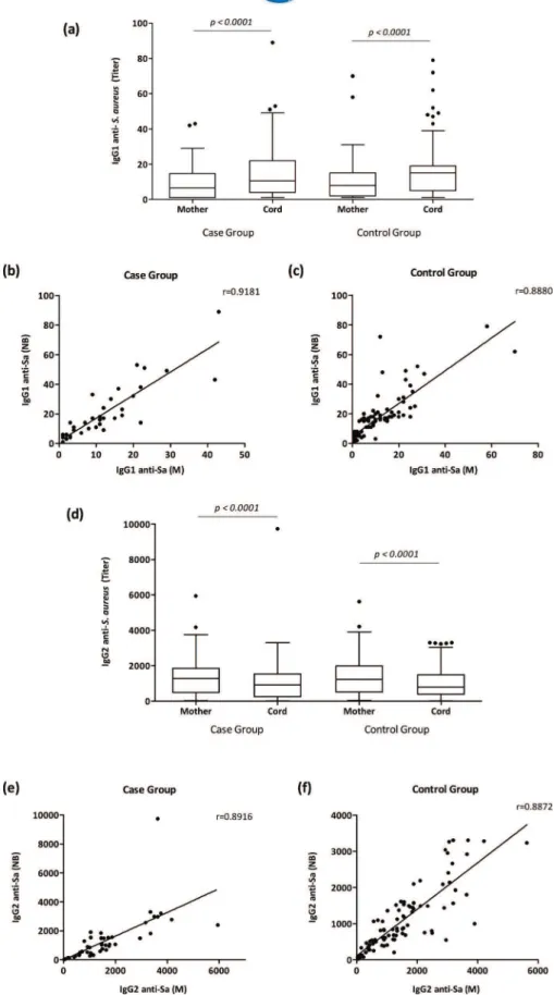
Anti-S. aureusIgG1 and IgG2 titers and placental transfer ratios
The Wilcoxon analyses demonstrated significantly higher anti-Sa IgG1 titers in newborns from the case and control
groups than those observed in their respective mothers (po0.0001), whereas the analysis of anti-Sa IgG2 showed
lower levels in the newborns from both groups than in their mothers (po0.0001) (Figure 1a and 1d). When the maternal
sera from the case group and the control group were compared, no differences were detected in the anti-Sa IgG1
or anti-SaIgG2 titers. The same result was observed between the newborns from both groups (Figure 1a and 1d).
As revealed by the Wilcoxon analyses, the comparison of the anti-SaIgG1 and IgG2 titers showed significantly higher IgG2 titers than anti-SaIgG1 levels in both the maternal and umbilical cord sera from both groups (po0.0001).
The Spearman correlation analysis revealed high correla-tion indexes of anti-Sa IgG1 and anti-Sa IgG2 between the paired maternal and newborn serum in both groups (po0.00001) (Figure 1b, c, e and f).
Interestingly, in the case group, the Spearman analysis showed a significant correlation index between the maternal anti-Sa IgG levels and the neonatal anti-Sa IgG2 titers
(r=0.601, po0.00001), and the opposite was also observed,
wherein the maternal anti-SaIgG2 titers were correlated with the neonatal anti-Sa IgG levels (r=0.544, po0.00001). The
same result was observed in the control group when the maternal anti-Sa IgG levels and the neonatal anti-Sa IgG2 titers were compared (r=0.343, po0.001) and when the
maternal anti-Sa IgG2 titers and the neonatal anti-Sa IgG
levels were compared (r=0.364, po0.001). Neither the
maternal nor neonatal anti-Sa IgG1 titers were correlated with the anti-SaIgG levels or the anti-SaIgG2 titers in either
group.
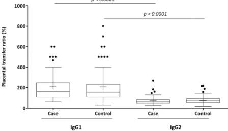
No statistically significant differences were detected in the medians of the anti-Sa IgG1 placental transfer ratios
between the case and control groups or when the placen-tal transfer ratios of anti-Sa IgG2 were analyzed. Alter-natively, the case and control groups revealed significantly higher anti-Sa IgG1 placental transfer ratios than IgG2
(Figure 2).
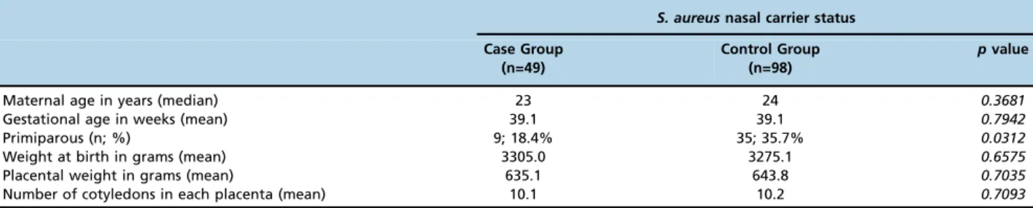
Table 1-Demographic and obstetric characteristics of the parturients (n=147) according to their nasalS. aureuscarrier status, Cuiaba´-MT.
S. aureusnasal carrier status Case Group
(n=49)
Control Group (n=98)
pvalue
Maternal age in years (median) 23 24 0.3681
Gestational age in weeks (mean) 39.1 39.1 0.7942
Primiparous (n; %) 9; 18.4% 35; 35.7% 0.0312
Weight at birth in grams (mean) 3305.0 3275.1 0.6575
Placental weight in grams (mean) 635.1 643.8 0.7035
Total and anti-S. aureusIgA concentrations in the colostrum
Although all of the colostrum samples were collected approximately 24 hours post-partum (range=17.5 - 24.6 hours), the total IgA in the colostra had a large range of variation. Significantly higher total IgA concentrations were observed in the colostrum samples from the case group than in those from the control group (po0.05). TheSa-specific IgA
concentrations in the maternal colostrum were equivalent between the groups (Table 3).
Avidity indexes of anti-S. aureusIgG and IgA
In both the case and control groups, the avidity indexes of the anti-Sa IgG antibodies did not differ between the
maternal and umbilical cord serum samples. The same result was observed for the avidity indexes of anti-Sa IgA in the colostrum samples, which did not show differences between the case and control groups (Table 4).
’ DISCUSSION
The present study revealed similar concentrations of anti-staphylococcal IgG antibodies in the sera of mothers with and without bacterial nasal carriage, suggesting that maternal anti-SaIgG antibodies are not related to colonization with this pathogen. The same effect was observed in the avidity indexes of anti-SaIgG between the maternal or neonatal sera from the case and control groups and between the paired maternal and cord sera. Avidity and antibody specificity are determined by the Fab’fragment of IgG, while placental transfer is Fc-related;
therefore, it was unexpected that antibody avidity could be different between the mother/newborn pairs (15).
S. aureusbecomes part of the host microbiota within the first hours of life (16), which allows healthy individuals to have high specific antibody levels independent of nasal colonization at the time of measurement (17). Furthermore, anti-staphylococcal IgG and IgA levels and Sa antigenic diversity show great individual variability in healthy individuals and in the presence of bacteremia (17,18). This variability has been attributed to strain characteristics, with
strain-specific arsenals of evasion mechanisms and differ-ences in the host immune response to this bacterium (19).
In the present study, the placental transfer of total IgG agrees with the current literature that describes positive IgG placental transfer ratios for newborns in healthy full-term pregnancies. However, when the mother has a high content of total IgG or of a specific antibody, the neonatal value usually remains below the maternal value due to the amount of cell surface receptors, and a lower placental transfer ratio is a result of the digestion of unbound IgG molecules by lysosomal enzymes within vesicles (20,21).
The finding that both groups had lower placental transfer ratios of anti-staphylococcal antibodies than expected in an assay performed with whole bacteria prompted us to evaluate specific IgG subclasses. Several studies have demo-nstrated that the immune response in adults is composed of antibodies reactive to antigenic determinants of bacterial surfaces related to different stages ofSapathogenesis;
never-theless, the highest levels of neutralizing antibodies are predominantly directed to non-protein antigens (17,22).
The higher anti-Sa IgG2 than IgG1 titers in maternal and
neonatal sera in both groups was unexpected because it is well known that preferential placental transport occurs for IgG1, followed by IgG4, IgG3 and IgG2, for which the FcRn receptors have the lowest affinity (23). In our study, to compare the anti-staphylococcal IgG1 results with those of IgG2 in each sample, the assays were performed under the same conditions, and the subclass results were expressed in titers. As expected, the placental transfer ratio of anti-SaIgG1 was higher than that of IgG2. However, as the maternal anti-staphylococcal IgG1 titers were much lower than those of IgG2, the anti-Sa IgG2 titers in neonatal serum were markedly higher than those of IgG1. The predominance of anti-SaIgG2 over IgG1 in the maternal sera could explain the
lower placental transfer ratio observed for IgG anti-Sa in both the case and control groups. This idea was further reinforced by the significant correlation indexes revealed when the maternal serum anti-Sa IgG and neonatal serum anti-Sa IgG2 were compared and vice-versa. A study with children with and without otitis media who were vaccinated
Table 2-Total IgG,S. aureus-specific IgG antibodies and placental IgG transfer ratios in the maternal and umbilical cord serum samples from the case (n=45) and control (n=98) groups.
Total IgG (mg/dL)
Case Group Control Group pvalue Maternal Serum 1069.0
[991.0-1116.0]
939.0* [907.0-996.0]
po0.01
Cord Serum 1034.0 [1027.0-1138.0]
1041.0* [1044.0-1146.0]
Cord/Maternal Ratio (%) 100.9 [98.9-112.6]
114.1 [112.6-125.9]
po0.05
Anti-S. aureusIgG (AU/mL)
Case Group Control Group pvalue Maternal Serum 317.0w
[366.6-678.8]
317.4w [409.4-579.9]
Cord Serum 224.7w [243.3-453.8]
264.7w [308.4-429.2]
Cord/Maternal Ratio (%) 69.5 [65.5-76.3]
81.3 [77.3-90.1]
po0.05
The values represent median and confidence intervals (95%).
* Higher total IgG concentrations in the newborns from the control group compared with their respective mothers (po0.0001).
Figure 1 -Anti-S. aureusIgG1 and IgG2 titers in paired maternal and cord serum samples from the case and control groups.(a)
withStreptococcus pneumoniae,another Gram-positive bacter-ium, showed no differences between groups, and strong correlations were demonstrated between anti-PCP (capsular antigen of S. pneumoniae) IgG and the anti-PCP IgG2, as observed in our study (24).
In a study that correlated the protective immune response to a conjugated pneumococcal vaccine, high levels of speci-fic IgG, IgG1 and IgG2 were found, and both IgG subclas-ses correlated significantly with the opsonophagocytic activity. No significant increases were found in the mean antibody avidities, which were already high in the pre-vaccination samples (25). However, they revealed that neu-tralization of antibodies directed to the cell wall polysac-charide had no influence on the opsonophagocytic activity of the sera. It has been reported that anti-pneumococcal cell wall polysaccharide antibodies have very little opsonic activity (26).
It is well known that complement poorly lyses the plasma membranes of Gram-positive bacteria because of their thick peptidoglycan layers (27). Thus, it has been argued that antibody-mediated opsonophagocytic bacterial killing is neces-sary to increase the bactericidal efficiency of MAC against Gram-positive bacteria (28). IgM antibodies and IgG1 and IgG3 subclasses are known to effectively act in these processes, whereas IgG2 shows a much weaker ability. According to our results, this could be an additional limiting factor for newborns to cope with these bacteria (29).
Vaccine trials using theSacapsular polysaccharide 5 and 8 conjugate vaccine revealed that, despite a substantial rise in the post-vaccination anti-CP5 and anti-CP8 antibody con-centrations, theSa nasal colonization rates did not signifi-cantly change (30).
These results highlight the importance of therapeutic strategies to address Gram-positive bacteria using the induction of Figure 2 -Placental transfer ratios (%) of anti-S. aureusIgG1 and IgG2 from case and control groups. Comparison of the data is presented as box and whisker plots. The box represents the 25th–75thpercentiles, and the median is represented by the line within the box. The whiskers represent the 5th–95thpercentiles.
Table 3-Total andS. aureus-specific IgA antibodies in the maternal colostrum samples from the case (n=45) and control (n=98) groups.
Case Group Control Group pvalue Total IgA (g/L) 163.3
[152.2 - 216.2]
125.7 [125.8 - 158.3]
po0.05
Anti-S. aureusIgA (AU/mL) 4019.0 [3747.0 - 7024.0]
5337.0 [6048.0 - 12423.0]
The values represent median and confidence intervals (95%).
Table 4-Avidity indexes of anti-S. aureus(Sa) IgG in the maternal and umbilical cord serum samples and IgA in the maternal colostrum from the case (n=17) and control (n=18) groups.
Serum Anti-SaIgG Avidity Index (M)* Colostrum Anti-SaIgA Avidity Index (M)
Case Group Control Group Case Group Control Group Maternal Cord Maternal Cord
Median 1.4 1.3 1.5 1.2 2.5 2.3
Min 0.9 0.9 0.7 0.8 1.3 1.2
Max 3.5 2.9 3.7 3.2 4.0 2.8
CI 95% 1.2-1.9 1.2-1.8 1.3-1.9 1.2-1.9 2.0-2.8 2.0-2.5
immunity by vaccination or passive immunization. These strat-egies should consider the subclass produced or administered because IgG1 is the main subclass involved in opsonophago-cytosis and the interaction of innate and acquired immunity.
Studies aimed at developing vaccines against Saare still performed because carriers have more infections, but these are less severe (31,32), which strongly suggests that some form of immunity has developed during prolonged colonization. The rationale for the development of those vaccines is based on evidence that antibodies directed to toxic shock toxin (33) and a-hemolysin (Hla) reduce the severity of the disease (34), and inhibitors of Hla receptor are also protective (35).
In pregnant women, due to the mucosal immune system and broncoenteromammary axis, secretory IgA produced by the mother against pathogens that colonize mucous mem-branes is offered to the newborn through the breast milk. This occurs because, once in the nasal site, Sa can interact
with the nasopharynx-associated lymphoid tissue (NALT), and anti-staphylococcal IgA-producing B lymphocytes can migrate to the mammary gland. The mammary gland then releases specific IgA in breast milk that may help to prevent nasal colonization, since the main characteristic of secretory IgA antibody is to perform immune exclusion by inhibiting colonization and invasion by pathogens (10).
Kawano and Emori (36) assessed total secretory IgA in human milk and found higher IgA concentrations in the colostrum samples at 72 hours post-partum in primiparous women. In the present study, there were more primiparous women in the control group, but we found higher total IgA concentrations in the case group. However, the total IgA concentrations in the colostrum at 24 hours did not differ between primiparous and multiparous women.
Because we found no differences in the anti-Sa IgA
con-centrations and avidity indexes between the groups, nasal colonization was not related to an increase in specific IgA antibodies or avidity indexes in the colostrum.
In conclusion, this study revealed that, in healthy pregnant women, nasal colonization by S. aureus at delivery is not associated with higher specific serum IgG concentrations in the mother or in newborns or with higher specific secretory IgA in the colostrum. The high titers of anti-SaIgG2 found in the umbilical cord serum suggest a greater reactivity with non-protein antigens, which may further contribute to the susceptibility to staphylococcal infections at birth. Because newborns are early exposed to S. aureus the detection of specific IgA with avidity for this pathogen in the colostrum reinforces the importance of encouraging breastfeeding shortly after birth.
’ ACKNOWLEDGMENTS
This work was supported by CAPES–Coordenac¸ão de Aperfeic¸oamento
de Pessoal de Nível Superior, Project number AUX-PE-DINTER/NF 2535/2009 Grant. The authors are grateful to the mothers who kindly agreed to participate in this study.
’ AUTHOR CONTRIBUTIONS
Nadaf MI, Takano OA and Palmeira P designed the overall study and wrote the manuscript. Nadaf MI and Lima L performed the study, analyzed and interpreted data. Nadaf MI and Takano OA contributed with
newborns’ samples and the data collection. Stranieri I performed the
S. aureusisolation and identification in nasal samples; Carneiro-Sampaio M
analyzed and discussed the results, read and discussed the paper. Palmeira P supervised the performance of experiments, oversaw data analysis and
interpretation, co-wrote and edited the manuscript. All authors contributed
to thefinal version of the manuscript and approved it.
’ REFERENCES
1. Jursa-Kulesza J, Kordek A, Kopron K, Wojciuk B, Giedrys-Kalemba S. Molecular studies of an impetigo bullosa epidemic in full-term infants. Neonatology. 2009;96(1):61-8, http://dx.doi.org/10.1159/000204961. 2. Mir F, Tikmani SS, Shakoor S, Warraich HJ, Sultana S, Ali SA, et al.
Incidence and etiology of omphalitis in Pakistan: a community-based cohort study. J Infect Dev Ctries. 2011;5(12):828-33.
3. Montgomery CO, Siegel E, Blasier RD, Suva LJ. Concurrent septic arthri-tis and osteomyeliarthri-tis in children. J Pediatr Orthop. 2013;33(4):464-7, http://dx.doi.org/10.1097/BPO.0b013e318278484f.
4. Downie L, Armiento R, Subhi R, Kelly J, Clifford V, Duke T. Community-acquired neonatal and infant sepsis in developing countries: efficacy of
WHO’s currently recommended antibiotics-systematic review and
meta-analysis. Arch Dis Child. 2013;98(2):146-54, http://dx.doi.org/10.1136/ archdischild-2012-302033.
5. Power Coombs MR, Kronforst K, Levy O. Neonatal host defense against staphylococcal infections. Clin Dev Immunol. 2013;2013:826303, http://dx.doi.org/10.1155/2013/826303.
6. Harrison LM, Morris JA, Lauder RM, Telford DR. The effect of
Staphylo-coccus aureuscarriage in late pregnancy on antibody levels to staphylo-coccal toxins in cord blood and breast milk. FEMS Immunol Med Microbiol. 2008;54(1):137-43, http://dx.doi.org/10.1111/j.1574-695X.2008. 00463.x.
7. Jimenez-Truque N, Tedeschi S, Saye EJ, McKenna BD, Langdon W, Wright JP, et al. Relationship between maternal and neonatalStaphylococcus aureus
colonization. Pediatrics. 2012;129(5):e1252-9, http://dx.doi.org/10.1542/ peds.2011-2308.
8. Leshem E, Maayan-Metzger A, Rahav G, Dolitzki M, Kuint J, Roytman Y,
et al. Transmission ofStaphylococcus aureusfrom mothers to newborns.
Pediatr Infect Dis J. 2012;31(4):360-3, http://dx.doi.org/10.1097/INF. 0b013e318244020e.
9. Rigby KM, DeLeo FR. Neutrophils in innate host defense against
Staphylococcus aureusinfections. Semin Immunopathol. 2012;34(2):237-59, http://dx.doi.org/10.1007/s00281-011-0295-3.
10. Brandtzaeg P. Secretory IgA: designed for anti-microbial defense. Front Immunol. 2013;4:222, http://dx.doi.org/10.3389/fimmu.2013.00222. 11. Corthésy B. Role of secretory IgA in infection and maintenance of
homeo-stasis. Autoimmunity Rev. 2013;12(6):661-5, http://dx.doi.org/10.1016/ j.autrev.2012.10.012.
12. Quinello C, Quintilio W, Carneiro-Sampaio M, Palmeira P. Passive acquisition of protective antibodies reactive with Bordetella pertussis in newborns via placental transfer and breast-feeding. Scand J Immunol. 2010;72(1):66-73.
13. Carbonare SB, Palmeira P, Silva ML, Carneiro-Sampaio MM. Effect of microwave radiation, pasteurization and lyophilization on the ability of human milk to inhibit Escherichia coli adherence to HEp-2 cells. J Diarrhoeal Dis Res. 1996;14(2):90-4.
14. Jones CL, Macdonald RA, Hosking CS, Roberton DM. Estimating the relative avidity of mucosal IgA for antigen. J Immunol Methods. 1987; 105(1):111-7, http://dx.doi.org/10.1016/0022-1759(87)90420-0. 15. Palmeira P, Costa-Carvalho BT, Arslanian C, Pontes GN, Nagao AT,
Carneiro-Sampaio MM. Transfer of antibodies across the placenta and in breast milk from mothers on intravenous immunoglobulin. Pediatr Allergy Immunol. 2009;20(6):528-35, http://dx.doi.org/10.1111/j.1399-3038.2008.00828.x.
16. Acton DS, Plat-Sinnige MJ, van Wamel W, de Groot N, van Belkum A. Intestinal carriage ofStaphylococcus aureus: how does its frequency com-pare with that of nasal carriage and what is its clinical impact? Eur J Clin Microbiol Infect Dis. 2009;28(2):115-27, http://dx.doi.org/10.1007/ s10096-008-0602-7.
17. Dryla A, Prustomersky S, Gelbmann D, Hanner M, Bettinger E, Kocsis B, et al. Comparison of antibody repertoires against Staphylococcus aureus in healthy individuals and in acutely infected patients. Clin Diagn Lab Immunol. 2005;12(3):387-98.
18. Verkaik NJ, Boelens HA, de Vogel CP, Tavakol M, Bode LG, Verbrugh HA, et al. Heterogeneity of the humoral immune response following Staphy-lococcus aureus bacteremia. Eur J Clin Microbiol Infect Dis. 2010; 29(5):509-18, http://dx.doi.org/10.1007/s10096-010-0888-0.
19. Bröker BM, van Belkum A. Immune proteomics of Staphylococcus
aureus. Proteomics. 2011;11(15):3221-31, http://dx.doi.org/10.1002/pmic. 201100010.
20. Englund JA. The influence of maternal immunization on infant immune responses. J Comp Pathol. 2007;137 Suppl 1:S16-9, http://dx.doi.org/ 10.1016/j.jcpa.2007.04.006.
21. Roopenian DC, Akilesh S. FcRn: the neonatal Fc receptor comes of age. Nat Rev Immunol. 2007;7(9):715-25, http://dx.doi.org/10.1038/nri2155. 22. Skurnik D, Merighi M, Grout M, Gadjeva M, Maira-Litran T, Ericsson M,
antigens mutually neutralize opsonic killing and protection in mice. J Clin Invest. 2010;120(9):3220-33, http://dx.doi.org/10.1172/JCI42748. 23. Palmeira P, Quinello C, Silveira-Lessa AL, Zago CA, Carneiro-Sampaio
M. IgG placental transfer in healthy and pathological pregnancies. Clin Dev Immunol. 2012;2012:985646, http://dx.doi.org/10.1155/2012/ 985646.
24. Drake-Lee AB, Hughes RG, Dunn C. Serum IgA and IgG functional antibodies and their subclasses to Streptococcus pneumoniae capsular antigen found in two aged-matched cohorts of children with and without otitis media with effusion. Clin Otolaryngol Allied Sci. 2003;28(4):335-40, http://dx.doi.org/10.1046/j.1365-2273.2003.00717.x.
25. Anttila M, Voutilainen M, Jäntti V, Eskola J, Käyhty H. Contribution of serotype-specific IgG concentration, IgG subclasses and relative antibody avidity to opsonophagocytic activity against Streptococcus pneumoniae. Clin Exp Immunol. 1999;118(3):402-7, http://dx.doi.org/10.1046/j.1365-2249.1999.01077.x.
26. Vitharsson G, Jónsdóttir I, Jónsson S, Valdimarsson H. Opsonization and antibodies to capsular and cell wall polysaccharides of Streptococcus pneumoniae. J Infect Dis. 1994;170(3):592-9, http://dx.doi.org/10.1093/ infdis/170.3.592.
27. Ricklin D, Hajishengallis G, Yang K, Lambris JD. Complement: a key system for immune surveillance and homeostasis. Nat Immunol. 2010; 11(9):785-97, http://dx.doi.org/10.1038/ni.1923.
28. Daha NA, Banda NK, Roos A, Beurskens FJ, Bakker JM, Daha MR, et al. Complement activation by (auto-) antibodies. Mol Immunol. 2011; 48(14):1656-65, http://dx.doi.org/10.1016/j.molimm.2011.04.024. 29. Bindon CI, Hale G, Brüggemann M, Waldmann H. Human monoclonal
IgG isotypes differ in complement activating function at the level of C4 as
well as C1q. J Exp Med. 1988;168(1):127-42, http://dx.doi.org/10.1084/ jem.168.1.127.
30. Creech CB 2nd, Johnson BG, Alsentzer AR, Hohenboken M, Edwards KM, Talbot TR 3rd. Vaccination as infection control: a pilot study to determine the impact ofStaphylococcus aureusvaccination on nasal carriage. Vaccine. 2009;28(1):256-60, http://dx.doi.org/10.1016/j.vaccine.2009.09.126. 31. Verkaik NJ, de Vogel CP, Boelens HA, Grumann D, Hoogenboezem T,
Vink C, et al. Anti-staphylococcal humoral immune response in persistent
nasal carriers and noncarriers of Staphylococcus aureus. J Infect Dis.
2009;199(5):625-32, http://dx.doi.org/10.1086/596743.
32. Verkaik NJ, van Wamel WJ, van Belkum A. Immunotherapeutic
approa-ches against Staphylococcus aureus. Immunotherapy. 2011;3(9):1063-73,
http://dx.doi.org/10.2217/imt.11.84.
33. Spaulding AR, Lin YC, Merriman JA, Brosnahan AJ, Peterson ML,
Schlievert PM. Immunity to Staphylococcus aureus secreted proteins
protects rabbits from serious illnesses. Vaccine. 2012;30(34):5099-109, http://dx.doi.org/10.1016/j.vaccine.2012.05.067.
34. Fritz SA, Tiemann KM, Hogan PG, Epplin EK, Rodriguez M, Al-Zubeidi DN, et al. A serologic correlate of protective immunity against
commu-nity-onset Staphylococcus aureus infection. Clin Infect Dis. 2013;
56(11):1554-61, http://dx.doi.org/10.1093/cid/cit123.
35. Foletti D, Strop P, Shaughnessy L, Hasa-Moreno A, Casas MG, Russell M, et al. Mechanism of action and in vivo efficacy of a human-derived
antibody against Staphylococcus aureus a-hemolysin. J Mol Biol. 2013;
425(10):1641-54, http://dx.doi.org/10.1016/j.jmb.2013.02.008.