C
a s eR
e p o Rt3 3 0 Arq Bras Oftalmol. 2016;79(5):330-2 http://dx.doi.org/10.5935/0004-2749.20160094
Toxic anterior segment syndrome following deep anterior lamellar keratoplasty
Síndrome tóxica do segmento anterior após transplante lamelar anterior profundo
NeslihaN sevimli1, Remzi KaRadag2, OzguR CaKiCi1, huseyiN BayRamlaR2, seydi OKumus3, uNsal saRi2
Submitted for publication: July 13, 2015 Accepted for publication: November 6, 2015
1 Department of Ophthalmology, Istanbul Medeniyet University Goztepe Research and Training
Hospital, Istanbul, Turkey.
2 Department of Ophthalmology, Istanbul Medeniyet University School of Medicine, Istanbul, Turkey. 3 Department of Ophthalmology, Gaziantep University School of Medicine, Gaziantep, Turkey.
Funding: No specific financial support was available for this study.
Disclosure of potential conflicts of interest: None of the authors have any potential conflicts of interest to disclose.
Corresponding author: Remzi Karadag. Department of Ophthalmology. School of Medicine. Istanbul Medeniyet University - Goztepe, Istanbul - Turkey - E-mail: [email protected]
ABSTRACT
We present the case of a 31-year-old patient with toxic anterior segment syn-drome (TASS) that developed after undergoing deep anterior lamellar keratoplasty (DALK). She had keratoconus, and despite wearing hard contact lenses for many years in the left eye, her vision had deteriorated; therefore, DALK was performed on this eye.The preoperative visual acuity (VA) was finger counting at 3 m. Routine DALK was performed using the “big-bubble” technique. The corneal entry incision was hydrated at the end of the surgery, which was terminated by air injection into the anterior chamber. On postoperative day 1, VA was at the level of hand movements, and the cornea was edematous. Topical high-dose dexamethasone and oral steroids were initiated considering the diagnosis of TASS. Subsequently, the patient’s VA increased, and the corneal edema decreased. We believe that the use of re-sterilized cannulas may have been the likely cause of TASS. Although DALK can be performed without interfering with the anterior chamber, one should keep in mind that TASS may occur in response to the solution used to hydrate the incision site and the air injected into the anterior chamber.
Keywords: Keratoplasty; Anterior eye segment/pathology; Keratoconus; Corneal transplantation
RESUMO
Apresentamos o relato de uma paciente com 31 anos de idade, que desenvolveu síndrome tóxica do segmento anterior ( TASS) após o procedimento de transplante lamelar anterior profundo (DALK). Ela apresentava ceratocone e, apesar de ter usado lentes de contato rígidas por muitos anos no olho esquerdo, apresentou deterioração da visão nesse olho que foi submetido a procedimento DALK. A acuidade visual (VA) era de conta dedos a três metros. O procedimento DALK de rotina foi realizado utilizando técnica de bolha grande (Big Bubble). A incisão de entrada da córnea foi hidratada ao final da cirurgia que foi terminada com a injeção de ar na câmara anterior. No primeiro dia de pós-operatório a VA era de percepção de movimentos da mão e a córnea estava edemaciada. Dexametasona tópica em alta dose e esteróides orais foram iniciadas ao se considerar o diagnóstico de TASS. Acreditamos que o uso de cânulas reesterilizadas podem ter sido a causa provável da TASS. A VA melhorou e o edema da córnea do diminuiu durante a evolução. Embora o procedimento DALK foi realizado sem interferir com câmara anterior, deve-se ter em mente que TASS pode ocorrer com a solução utilizada para hidratar o local da incisão e o ar injetado na câmara anterior.
Descritores: Ceratoplastia; Segmento anterior do olho/patologia; Ceratocone;
Trans-plante de córnea
INTRODUCTION
Toxic anterior segment syndrome (TASS) is an acute and nonin-fectious inflammation of the anterior segment. Most cases have been reported as occurring after cataract surgery(1). Anterior segment inflammation usually occurs within 12-48 h after surgery, and the symptoms include decreased visual acuity, increased intraocular pressure, corneal edema, anterior chamber (AC) inflammation, fibrin formation, hypopyon, and fixed pupils. The vitreous body is not in-fected in this syndrome(2).
Various contaminants, usually from surgical equipment or sup-plies, including denatured ophthalmic viscosurgical devices (OVDs), pre servatives, talc material in surgical gloves, topical ophthalmic ointment, inappropriately reconstituted intraocular preparations, heat-stable endotoxins, and detergents have all been suspected as causes of TASS(1-3). Further, cataract surgery(1-3), iris-supported phakic in-traocular lenses (IOLs)(4), penetrating keratoplasty(5), and Descemet’s stripping automated endothelial keratoplasty (DSAEK)(6) have all been speculated as causing TASS. Most cases of TASS can be success-fully treated with topical steroids and nonsteroidal anti-inflammatory agents(3). To our knowledge, TASS the following DALK case has not been previously reported in the literature.
In this case report, we present a case of TASS following uncom-plicated DALK.
CASE REPORT
Se v i m l N, e ta l.
3 3 1 Arq Bras Oftalmol. 2016;79(5):330-2 was topically or intracamerally applied, and moxifloxacin eye drops
(0.1 ml) and dexamethasone solution (0.4 ml) were subconjunctivally injected. Ointments were not used at the end of the surgery. Up to this point, we had been using the cannulas that have also been used for viscoelastic injection, a couple of times. To clean them, we routinely wash out the cannulas with distilled water, pass 10 ml of air with an injector through the lumen, and put them in the autoclave immediately after use to avoid waste sedimentation in the lumen. We do not use enzymatic or other cleaning detergents or an ultrasonic bath. During the postoperative course, topical loteprednol etabo-nate, antibiotic eye drops, and artificial tears drops were initiated. One day postoperatively, the patient had a moderately painful eye; her visual acuity was at the level of hand movements, and there was edema in the cornea. The pupil was mid-dilated, and light reactions were weak. There was no hypopyon in the AC, and no vitreous pa-thology on ultrasonography. Topical high-dose dexamethasone was initiated, and samples from the solution and donor cornea were sent to a microbiology laboratory. Oral steroids were started because the symptoms did not regress with treatment, and pupillary membrane formation occurred despite treatment (Figure 1 A, B). There were no microorganisms in the cultures. At the follow-up visits, visual acuity increased, and the corneal edema and membrane formation decrea-sed. Treatment was gradually discontinued. At the 18-month follow up, spectacle-corrected visual acuity was 4/10 with a Snellen chart, the cornea was clear, the pupil was mid-dilated, and iris pigments were present on the crystalline lens (Figure 2).
We investigated the medical records and found that there had been no other cases of TASS at our institute over the same period (1 month before and after). In fact, we have not encountered more than three cases of TASS over the last year.
DISCUSSION
TASS is an acute inflammation in the AC, and increasing rates of TASS diagnosis have recently been found(1-3). It is important to differentiate TASS from postoperative endophthalmitis(7). Clinically, TASS occurs during the early postoperative period, with symptoms usually beginning within 12-48 h after surgery(1-3,7). However, it has been reported that early-onset endophthalmitis cases may be seen due to the presence of Staphylococcusepidermidis and Bacillus cereus
similarly, which are also seen in TASS(3). Inflammation is restricted to the AC in TASS, and AC reaction, fibrin formation, hypopyon, and corneal edema are observed in this syndrome(8).
TASS often occurs due to contamination of the surgical instru-ments used during surgery, OVDs, improper intraoperative drugs, or talc material in gloves(1-3,7). The most widely known risk factor is the procedures applied during sterilization(9). Maier et al. detected sterile keratitis in 24 patients following penetrating keratoplasty during the postoperative period. TASS was diagnosed, and they reported that the trephine system was responsible for the development of TASS(5).
In a previous study, TASS was detected in three patients with foldable iris-fixated phakic IOLs. These patients were successfully treated with intensive topical steroids. They also reported that endo-toxins in OVDs may cause TASS(4). Sorkin and Varssano reported a case of TASS following DSAEK, phacoemulsification, and IOL implantation. The patient was successfully treated with cycloplegic and topical steroids(6).
There have been reports of TASS following penetrating kerato-plasty(5) and DSAEK(6) in the literature, but there has never been a reported case of TASS following DALK. In our case, TASS occurred following uncomplicated DALK. In contrast to penetrating kerato-plasty and DSAEK, entry into the AC is extremely rare in DALK. The purpose of entering the AC is to open a side-port with an MVR knife to reduce the AC pressure, thus allowing the side-port to be hydrated with BSS solution and letting some air into the AC. In our case, we used disposable MVR blades. Therefore, we do not believe that TASS
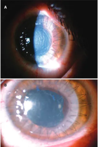
Figure 1. A) Severe postoperative corneal edema; B) 5 days postoperatively, the corneal edema and pupillary membrane formation decreased.
A
B
Figure 2. Slit-lamp image at 18 months follow up.
To x i ca n T e r i o rs e g m e n Ts y n d r o m ef o l l o w i n gd e e pa n T e r i o rl a m e l l a rk e r aT o p l a s T y
3 3 2 Arq Bras Oftalmol. 2016;79(5):330-2
particles enter the AC during these processes, they will not be remo-ved, and may lead to a reaction. TASS is mostly reported after cataract surgery(1-3). There is plenty of irrigation from phaco equipment during phacoemulsification, and more fluid is used during this surgery. The-refore, even if there are particles in the cannula, they may be cleaned or diluted with more liquid, thus lowering the risk of a reaction.
Contact with the AC is relatively lesser during DALK than during other similar procedures. This contact is during the final stages of the operation, and thus, it is important to use a new cannula, or if it is absolutely necessary to re-use it, it should be cleaned by passing plenty of fluids through it.
REFERENCES
1. Mamalis N, Edelhauser HF, Dawson DG, Chew J, LeBoyer RM, Werner L. Toxic anterior segment syndrome. J Cataract Refract Surg. 2006;32(2):324-33.
2. Centers for Disease Control and Prevention (CDC). Toxic anterior segment syndrome after cataract surgery--Maine, 2006. MMWR Morb Mortal Wkly Rep. 2007;29;56(25): 629-30.
3. Holland SP, Morck DW, Lee TL. Update on toxic anterior segment syndrome. Curr Opin Ophthalmol. 2007;18(1):4-8.
4. van Philips LA. Toxic anterior segment syndrome after foldable artiflex iris-fixated phakic intraocular lens implantation. J Ophthalmol. 2011;2011:982410.
5. Maier P, Birnbaum F, Böhringer D, Reinhard T. Toxic anterior segment syndrome following penetrating keratoplasty. Arch Ophthalmol. 2008;126(12):1677-81. 6. Sorkin N, Varssano D. Toxic anterior segment syndrome following a triple descemet’s
stripping automated endothelial keratoplasty procedure. Case Rep Ophthalmol. 2012;3(3):406-9.
7. Gopal L, Vijaya L. Toxic anterior segment syndrome. Br J Ophthalmol. 2013;97(8):953. 8. Choi JS, Shyn KH. Development of toxic anterior segment syndrome immediately
after uneventful phaco surgery. Korean J Ophthalmol. 2008;22(4):220-7.
9. Cutler Peck CM, Brubaker J, Clouser S, Danford C, Edelhauser HE, Mamalis N. Toxic anterior segment syndrome: Common causes. J Cataract Refract Surg. 2010;36(7): 1073-80.