online | memorias.ioc.fiocruz.br
Hepatitis C virus (HCV) is estimated to infect around 170 million people worldwide and is one of the main causes of chronic liver failure (Brown & Gaglio 2003). Combined treatment with interferon and ribavirin has been shown to interrupt fibrogenesis and can even reverse liver fibrosis when a sustained virological response (SVR) is obtained (Poynard et al. 2002). Unfortunately, the response rates are low, especially among genotype 1 infected patients, older patients and patients with advanced liver fibrosis (Manns et al. 2001, Fried et al. 2002, Lee et al. 2002, Mihm et al. 2006, Yamada et al. 2008). Identifying appropriate candi-dates for treatment is desirable due to the high cost and potentially serious adverse effects of the current therapy.
Treatment for chronic hepatitis C (CHC) patients cur-rently consists of a combination of interferon-α (IFN-α) and ribavirin for 24 or 48 weeks depending on the viral genotype (Strader et al. 2004, de Araújo et al. 2007). Ex-ogenous IFN-α acts similarly to its endEx-ogenous coun -terpart through induction of the IFN-stimulated genes responsible for establishing an antiviral state within the
cell (Tilg 1997, Feld & Hoofnagle 2005). In addition to its direct antiviral activity, IFN-α strengthens both in -nate and adaptive immune responses through interac-tions with T lymphocytes, natural killer cells and den-dritic cells (Tilg 1997). Ribavirin, a guanosine-analogue, seems to work as an antiviral agent mainly by inducing HCV mutagenesis, which results in a lower replicative profile (Feld & Hoofnagle 2005). Ribavirin also acts as an immunomodulator, enhancing type 1 T helper cell (Th)-1 cytokine secretion and altering the Th1/Th2 bal-ance in favor of a Th1 response (Tam et al. 1999).
SVRs [i.e., negative HCV qualitative polymerase chain reaction (PCR) 24 weeks after the end of treatment] are observed in approximately half of the treated patients and many factors, such as age, genotype, viral load and body weight, are related to treatment outcome (Manns et al. 2001, Friedet al. 2002, Asselah et al. 2010). The outcome of HCV treatment seems also to depend on the ability of host cellular immune responses to control viral replica-tion. An early Th1 response is key for viral clearance dur-ing acute HCV infection (Guidotti & Chisari 2001, Ka-mal et al. 2004, Rehermann & Nascimbeni 2005) and an enhanced HCV-specific T-cell response is also associated with treatment response in chronically infected patients (Nelson et al. 1998, Cramp et al. 2000, Kamal et al. 2002, 2004). Therefore, it is reasonable to suppose that pretreat-ment levels of cytokines, especially those involved in the Th1 response, may predict treatment outcome.
Soluble inflammatory markers as predictors of virological
response in patients with chronic hepatitis C virus
infection treated with interferon-
α
plus ribavirin
Alexandre Sampaio Moura1/+,Ricardo Andrade Carmo2,Antonio Lucio Teixeira1, Mauro Martins Teixeira1,Manoel Otávio da Costa Rocha1
1Programa de Pós-graduação em Infectologia e Medicina Tropical
2Centro de Treinamento e Referência em Doenças Infecciosas e Parasitárias Orestes Diniz, Faculdade de Medicina,
Universidade Federal de Minas Gerais, Av. Alfredo Balena 190, 30130-100 Belo Horizonte, MG, Brasil
The host immune response plays an important role in viral clearance in patients who are chronically infected with hepatitis C virus (HCV) and are treated with interferon and ribavirin. Activation of the immune system involves the release of pro and anti-inflammatory molecules that can be measured in plasma samples. The present study aimed to evaluate the association between pretreatment plasma levels of chemokines and soluble tumor necrosis factor recep-tors (sTNF-R) and the virological response in treated patients with chronic hepatitis C infection. Forty-one chronically-infected HCV patients that were being treated with interferon-α (IFN-α) plus ribavirin were included in the study. Socio-demographic, clinical and laboratory data were collected and pretreatment plasma levels of chemokine CCL2, CCL3, CCL11, CCL24, chemokine CXCL9, CXCL10, sTNF-R1 and sTNF-R2 were measured. The virological response was assessed at treatment week 12, at the end of treatment and 24 weeks after treatment. Pretreatment CXCL10 levels were significantly higher in patients without an early virological response (EVR) or sustained virological response (SVR) compared to responders [512.9 pg/mL vs. 179.1 pg/mL (p = 0.011) and 289.9 pg/mL vs. 142.7 pg/mL (p = 0.045), respectively]. The accuracy of CXCL10 as a predictor of the absence of EVR and SVR was 0.79 [confidence interval (CI) 95%: 0.59-0.99] and 0.69 (CI 95%: 0.51-0.87), respectively. Pretreatment plasma levels of the other soluble inflamma-tory markers evaluated were not associated with a treatment response. Pretreatment CXCL10 levels were predictive of both EVR and SVR to IFN-α and ribavirin and may be useful in the evaluation of candidates for therapy.
Key words: hepatitis C - chemokines - tumour necrosis factor receptors - interferon-alpha
Financial support: CNPq, FAPEMIG
+ Corresponding author: alexandresmoura@gmail.com Received 15 May 2010
Chemokines, a subgroup of small cytokines, are in-volved in leukocyte trafficking through a process called haptotaxis, in which leukocytes move towards higher con-centrations of chemokines (Baggiolini et al. 1997, Mackay 1997, Luster 1998, Kunkel 1999, Gerard & Rollins 2001, Ono et al. 2003, Charo & Ransohoff 2006). Chemokines are also involved in leukocyte activation, lymphocyte dif-ferentiation, regulation of the Th1/Th2 balance, angiogen-esis and fibrogenangiogen-esis (Charo & Ransohoff 2006).
As the Th1 response is particularly involved in treat-ment responses of CHC patients, there is special interest in chemokines that are responsible for the recruitment of Th1 cells into the liver (Moura et al. 2009). The most important of these chemokines are CCL2 (monocyte chemotactic protein-1), CCL3 (macrophagic inflamma-tory protein 1 alpha), CCL4 (macrophagic inflammainflamma-tory protein 1 beta), CCL5 (regulated upon activation, normal T cell expressed and secreted), CXCL9 (monokine in-duced by IFN-gamma) and CXCL10 (interferon-gamma inducible protein). The above mentioned CCL chemokines bind to C-C chemokine receptor type 5 while the CXCL chemokines bind to CXC chemokine receptor type 3 (Bonecchi et al. 1998). In the liver, chemokines are mainly produced by activated monocytes, Kupffer cells, endothelial cells and hepatocytes (Koziel 1999, Apoli-nario Fernández de Sousa & García Monzón 2003).
Another cytokine, tumor necrosis factor (TNF)-α, does not affect HCV RNA or protein synthesis and, therefore, does not seem to be directly involved in HCV clearance (Frese et al. 2003). TNF synthesis is sup -pressed by IFN-α during HCV treatment (Abu-Khabar et al. 1992) and among patients treated with IFN-α and ribavirin, serum TNF-α levels significantly decreased at the end of treatment (Neuman et al. 2001). Soluble TNF-α receptors (sTNF-R), which are released by ac -tivated neutrophils, mononuclear blood cells and fibro-blasts (Porteu et al. 1991, Lien et al. 1995) in response to mediators, such as interferon and TNF-α itself (Lantz et al. 1990, Joyce et al. 1994, Lien et al. 1995, Tilg et al. 1995), retain their ability to bind circulating TNF-α and are important in regulating its activity. These sTNF-R may contribute to the anti-inflammatory action of IFN-α. In hepatitis B virus infection, elevated serum levels of sTNF-R before interferon therapy were found to predict a successful response to treatment (Marinos et al. 1995). In hepatitis C, the role of sTNF-R in pre -dicting treatment responses is still not clear. Consider-ing the characteristics of sTNF-R, includConsider-ing its greater stability in peripheral circulation, further studies are warranted to better understand the role of sTNF-R in predicting treatment responses.
We sought to investigate the association between peripheral pretreatment levels of several inflammatory markers, such as sTNF-R1 and sTNF-R2 and chemokines (CCL2, CCL3, CCL11, CCL24, CXCL9, CXCL10) and the virological response to IFN-α and ribavirin among patients with chronic HCV infection.
PATIENTS, MATERIALS AND METHODS
Patients - Between June 2005-December 2007, 41 con-secutive patients with chronic hepatitis C infection who were submitted for treatment at the Orestes Diniz
Cen-ter, a public university-based referral service for chronic hepatitis patients in Belo Horizonte, Minas Gerais, Brazil, were recruited for the study. All included patients were adults, had a positive anti-HCV antibody test [enzyme linked immunosorbent assay (ELISA)-3, Ortho Diag-nostic Systems] and had HCV RNA that was detectable by PCR (AMPLICOR®, Roche Molecular Systems) for
more than six months. All patients had available liver bi-opsy samples with a length ≥ 1 cm and that contained at least five portal tracts, as evaluated by an independent, experienced liver pathologist using the METAVIR scor-ing system (Bedossa & Poynard 1996). Patients were also negative for auto-antibodies (ANA, anti-mitochondria, anti-smooth muscle) and had negative results for Schis-tosoma mansoni ova on three stool samples. The mean age of the included patients was 44.4 (± 11.11) years and 23 (56.1%) were male. Moderate/severe liver inflamma-tory activity was present in 20 (48.8%) patients, moder-ate/severe liver fibrosis (METAVIR F ≥ 2) was present in 28 (68.3%) and cirrhosis (METAVIR F4) was present in 10 (24.4%). Patients were excluded if they had previ-ously used IFN-α with or without ribavirin or had any of the following: coinfection with HBV or HIV, chronic use of steroids or immunosuppressant drugs or renal failure. Socio-demographic, clinical and laboratory data were ob-tained through chart review and patient interview.
Treatment of CHC virus infection - The study treat-ment protocol followed Brazilian national guidelines for treatment of HCV infection that were available at the time (MS 2002). According to the guidelines, genotype 1-in-fected patients received a 48-week regimen of subcuta-neous peg-IFN-α [either peg-IFN-α-2a (180 mcg/week) or peg- IFN-α-2b (1.5 mcg/kg/week)], at the physician’s discretion) plus weight-based oral ribavirin (1000 mg/day for patients weighing less than 75 kg and 1250 mg/day for those weighting 75 kg or more). Patients with genotype 2 or 3 HCV infection received a 24-week regimen of sub-cutaneous IFN-α (3 mega units 3 times a week) plus the same weight-based oral ribavirin mentioned previously.
To assess early virological response (EVR), all geno-type 1 patients had their HCV RNA quantified in plasma samples before treatment and at treatment week 12 using a PCR assay (AMPLICOR®, Roche Molecular Systems,
with detection limits of 600 IU/mL and 850,000 IU/mL). Those patients without an EVR (i.e., without a drop in viral load of at least 2 logs at treatment week 12) had their treatment interrupted. Patients with genotypes 2 and 3 were treated for 24 weeks with no interim quanti-tative assessment of HCV viral load.
for 5 min at 3000 g and the supernatants were adjust-ed for salt content (0.14 M sodium chloride and 0.01 M sodium phosphate) and pH (7.4) for the determination of chemokine and sTNF-R levels. For sTNF-R measure -ment, samples were diluted in phosphate buffered saline.
Plasma concentrations of chemokines and sTNF-R were measured using sandwich ELISA kits for CCL2, CCL3, CCL11, CCL24, CXCL9, CXCL10, sTNF-R1 and sTNF-R2 (DuoSet, R&D Systems, Minneapolis, MN, USA), according to the manufacturer’s instructions. The detection limits were 10 pg/mL for chemokines and 5 pg/mL for sTNF-R. All samples were assayed in dupli -cate on the same plate.
Statistical analysis - Non-parametric analyses were performed using the Kruskal-Wallis test to compare me-dian levels of soluble inflammatory markers between patients with or without SVRs. For those with genotype 1 infection, a comparison was also made between those with and without EVR. The receiver operator character-istic curve was used to evaluate the accuracy of soluble inflammatory markers to predict a virological response. The outcomes assessed were both EVR and SVR for pa-tients with genotype 1 infection, whereas only the SVR was assessed for those with genotype 2 or 3 infection.
Statistical analysis was performed using the SPSS software package (version 12.0, SPSS Inc, Chicago, IL, USA). All reported p values are 2-sided and statistical significance was set at p < 0.05.
Ethical approval - The study was approved by ethical committees at the Federal University of Minas Gerais and was performed in accordance with the ethical stan-dards outlined in the Declaration of Helsinki.
RESULTS
Of the 41 patients included in the study, 29 (70.7%) had a genotype 1 infection, 29 (70.7%) had alanine amin-otransferase greater than 1.5 times the normal upper limit and 28 (68.3%) had moderate/severe liver fibrosis, includ-ing 10 (24.4%) with cirrhosis. Pretreatment HCV viral load was assessed among those with genotype 1 infections and 16 (57.1%) had levels greater than 600,000 IU/mL. Of patients with genotype 1 infection, eight were treated with peg-IFN-α-2a and 21 with peg-IFN-α-2b and both treatments included weight-based ribavirin. All patients with genotype 2 or 3 infections were treated with conven-tional IFN-α combined with weight-based ribavirin. EVR was observed in 69% of patients infected with genotype 1. Global SVR was 55.3% and was greater among those infected with genotype 2 compared to those infected with genotype 1 or 3 (100%, 57.1% and 46.2%, respectively). Three patients were excluded from the SVR analysis: two abandoned treatment after week 12 and one interrupted treatment at week 16 due to severe anemia.
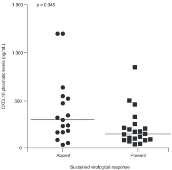
Analysis of the association between pretreatment levels of soluble inflammatory markers and virological response showed that elevated CXCL10 levels were associated with a lack of both EVR (p = 0.011) (Table I) and SVR (p = 0.045) (Figs 1, 2, Tables I, II). The accuracy of plasma CXCL10 levels to predict a lack of EVR was 0.79 [confidence inter-val (CI) 95%: 0.59-0.99], with a sensitivity of 78% and 65% specificity of lack of EVR using 220 pg/mL as the cut-off
point. Using a lower cut-off point of 150 pg/mL and an up-per cut-off level of 450 pg/mL for predicting EVR yielded a positive predictive level and a negative predictive level of 85.7% and 71.4%, respectively; in this setting, 15 (51.7%) patients presented with CXCL10 plasma levels between 150-450 pg/mL and could not be classified.
1.000 1.500
500
0
C
X
C
L
1
0
p
la
s
m
a
tic
le
v
e
ls
(
p
g
/m
L
)
p = 0.045
Sustained virological response Present Absent
Fig. 2: pretreatment plasma levels of CXCL10 among patients with chronic hepatitis C infection treated with interferon-α plus ribavirin stratified by sustained virological response. p values presented as cal-culated with Mann-Whitney U-test.
1.000 1.500
500
0
C
X
C
L
1
0
p
la
s
m
a
tic
le
v
e
ls
(
p
g
/m
L
)
p = 0.011
Early virological response Present Absent
The accuracy of CXCL10 plasma levels was lower for predicting SVR (0.69; CI 95%: 0.51-0.87). Applying the same cut-off values used in the evaluation of EVR (i.e., 150 pg/mL and 450 pg/mL), positive and negative predictive values for achieving SVR were 78.6% and 66.7%, respectively. Fifteen (39.5%) patients presented with CXCL10 plasma levels between 150-450 pg/mL and could not be classified using these cut-off points.
Only four patients had CXCL10 plasma levels great-er than 600 pg/mL. Among this small group of patients, global SVR was 25% and the only responder had a gen-otype 2 infection.
The other soluble inflammatory markers assessed were not associated with therapeutic response (Tables I, II).
DISCUSSION
We showed that pretreatment plasma levels of CXCL10 were inversely correlated with response to treatment with IFN-α and ribavirin in patients with hepatitis C infection.
Although this finding has been reported by other authors (Narumi et al. 1997, Apolinario et al. 2004, Butera et al. 2005, Diago et al. 2006, Lagging et al. 2006, Romero et al. 2006), their results were based on the analysis of pa-tients treated in the well-controlled context of clinical tri-als; our study strengthens the evidence for an association between CXCL10 levels and virological response among patients with CHC infection by showing an elevated neg-ative predictive value in a real-life clinical setting.
In addition, our study did not show an association between plasma levels of the CC chemokines evaluated (CCL2, CCL3, CCL11, CCL24) and of sTNF-R with vi -rological response. We did not find studies in the litera-ture evaluating the association between peripheral levels of these other inflammatory markers and treatment re-sponse among patients with CHC.
Apolinario et al. (2004) were the first to point out the association between serum levels of CXCL10 and thera-peutic response. Accuracy of CXCL10 in predicting lack
TABLE I
Comparison of median and interquartile range pretreatment levels of soluble inflammatory markers (pg/mL) among treated chronic hepatitis C patients with and without early virological response (EVR)
Inflammatory marker
Without EVR (n = 9)
With EVR
(n = 20) p value
CCL2 124.5 (85.5-177.8) 122.7 (36.2-133.3) 0.627
CCL3 141.1 (89.1-207.6) 100.9 (5.0-126.9) 0.340
CCL11 259.1 (239.4-304.9) 216.6 (168.0-273.0) 0.183
CCL24 690.4 (346.2-1050.1) 686.6 (429.5-1228.6) 0.945
CXCL9 434.4 (102.5-1711.0) 637.2 (2.5-1208.4) 0.729
CXCL10 512.9 (225.5-630.3) 179.1 (107.2-268.9) 0.011
sTNF-R1 721.7 (636.7-996.7) 824.6 (537.3-1064.9) 0.982
sTNF-R2 2078.0 (1992.8-2947.1) 2411.0 (1837.6-2657.8) 0.594
CCL2: monocyte chemotactic protein-1; CCL3: macrophagic inflammatory protein 1 alpha; CCL11: eotaxin-1; CCL24: eotaxin-2; CXCL9:
monokine induced by IFN-gamma; CXCL10: interferon-gamma inducible protein; sTNF-R: soluble tumor necrosis factor receptors.
TABLE II
Comparison of median and interquartile range pretreatment levels of soluble inflammatory markers (pg/mL) among treated chronic hepatitis C patients with and without sustained virological response (SVR)
Inflammatory marker
With SVR (n = 17)
Without SVR
(n = 21) p value
CCL2 120.9 (85.5-177.8) 124.5 (53.8-135.1) 0.728
CCL3 103.3 (89.1-207.6) 108.0 (0-169.5) 0.561
CCL11 239.4 (174.4-273.1) 220.1 (167.9-265.6) 0.504
CCL24 806.5 (610.4-1050.1) 690.5 (390.0-1132.0) 0.581
CXCL9 626.5 (102.5-1273.6) 583.7 (158.7-884.0) 0.486
CXCL10 289.9 (157.7-512.9) 142.7 (86.7-206.6) 0.045
sTNF-R1 721.7 (636.7-996.7) 613.4 (498.3-1049.6) 0.622
sTNF-R2 2078.0 (1808.5-2683.4) 2328.7 (1910.0-2632.2) 1.0
of SVR was 0.74, which is slightly higher than that found in our study (0.69; CI 95%: 0.51-0.87). Using a cut-off point of 299 pg/mL for predicting lack of virological response, Apolinario et al. (2004) found a sensitivity and specificity of 80% and 63%, respectively, which was very similar to that found in our study (78% and 65%, respectively).
Lagging et al. (2006) stratified patients into three groups according to pretreatment CXCL10 levels (0-150 pg/mL; 150-600 pg/mL; > 600 pg/mL). Levels greater than 600 pg/mL had a negative predictive value for SVR of 79%. In a subanalysis including only diffi-cult to treat patients (i.e., elevated body mass index or high viral load) with high CXCL10 levels, none of the seven patients achieved SVR. In our study, of the four patients with levels greater than 600 pg/mL, only one genotype 2 infected patient achieved SVR.
Different theories have been presented to explain the relationship between elevated levels of CXCL10 and poor response rates. High peripheral levels of CXCL10 could impair local CCL5 or CXCL10 gradient signaling (Butera et al. 2005) or could downregulate CXCR5 receptors in circulating CTL (Larrubia et al. 2007). Other authors (Di-agoet al. 2006) propose that elevated CXCL10 levels may result in an accumulation of effector T cells in the liver and the selective pressure imposed by this accumulation may foster outgrowth of immune escape HCV mutants that would be more difficult to eradicate with combined therapy (Diagoet al. 2006). The observed association be-tween CXCL10 levels and therapeutic response was not mediated by liver histological changes, as plasma levels of this chemokine were not associated with liver inflam-matory activity or fibrosis in a subanalysis of the dataset (Moura et al. 2010).
CXCL9, which binds the same chemokine receptor as CXCL10 (i.e., CXC3), did not show an association with virological response in our study, in accordance with findings from a previous study conducted by Butera et al. (2005). Although acting in similar cells, a different regu-lation mechanism for this chemokine may explain this ap-parent inconsistency.
The small number of treated patients in our study lim-its the interpretation of the lack of an association between some of the inflammatory markers evaluated and viro-logical response. Another limitation of our study was the evaluation of EVR only for genotype 1 infected patients; therefore, the association found between CXCL10 levels and EVR cannot be extrapolated to patients infected by other HCV genotypes.
In summary, we have shown that pretreatment plasma levels of CXCL10 were associated with virological response among patients with CHC infection. Due to the high nega-tive predicnega-tive value of elevated pretreatment CXCL10 lev-els, their assessment may be useful in the evaluation of po-tential candidates for therapy with interferon and ribavirin.
ACKNOWLEDGEMENTS
To Dr Dora Mendez del Castillo and Nara de Oliveira Car
-valho, from the Molecular Biology Laboratory at the Núcleo de Ações e Pesquisa em Apoio Diagnóstico (NUPAD), Fed -eral University of Minas Gerais, for storing and processing the plasma samples.
REFERENCES
Abu-Khabar KS, Armstrong JA, Ho M 1992. Type I interferons (IFN-alpha and -beta) suppress cytotoxin (tumor necrosis factor-(IFN-alpha and lymphotoxin) production by mitogen-stimulated human pe-ripheral blood mononuclear cell. J Leukoc Biol52: 165-172.
Apolinario A, Diago M, Lo Iacono O, Lorente R, Pérez C, Majano PL, Clemente G, García-Monzón C 2004. Increased circulating and intrahepatic T-cell-specific chemokines in chronic hepatitis C: relationship with the type of virological response to peginter-feron plus ribavirin combination therapy. Aliment Pharmacol Ther19: 551-562.
Apolinario Fernández de Sousa A, García Monzón C 2003. Role of chemokines in the pathogenesis of liver diseases. Rev Esp Enferm Dig95: 614-620.
Asselah T, Estrabaud E, Bieche I, Lapalus M, De Muynck S, Vidaud M, Saadoun D, Soumelis V, Marcellin P 2010. Hepatitis C: viral and host factors associated with non-response to pegylated inter-feron plus ribavirin. Liver Int30: 1259-1269.
Baggiolini M, Dewald B, Moser B 1997. Human chemokines: an up-date. Annu Rev Immunol15: 675-705.
Bedossa P, Poynard T 1996. An algorithm for the grading of activity in chronic hepatitis C. The METAVIR Cooperative Study Group.
Hepatology 24: 289-293.
Bonecchi R, Bianchi G, Bordignon PP, D’Ambrosio D, Lang R, Bor -satti A, Sozzani S, Allavena P, Gray PA, Mantovani A, Siniga-glia F 1998. Differential expression of chemokine receptors and chemotactic responsiveness of type 1 T helper cells (Th1s) and Th2s. J Exp Med187: 129-134.
Brown RS Jr, Gaglio PJ 2003. Scope of worldwide hepatitis C prob-lem. Liver Transpl9: S10-13.
Butera D, Marukian S, Iwamaye AE, Hembrador E, Chambers TJ, Di Bis-ceglie AM, Charles ED, Talal AH, Jacobson IM, Rice CM, Dustin LB 2005. Plasma chemokine levels correlate with the outcome of antiviral therapy in patients with hepatitis C. Blood106: 1175-1182.
Charo IF, Ransohoff RM 2006. The many roles of chemokines and chemokine receptors in inflammation. N Engl J Med354: 610-621. Cramp ME, Rossol S, Chokshi S, Carucci P, Williams R, Naoumov
NV 2000. Hepatitis C virus-specific T-cell reactivity during in -terferon and ribavirin treatment in chronic hepatitis C. Gastroen-terology118: 346-355.
de Araújo ES, Mendonça JS, Barone AA, Gonçales FL Jr, Ferreira MS, Focaccia R, Pawlotsky JM 2007. Consensus of the Brazilian Society of Infectious Diseases on the management and treatment of hepatitis C. Braz J Infect Dis11: 446-450.
Diago M, Castellano G, García-Samaniego J, Pérez C, Fernández I, Romero M, Iacono OL, García-Monzón C 2006. Association of pretreatment serum interferon gamma inducible protein 10 levels with sustained virological response to peginterferon plus ribavi-rin therapy in genotype 1 infected patients with chronic hepatitis C. Gut55: 374-379.
Feld JJ, Hoofnagle JH 2005. Mechanism of action of interferon and ribavirin in treatment of hepatitis C. Nature436: 967-972.
Frese M, Barth K, Kaul A, Lohmann V, Schwärzle V, Bartenschlager R 2003. Hepatitis C virus RNA replication is resistant to tumour necrosis factor-alpha. J Gen Virol84: 1253-1259.
Fried MW, Shiffman ML, Reddy KR, Smith C, Marinos G, Gonçales FL Jr, Häussinger D, Diago M, Carosi G, Dhumeaux D, Craxi A, Lin A, Hoffman J, Yu J 2002. Peginterferon alfa-2a plus ribavirin for chronic hepatitis C virus infection. N Engl J Med347: 975-982.
Gerard C, Rollins BJ 2001. Chemokines and disease. Nat Immunol
Guidotti LG, Chisari FV 2001. Noncytolytic control of viral infec -tions by the innate and adaptive immune response. Annu Rev Im -munol19: 65-91.
Joyce DA, Gibbons DP, Green P, Steer JH, Feldmann M, Brennan FM 1994. Two inhibitors of pro-inflammatory cytokine release, interleukin-10 and interleukin-4, have contrasting effects on re-lease of soluble p75 tumor necrosis factor receptor by cultured monocytes. Eur J Immunol24: 2699-2705.
Kamal SM, Fehr J, Roesler B, Peters T, Rasenack JW 2002. Peginter-feron alone or with ribavirin enhances HCV-specific CD4 T-helper 1 responses in patients with chronic hepatitis C. Gastro-enterology123: 1070-1083.
Kamal SM, Ismail A, Graham CS, He Q, Rasenack JW, Peters T, Tawil AA, Fehr JJ, Khalifa Kel S, Madwar MM, Koziel MJ 2004. Pegylated interferon alpha therapy in acute hepatitis C: relation to hepatitis C virus-specific T cell response kinetics. Hepatology 39: 1721-1731.
Koziel MJ 1999. Cytokines in viral hepatitis. Semin Liver Dis19: 157-169.
Kunkel SL 1999. Through the looking glass: the diverse in vivo activi-ties of chemokines. J Clin Invest104: 1333-1334.
Lagging M, Romero AI, Westin J, Norkrans G, Dhillon AP, Pawlotsky JM, Zeuzem S, von Wagner M, Negro F, Schalm SW, Haagmans BL, Ferrari C, Missale G, Neumann AU, Verheij-Hart E, Hellstrand K 2006. IP-10 predicts viral response and therapeutic outcome in difficult-to-treat patients with HCV genotype 1 infection. Hepatology44: 1617-1625.
Lantz M, Malik S, Slevin ML, Olsson I 1990. Infusion of tumor necrosis factor (TNF) causes an increase in circulating TNF-binding protein in humans. Cytokine2: 402-406.
Larrubia JR, Calvino M, Benito S, Sanz-de-Villalobos E, Perna C, Pérez-Hornedo J, González-Mateos F, García-Garzón S, Bien-venido A, Parra T 2007. The role of CCR5/CXCR3 expressing CD8+ cells in liver damage and viral control during persistent
hepatitis C virus infection. J Hepatol47: 632-641.
Lee SS, Heathcote EJ, Reddy KR, Zeuzem S, Fried MW, Wright TL, Pockros PJ, Häussinger D, Smith CI, Lin A, Pappas SC 2002. Prognostic factors and early predictability of sustained viral response with peginterferon alfa-2a (40KD). J Hepatol37: 500-506. Lien E, Liabakk NB, Johnsen AC, Nonstad U, Sundan A, Espevik T
1995. Polymorphonuclear granulocytes enhance lipopolysaccha-ride-induced soluble p75 tumor necrosis factor receptor release from mononuclear cells. Eur J Immunol25: 2714-2717. Luster AD 1998. Chemokines-chemotactic cytokines that mediate
inflammation. N Engl J Med338: 436-445.
Mackay CR 1997. Chemokines: what chemokine is that? Curr Biol 7: R384-386.
Manns MP, McHutchison JG, Gordon SC, Rustgi VK, Shiffman M, Reindollar R, Goodman ZD, Koury K, Ling M, Albrecht JK 2001. Peginterferon alfa-2b plus ribavirin compared with interferon al-fa-2b plus ribavirin for initial treatment of chronic hepatitis C: a randomised trial. Lancet358: 958-965.
Marinos G, Naoumov NV, Rossol S, Torre F, Wong PY, Gallati H, Portmann B, Williams R 1995. Tumor necrosis factor receptors in patients with chronic hepatitis B virus infection. Gastroen-terology108: 1453-1463.
Mihm U, Herrmann E, Sarrazin C, Zeuzem S 2006. Review article: predicting response in hepatitis C virus therapy. Aliment Phar-macol Ther23: 1043-1054.
Moura AS, Carmo RA, Teixeira AL, Leite VH, Rocha MO 2010. Soluble inflammatory markers as predictors of liver histological changes in patients with chronic hepatitis C virus infection. Eur
J Clin Microbiol Infect Dis 29: 1153-1161.
Moura AS, Carmo RA, Teixeira AL, Rocha MO 2009. Soluble in-flammatory markers as predictors of hepatocellular damage and therapeutic response in chronic hepatitis C. Braz J Infect Dis
13: 375-382.
MS - Ministério da Saúde/Secretaria de Atenção à Saúde 2002. Por-taria 863 de 04 de novembro de 2002. Protocolo clínico e dire-trizes terapêuticas para o tratamento da hepatite viral crônica C. Available from: http://www.saude.ms.gov.br/controle/ShowFile. php?id=2227.
Narumi S, Tominaga Y, Tamaru M, Shimai S, Okumura H, Nishioji K, Itoh Y, Okanoue T 1997. Expression of IFN-inducible pro -tein-10 in chronic hepatitis. J Immunol158: 5536-5544. Nelson DR, Marousis CG, Ohno T, Davis GL, Lau JY 1998. Intrahe
-patic hepatitis C virus-specific cytotoxic T lymphocyte activity and response to interferon alfa therapy in chronic hepatitis C. He-patology28: 225-230.
Neuman MG, Benhamou JP, Malkiewicz IM, Akremi R, Shear NH, Asselah T, Ibrahim A, Boyer N, Martinot-Peignoux M, Jacob -son-Brown P, Katz GG, Le Breton V, Le Guludec G, Suneja A, Marcellin P 2001. Cytokines as predictors for sustained response and as markers for immunomodulation in patients with chronic hepatitis C. Clin Biochem34: 173-182.
Ono SJ, Nakamura T, Miyazaki D, Ohbayashi M, Dawson M, Toda M 2003. Chemokines: roles in leukocyte development, trafficking, and effector function. J Allergy Clin Immunol111: 1185-1199. Porteu F, Brockhaus M, Wallach D, Engelmann H, Nathan CF 1991.
Human neutrophil elastase releases a ligand-binding fragment from the 75-kDa tumor necrosis factor (TNF) receptor. Com -parison with the proteolytic activity responsible for shedding of TNF receptors from stimulated neutrophils. J Biol Chem266: 18846-18853.
Poynard T, McHutchison J, Manns M, Trepo C, Lindsay K, Goodman Z, Ling MH, Albrecht J 2002. Impact of pegylated interferon alfa-2b and ribavirin on liver fibrosis in patients with chronic hepatitis C. Gastroenterology122: 1303-1313.
Rehermann B, Nascimbeni M 2005. Immunology of hepatitis B virus and hepatitis C virus infection. Nat Rev Immunol5: 215-229. Romero AI, Lagging M, Westin J, Dhillon AP, Dustin LB, Pawlotsky
JM, Neumann AU, Ferrari C, Missale G, Haagmans BL, Schalm SW, Zeuzem S, Negro F, Verheij-Hart E, Hellstrand K 2006. Interferon (IFN)-gamma-inducible protein-10: association with histological results, viral kinetics, and outcome during treatment with pegylated IFN-alpha 2a and ribavirin for chronic hepatitis C virus infection. J Infect Dis194: 895-903.
Sousa-Pereira SR, Teixeira AL, Silva LC, Souza AL, Antunes CM, Teixeira MM, Lambertucci JR 2006. Serum and cerebral spinal fluid levels of chemokines and Th2 cytokines in Schistosoma mansoni myeloradiculopathy. Parasite Immunol28: 473-478. Strader DB, Wright T, Thomas DL, Seeff LB 2004. Diagnosis,
man-agement, and treatment of hepatitis C. Hepatology39: 1147-1171.
Tam RC, Pai B, Bard J, Lim C, Averett DR, Phan UT, Milovanovic T 1999. Ribavirin polarizes human T cell responses towards a Type 1 cytokine profile. J Hepatol30: 376-382.
Tilg H 1997. New insights into the mechanisms of interferon alfa: an immunoregulatory and anti-inflammatory cytokine. Gastroen-terology112: 1017-1021.
Tilg H, Vogel W, Dinarello CA 1995. Interferon-alpha induces circulating tumor necrosis factor receptor p55 in humans. Blood85: 433-435.