811
DOI: 10.1590/0004-282X20130127
VIEW AND REVIEW
Traumatic injuries of peripheral nerves:
a review with emphasis on surgical indication
Lesões traumáticas de nervos periféricos: uma revisão com ênfase na indicação cirúrgica
Roberto Sergio Martins1,2, Dhiego Bastos3, Mario Gilberto Siqueira1, Carlos Otto Heise1,
Manoel Jacobsen Teixeira1
Acute peripheral nerve injuries are one of the
complica-tions of trauma afecting the extremities, and is present in 3–10% of patients, depending on the mechanism of trauma1–3. hese traumatic injuries are a signiicant cause of physi cal disability that afects mainly young adults of working age. Although some nerve injuries recover spontaneously, in some cases surgery is the only therapeutic option for the improve
-ment of neurological deicits or control of neuropathic pain. Indications for surgery in patients with peripheral nerve in
-jury depends on several variables including mechanism of injury, interval between injury and treatment, lesion severity, indings of the clinical examination, and intensity of neuro -pathic pain4.
In this article we review the classiication, mechanisms and evaluation of peripheral nerve injuries, with emphasis on indications for surgical treatment.
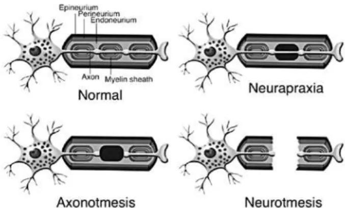
CLASSIFICATION OF PERIPHERAL NERVE INJURY
A key issue in deining surgical treatment for patients with peripheral nerve injury is to determine whether the
injury results in an open or closed lesion5. he severity of the injury is variable and can be classiied according to Seddon’s classiication in three types: neuropraxia, axonotmesis, and
neurotmesis4 (Figure 1).
1Peripheral Nerve Surgery Unit, Department of Neurosurgery, University of São Paulo School of Medicine, São Paulo SP, Brazil; 2Hospital do Servidor Público do Estado de São Paulo, São Paulo SP, Brazil;
3Universidade Estadual de Campinas, Campinas SP, Brazil.
Correspondence: Roberto S. Martins; Rua Oscar Freire 2250 / cj. 18 térreo; 05409011 São Paulo SP - Brasil; E-mail: [email protected]
Conflict of interest: There is no conflict of interest to declare.
Received 16 May 2013; Received in final form 20 May 2013; Accepted 27 May 2013. ABSTRACT
Traumatic peripheral nerve injury is a dramatic condition present in many of the injuries to the upper and lower extremities. An understanding of its physiopathology and selection of a suitable time for surgery are necessary for proper treatment of this challenging disorder. This article reviews the physiopathology of traumatic peripheral nerve injury, considers the most used classification, and discusses the main aspects of surgical timing and treatment of such a condition.
Keywords: peripheral nerve, peripheral nerve surgery, peripheral nerve injury, nerve.
RESUMO
Traumatismos dos nervos periféricos resultam em lesões incapacitantes e estão presentes em muitas das lesões dos membros. A com-preensão da fisiopatologia dessas lesões e a seleção do momento operatório mais adequado são imprescindíveis para que o tratamento seja adequado. Neste artigo revisamos a fisiopatologia das lesões traumáticas dos nervos periféricos, apresentamos a classificação mais utilizada dessas lesões e discutimos os principais aspectos relacionados ao momento da cirurgia e às formas de reparo cirúrgico.
Palavras-chave: nervo periférico, cirurgia de nervo periférico, traumatismo de nervo periférico, enxerto de nervo, reparo do nervo.
Figure 1. Schematic drawing of a normal nerve fiber and
812 Arq Neuropsiquiatr 2013;71(10):811-814
Neurapraxia represents the mildest type of nerve injury
and is deined by a temporary blockage of nervous conduc
-tion caused by a segmental demyeliniza-tion5. he large ibers are more selectively and severely afected than the small i
-bers, leading to motor paralysis, and some proprioceptive and tactile sensitivity loss, but with maintenance of thermal and pain sensitivity in most cases. he prognosis is excellent since there is no distal axonal degeneration; the blockage re
-solves through remyelinization and the nerve function is re
-covered in a matter of days or weeks4.
Axonotmesis occurs when the injury is suicient to de
-termine the loss of axonal continuity, but most of nerve con
-nective tissue framework is preserved, including the tubular endoneural support that surrounds each axon5. Despite the damage being more extensive in axonotmesis than in neura
-praxia, spontaneous regeneration is still possible, although longer, taking up weeks to months after the injury. In axo
-notmesis, as well as in neurotmesis, a sequence of patho
-logical events known as wallerian degeneration occurs in the nerve segment distal to the injury. his process includes fragmentation and degeneration of the axon distal to the le
-sion and phagocytosis of the myelin sheath by Schwann cells and macrophages6. In a phase of the process, the distal endo -neural tubes are illed by Schwann cells forming longitudinal lines inside the tubes known as Büngner’s bands7. he reco very depends on the axonal sprout from the proximal stump that must cross the lesion site and reach the correspondent endoneural tubes in the distal stump in order to reinnervate the target organ. Once the Büngner’s band is reached, the axon grows 1–3 mm a day8,9. hereby, reinnervation of the
target organ can take several months depending on its dis
-tance from the lesion. he reestablishment of the neuromus
-cular junctions depends on the interaction between regene rating axons and basal membranes of the myoibrils. Soon after nerve injury, degeneration of myoibers occurs and 18– 24 months after the injury muscle ibers are replaced by fat and ibrous connective tissue, which makes the muscle pro
-gressively refractory to reinnervation4. herefore, the sooner the axons reach the muscle ibers, the more efective reinnervation can be expected and that is the reason why surgery for nerve in
-jury, when indicated, must be performed as soon as possible10. In neurotmesis, besides the loss of axonal continuity and of the internal nerve connective tissue framework, a rupture
occurs in the epineurium with macroscopic loss of nerve con
-tinuity or interposition of scar tissue between the interrupted ibers, which prevents spontaneous regeneration and requires surgical treatment4. he correct identiication of these lesions is the main objective of the surgeon dealing with such event.
OPEN VERSUS CLOSED INJURIES
Nerve injuries can be classiied as closed and open de
-pending on whether the cutaneous integrity has been dis
-rupted or not. Closed injuries are more frequently asso ciated
with nerve injuries in continuity, characterized by absence of nerve rupture and by occurrence of neuropraxis and axonot
-mesis as the predominant mechanisms of injury11. herefore, spontaneous recovery is possible and surgery is indicated only after 3 months if no recovery is identiied. his period is arbitrated based on axonal growth rate (1–3 mm/day) and improvement identiied on clinical or electromyographic evaluation. Classical examples of closed injuries are those re
-sulting from stretching after brachial plexus injuries secon dary to motorcycle falls and peroneal nerve injuries associa ted with knee dislocation and concomitant ligament lesion12. Conversely, the occurrence of an open injury related to a nerve course has been more frequently related to neurotme tic injuries and must be treated with early surgery12. Examples of these injuries include those provoked by knives, propellers, piece of glass, and scalpel iatrogenic lesions. Within this con
-text, it is important to keep in mind that the distal portion of the nerve undergoes wallerian degeneration that occurs up 2 to 3 weeks after the injury8. So, electrophysiological assess -ment is not indicated in these cases before 3–4 weeks, since false results may compromise the evaluation.
SHARP VERSUS BLUNT INJURIES
he aspect of the nerve stumps identiied during surge ry is another important factor to be considered for the de
-initive treatment11,13. Two situations can be distinguished:
identiication of a sharp stump with homogeneous aspect
and no signiicant inlammation; or inding a blunt or rug
-ged stump, associated with signiicant inlammatory pro
-cess, heterogeneous aspect, and contusion (Figure 2). Sharp instruments like knives or scalpels have been identiied as a frequent causative factors resulting in sharp stumps. In these cases, the repair should be done promptly, if possible within
Figure 2. Intraoperative view of ulnar nerve approach in the right forearm. This 22-year-old patient suffered a stab wound 4 months before his presentation at our center. A complete palsy of the ulnar nerve was identified at physical examination and a neurotmesis was demonstrated during surgical
813 Roberto Sergio Martins et al. Peripheral nerves: traumatic injuries
the irst 3 days after the injury. Usually a direct coaptation of the nerve ends can be performed with a terminoterminal
tension-free suture12.
Technical conditions in performing surgery is another important issue that must be taken into consideration when deciding on an early repair14, as an adequate surgical tech -nique has been accepted as one of the factors that inluence the inal result after a nerve surgery15. his implies the use of microscope magniication, 9.0 or 10.0caliber sutures, and a careful manipulation of nerve structures using microsurgical
instrumental16. If there are no such conditions for surgery, the epineurium of each nerve stump should be sutured to some adjacent structure, such as a tendon or fascia, in order to avoid excessive retraction of the stumps and to facilitate its identiication in a second surgical procedure14. Any attempt to suture the nerve beyond these conditions will result in un
-necessary damage to nerve tissue, increase in local ibrosis, and worse functional results at longterm followup.
When blunt stumps are identiied during surgery (Figure 2), the repair should not be performed immediately because the inlammatory process that takes place extends for up to 3 weeks after the injury11. If repair is performed within this pe -riod there is a risk to connect nerve stumps still involved in an ongoing inlammatory process that results in ibrosis and pre
-vents progression of the regenerated axons. When blunt nerve stumps are identiied, the surgeon should interrupt the pro
-cedure and perform the deinitive repair 3 weeks after the in
-jury11. During the deinitive repair the inlammatory tissue and ibrosis must be resected by trimming the nerve ends with a scalpel blade until viable fascicles have being exposed17.
THE “RULE OF THREE”
In summary, surgical timing in a traumatic peripheral nerve injury is deined by the “rule of three”: immediate sur
-gery within 3 days for clean and sharp injuries; early sur-gery within 3 weeks for blunt/contusion injuries; and delayed sur
-gery, performed 3 months after injury, for closed injuries.
SPECIAL SETTINGS
Nerve injuries due to gunshot wounds have been consi dered closed injuries since there is no tissue exposure. Most lesions are caused by indirect heat and by the shock wave from the bullet. Usually the projectile does not transect the nerve so continuity is preserved and there is potential for at least partial spontaneous recovery. herefore, surgery for pa
-tients with nerve injuries due to gunshot wounds should be performed 3–4 months after the injury18.
Another condition that does not follow the “rule of three” occurs when an injured nerve is located in an area where nonrelated surgery had been performed previously. An emergency vascular intervention nearby the nerve is an exam ple of such a condition19. Another example is an ortho
-pedic exploration of an open humerus fracture exposing the
adjacent radial nerve20. In both situations the lesion originally classiied as closed may result in nerve transection, and the early exploration allows performing nerve surgery before the usual 3month period of observation.
SURGICAL CONSIDERATIONS
Classically surgery for treatment of peripheral nerve in
-juries should be considered in patients demonstrating com
-plete palsy after the traumatism. Persistent neuropathic pain besides medical treatment is another indication, and, in these cases, neurolysis, which consists in the removal of a i
-brotic hypertrophic epineurium and adherent adjacent tissue to the nerve, should result in partial or total pain relief14.
Adequate surgical management of peripheral nerve inju
-ries requires that the surgeon, beyond knowing precisely the anatomical details of the region to be assessed, also be fa
-miliarized with microsurgical techniques and dispose of the necessary equipment to perform the surgery14. he basic pro -cedure in peripheral nerve surgery is the reestablishment of nerve continuity, which can be obtained by direct coaptation between the two stumps of the ruptured nerve or by inter
-position of nerve grafts21. he best results are achieved with endtoend nerve repair without tension, as the regenerating axons need to cross just one site of coaptation. In contrast, when using nerve graft, the regenerating axons need to cross two sites of repair, which may have a distinct inlammatory process, resulting in higher axonal loss13. However, in many cases approximation of the nerve stumps results in tension
on the suture line. Tension at the site of repair results in
ische mia, connective tissue proliferation, and scar formation that impair or prevent the regenerating axons to progress21. In these cases the reconstruction of nerve continuity is ac
-complished by the interposition of autologous nerve grafts, usually from the sural nerve.
Intraoperative eletrophysiologic evaluation has been ac
-cepted as an important tool in the management of lesions in continuity (Figure 3A)22. In this type of lesion, also named neuroma in continuity, it is diicult to deine the extent of internal nerve damage by macroscopic inspection only. In some cases, the presence of healthy axons inside the neuro
-ma allows spontaneous regenerations, but in others the scar tissue represents an obstacle to the regenerating axons. In this last situation, the scar tissue needs to be resected and substituted by normal nervous tissue usually by interposi
-tion of autologous nerve graft. hese speciics cases should be evaluated through nerve action potential (NAP) measure
-ment (Figure 3B). his evaluation is performed using a porta
-ble electromyography device and two electrodes. With a hook form, the stimulating and the recording electrodes are posi
-tioned under and around the nerve, proximal and distal to the neuroma, respectively, elevating and isolating the nerve. A supramaximal stimulus is then applied to generate an ac
814 Arq Neuropsiquiatr 2013;71(10):811-814
is identiied there are regenerating axons pas sing through the neuroma, regeneration will likely occur, and an external neu
-rolysis is the only surgical procedure to be done. When there are no regenerating axons crossing the lesion no NAP will be recorded and resection of the neuroma followed by nerve re
-construction, usually with grafts, is performed (Figure 3C, D). In conclusion, the social cost of traumatic peripheral nerve injuries is signiicant since it has a higher incidence on
Figure 3. Intraoperative photograph showing a sciatic nerve injury at the middle third of the thigh due to a gunshot wound. Since there was no recovery of a complete sciatic nerve palsy the patient was operated 3 months after injury. (A) Initial exposure showing a neuroma (N) in continuity of the sciatic nerve (SN) just before its division. (B) An intraoperative nerve action potential evaluation was performed in order to define if the lesion should be resected. No recordable response was obtained so the neuroma was resected. (C) Surgical view after neuroma resection. (D) Both sural nerves were used to re-establish nerve continuity with nine nerve grafts interposed between proximal and distal stumps. P: proximal; PN: peroneal nerve; R: recording electrode; S: stimulation electrode; TN: tibial nerve.
A
C
B
D
References
1. Hudson AR, Hunter D. Timing of peripheral nerve repair: important local and neuropathological factors. Clin Neurosurg 1977;24:391-405.
2. Deitch EA, Grimes WR. Experience with 112 shotgun wounds of the extremities. J Trauma 1984;24:600-603.
3. Noble J, Munro CA, Prasad VS, Midha R. Analysis of upper and lower extremity peripheral nerve injuries in a population of patients with multiple injuries. J Trauma 1998;45:116-122.
4. Campbell WW. Evaluation and management of peripheral nerve injury. Clin Neurophysiol 2008;119:1951-1965.
5. Grant GA, Goodkin R, Kliot M. Evaluation and surgical management of peripheral nerve problems. Neurosurgery 1999;44:825-840.
6. Schmid DB, Salyapongse N. Nerve injury and repair. Curr Orthop Pract 2008;19:475-480.
7. Torigoe K, Tanaka H, Takahashi A, Awaya A, Hashimoto K. Basic behavior of migratory Schwann cells in peripheral nerve regeneration. Exp Neurol 1996;137:301-308.
8. Hall S. Nerve repair: a neurobiologist’s view. J Hand Surg (Br) 2001;26:129-136.
9. Gordon T, Chan KM, Sulaiman OAR, Udina E, Amirjani N, Brushart TM. Accelerating axon growth to overcome limitations in functional recovery after peripheral nerve injury. Neurosurgery 2009;65:132-44.
10. Robinson LR. Traumatic injury to peripheral nerve. Muscle Nerve 2000;23:863-873.
11. Spinner RJ, Kline DG. Surgery for peripheral nerve and brachial plexus injuries or other nerve lesions. Muscle Nerve 2000;23:680-695.
12. Weber RV, MacKinnon SE. Bridging the neural gap. Clin Plast Surg 2005;32:605-616.
13. Isaacs J. Treatment of acute peripheral nerve injuries: current concepts. J Hand Surg (Am) 2010;35:491-497.
14. Harris ME, Tindall SC. Techniques of peripheral nerve repair. Neurosurg Clin North Am 1991;2:93-104.
15. Rowshan K, Jones NF, Gupta R. Current surgical techniques of peripheral nerve repair. Operat Tech Ortop 2004;14:63-170.
16. Sunderland S. The anatomic foundation of peripheral nerve repair techniques. Orthop Clin North Am 1981;12:245-266.
17. Dahlin LB. Nerve injuries. Curr Orthop 2008;22:9-16.
18. Katzman BM, Bozentka DJ. Peripheral nerve injuries secondary to missiles. Hand Clin 1999;15:233-244.
19. Manord JD, Garrard CL, Kline DG, Sternbergh WC 3rd, Money SR. Management of severe proximal vascular and neural injury of the upper extremity. J Vasc Surg 1998;27:43-47.
20. Thomsen NOB, Dahlin LB. Injury to the radial nerve caused by fracture of the humeral shaft: timing and neurobiological aspects related to treatment and diagnosis. Scand J Plast Reconstr Surg Hand Surg 2007;41:153-157.
21. Dvali L, Mckinnon S. The role of microsurgery in nerve repair and nerve grafting. Hand Clin 2007;21:73-81.
22. Sulaiman WAR. Kline DG. Nerve surgery: a review and insights about its future. Clin Neurosurg 2006;53:38-47.
young, previously healthy, and economically active people. A prompt and adequate handle of these lesions can result in the recovery, at least partially, of the lost function. herefore
it is fundamental to understand the mechanisms and pecu
-liarities of these lesions in order to deine an acceptable time for surgical intervention. Timely nerve reconstruction per