Echocardiographic Parameters of Epicardial Fat Deposition and its
Relation to Coronary Artery Disease
Juan Valiente Mustelier
1, Julio Oscar Cabrera Rego
1, Angela Gala González
2, Júlio César Gandarilla Sarmiento
1,
Beatriz Vega Riverón
2National Cardiology and Cardiovascular Surgery Institute1; Institute of Tropical Medicine ´Pedro Kourí´2 - Havana - Cuba
Abstract
Background: Epicardial fat has been associated to the presence of significant coronary artery disease (CAD). However, the association of lipomatous infiltration of the atrial septum and fat infiltration of the right ventricle remains uncertain. None of these parameters has been thoroughly studied in Hispanic patients.
Objective: To determine the association between epicardial fat, lipomatous infiltration of the atrial septum and fat infiltration of the right ventricle with the presence of CAD.
Methods: Two hundred and fifty Hispanic patients (86 women and 164 men, mean age 61.5 ± 8 vs 62 ± 10 years respectively), undergoing their first invasive coronary angiography (ICA) were studied. The day after the ICA, parameters of cardiac fat deposition were evaluated using bidimensional echocardiography. Clinical (age, sex, personal antecedents of smoking habit, hypertension and diabetes mellitus, as well as clinical presentation of CAD) and anthropometric (waist circumference and body mass index [BMI]) variables were also collected.
Results: Epicardial fat (OR 1.27 p = 0.009), as well as fat infiltration of the right ventricle (OR 2.94 p = 0.027), had a significant and independent association with the presence, but not the extent (p = 0.516) and clinical presentation (p = 0.153) of CAD. The extent of epicardial fat deposition showed a proportional and significant association (p = 0.001) with the presence of CAD.
Conclusion: Epicardial fat and fat infiltration of the right ventricle were both significant and independent factors associated to the presence of CAD, which was proportionally increased according to the extent of cardiac fat deposition. (Arq Bras Cardiol 2011; 97(2) : 122-129)
Keywords: Echocardiography; subcutaneous fat; pericardium; coronary disease.
Mailing address: Julio Oscar Cabrera Rego •
Dstrampes No.5 - Stos Suarez - 10400 - Havana - Cuba E-mail: [email protected]
Manuscript received May 10, 2010; revised manuscript received December 10, 2010; accepted February 23, 2011.
Introduction
The echocardiographic measurement of epicardial fat is a noninvasive and objective quantification method with high availability that has shown clear advantages as a marker of cardiometabolic risk, even superior to subcutaneous fat and total body adiposity1. Recently, some studies have
shown the association between epicardial fat and subclinical atherosclerosis2-4, the presence, extension and severity of
significant coronary artery disease (CAD)5-7 and coronary flow
reserve in women8.
In comparison with epicardial fat, other manifestations of cardiac fat deposition have received less attention in the literature. Epicardial fat is also the main determinant of the lipomatous infiltration of the atrial septum, which occasionally has been reported as lipomatous hypertrophy in several case reports9, a benign entity (defined as an atrial septum thickness
> 20 mm) in association with supraventricular arrhythmias and sudden death. Regarding this subject, Chaowalit et al recently reported its association with the presence of significant coronary artery disease10. On the other hand, fat infiltration of
the right ventricle (RV) is a relatively frequent phenomenon, found mainly in older subjects and females. The relationship between RV fat and CAD has not been adequately evaluated11.
There are no studies assessing the association among these manifestations of cardiac fat deposition (epicardial fat, lipomatous infiltration of the atrial septum and fat infiltration of the right ventricle) and its relationship to coronary artery disease, especially in Hispanic subjects.
Methods
Study population
(age, sex, personal antecedents of smoking habit, hypertension and diabetes mellitus, as well as clinical presentation of CAD) were collected. Dyslipidemia was defined as total
cholesterol ≥ 200 mg/dl or triglyceride levels ≥ 150 mg/dl.
Anthropometric measures such as weight, height and waist circumference weremeasured. Body mass index (BMI) in kg/ m2 was calculated by the formula: weight/(height)2.
The study protocol was in accordance to the ethical guidelines of the 1975 Declaration of Helsinki and it was approved by the ethics review board of our institution. All participants signed an informed consent.
Measurement of echocardiographic parameters of cardiac fat deposition
Echocardiographic examinations were performed using a Philips iE33 2006 (USA) cardiac ultrasound machine (version 2.0.1.420, S5-1 transducer with 1.3-3.6 MHz phase array) by an echocardiographist blinded to clinical and coronary angiography data, with patients in the left lateral decubitus position.
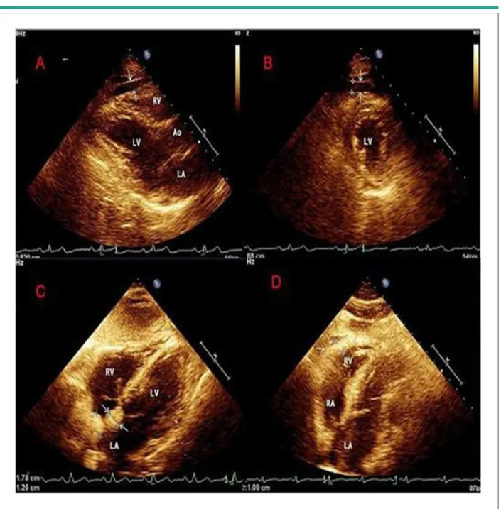
Epicardial fat was defined as the relatively echo-free space between the outer wall of the myocardium and the visceral layer of pericardium, and its thickness was measured from parasternal long- and short-axis bidimensional mode still-frame images perpendicularly to the free wall of the right ventricle at end-systole in 3 cardiac cycles, using the aortic annulus as anatomic reference for the parasternal long-axis view and the papillary muscles level for the short-axis view (Fig. 1 A, B). As Iacobellis and Willens12 suggested, epicardial
fat thickness is best measured at end-systole, because it is compressed during diastole. These values were averaged to obtain the mean thickness. Lipomatous infiltration of the
atrial septum was defined as a thickening ≥ 10 mm of both
poles of the atrial septum, with the fossa ovalis generally spared, giving it a characteristic “dumbbell” shape and perpendicularly measured in the subcostal four-chamber view at end-diastole with the patient in maximum inspiration (Fig. 1C). Fat infiltration of the right ventricle was defined as a
thickening ≥ 5 mm of the right ventricle wall in the absence of volume/pressure overload, with evidence of increase of the
fatty content (brighter pattern) in the subcostal four-chamber view at end-diastole with the patient in maximum inspiration (Fig. 1D). With the objective of diminishing the subjective component in the determination of fat infiltration of the right ventricle all the exams were displayed with fixed machine settings (general harmonic imaging, acquisition frequency of 39 Hz, field depth of 15 cm and gain setting of 65%).
Coronary angiography data
Coronary angiographic analysis was performed by two experienced invasive cardiologists using Judkin’s method,
following the percutaneous puncturing of the femoral artery
Statistics
Statistical analysis was performed using SPSS 13.0 for Windows. Continuous variables are expressed as means ± SD and categorical variables as absolute numbersand percentages. Comparisons of continuous variables were performed using theunpaired Student t test and categorical variables were compared with the chi-square test. To assess the reproducibility of the echocardiographic measurements of epicardial fat, 20 patients were randomly selected for the analysis by two independent observers who were blinded to clinical and angiographic data. Inter-observer and intra-observer correlation coefficients were calculated. Variabilities of measurements were also calculated as the mean of differences in measurements.
We first compared clinical, anthropometric and echocardiographic parameters according to the presence of significant CAD. A comparison between clinical and anthropometric variables with each one of the echocardiographic parameters of cardiac fat deposition was also performed. Mean comparison of epicardial fat thickness regarding anthropometric (according to sex), clinical presentation and extent of CAD was also made, using ANOVA test to assess differences among the groups. Multivariate analysis was performed using a multiple linear regression model including potential confounders (variables with p
value < 0.25 in the univariate analysis). The cut-off value of epicardial fat thickness for predicting CAD with corresponding specificity and sensitivity was estimated by receiver operating characteristic (ROC) curve analysis. Finally, we compared the extent of cardiac fat deposition with the presence of CAD.
Results
Clinical, anthropometric and cardiac fat deposition parameters in the study population
Our study group consisted of 86 (34%) women and 164 (66%) men, with a mean age of 61.5 ± 8 vs 62 ± 10 respectively. Epicardial fat thickness ranged from 1 mm to 18 mm, with a mean(SD) value of 6.1 ± 2.8 mm. Inter-observer and intra-observer correlation coefficients and variability of measurements of epicardial fat thickness were found to be 0.94, 0.92 and 0.5 ± 0.4 mm, 0.6 ± 0.5 mm, respectively, indicating good reproducibility.
There was no difference in epicardial fat thickness between men and women (5.85 ± 2.8 mm vs 6.25 ± 2.8 mm, p = 0.283). Lipomatous infiltration of the atrial septum was present in 141 patients (56.4%) and fat infiltration of the right ventricle in 71 (28.4%).
Figure 1 -Echocardiographic evaluation of cardiac fat deposition parameters. A) Parasternal long-axis view using the aortic annulus as anatomic reference. B) Parasternal short-axis view at the mitral papillary muscles level. Epicardial fat (arrows) A) and B). C) Subcostal four-chamber view. Lipomatous iniltration of the atrial septum (arrows). D) Subcostal four-chamber view. Fat iniltration of the right ventricle (arrows). AO - aorta; LV - left ventricle; RV - right ventricle; LA - left atrium; RA - right atrium.
Cardiac fat deposition parameters according to the presence and severity of CAD
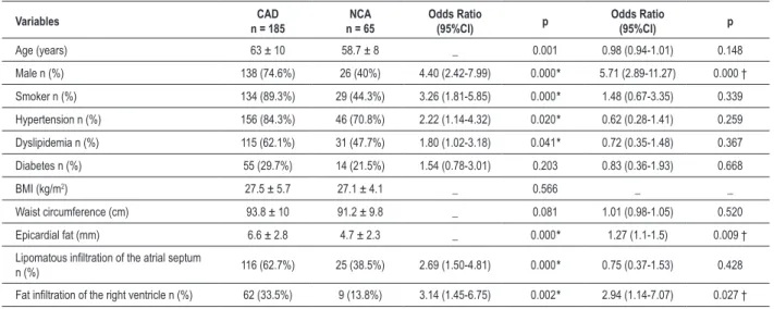
Epicardial fat thickness was increased in patients with significant CAD compared to those with NCA. All the cardiac fat deposition parameters showed a significant association with the presence of significant CAD in the univariate analysis. Sex, epicardial fat and fat infiltration of the right ventricle were the final variables that showed a significant and independent association with the presence of CAD in the multivariate analysis (Table 2).
Epicardial fat was not significantly thicker in patients with multivessel CAD than in those with two-vessel or
single-vessel CAD (7.0 ± 3 mm vs 6.6 ± 2.8 mm vs 6.4 ± 2.6 mm respectively, p = 0.516) (Fig. 2a). Although epicardial fat was greater in patients with acute coronary syndrome compared to those with stable angina, there was no significant association between both groups regarding differences in epicardial fat thickness (6.45 ± 2.9 mm vs
5.91 ± 1.7 mm, respectively, p= 0.153) (Fig. 2b).
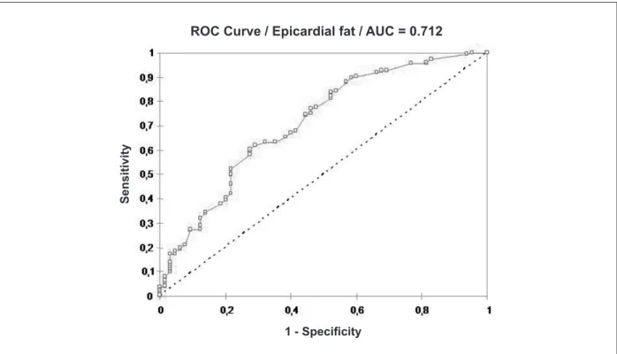
Epicardial fat thickness ≥ 5.2 mm had 65.4% sensitivity and
61.5% specificity (ROC area 0.712, 95% CI [0.640-0.784]) for predicting CAD (Fig. 3).
Table 1 - Clinical and anthropometric variables according to sex in the study population
Variables Female n = 86 (34%)
Male n = 164 (66%)
Age (years) 61.5 ± 8 62 ± 10
Smoker n (%) 63 (73.2%) 127 (77.4%)
Hypertension n (%) 68 (79%) 126 (76.8%)
Dyslipidemia n (%) 55 (63.9%) 88 (53.6%)
Diabetes 30 (34.8%) 38 (23.1%)
BMI (kg/m2) 27.3 ± 5.3 26.7 ± 4.1
Waist circumference (cm) 90.8 ± 10.1 94.3 ± 9.7
BMI - body mass index.
Table 2 - Association of clinical, anthropometric and cardiac fat deposition parameters with the presence of CAD
Variables n = 185CAD n = 65NCA Odds Ratio(95%CI) p Odds Ratio(95%CI) p
Age (years) 63 ± 10 58.7 ± 8 _ 0.001 0.98 (0.94-1.01) 0.148
Male n (%) 138 (74.6%) 26 (40%) 4.40 (2.42-7.99) 0.000* 5.71 (2.89-11.27) 0.000 †
Smoker n (%) 134 (89.3%) 29 (44.3%) 3.26 (1.81-5.85) 0.000* 1.48 (0.67-3.35) 0.339
Hypertension n (%) 156 (84.3%) 46 (70.8%) 2.22 (1.14-4.32) 0.020* 0.62 (0.28-1.41) 0.259
Dyslipidemia n (%) 115 (62.1%) 31 (47.7%) 1.80 (1.02-3.18) 0.041* 0.72 (0.35-1.48) 0.367
Diabetes n (%) 55 (29.7%) 14 (21.5%) 1.54 (0.78-3.01) 0.203 0.83 (0.36-1.93) 0.668
BMI (kg/m2) 27.5 ± 5.7 27.1 ± 4.1 _ 0.566 _ _
Waist circumference (cm) 93.8 ± 10 91.2 ± 9.8 _ 0.081 1.01 (0.98-1.05) 0.520
Epicardial fat (mm) 6.6 ± 2.8 4.7 ± 2.3 _ 0.000* 1.27 (1.1-1.5) 0.009 †
Lipomatous iniltration of the atrial septum
n (%) 116 (62.7%) 25 (38.5%) 2.69 (1.50-4.81) 0.000* 0.75 (0.37-1.53) 0.428
Fat iniltration of the right ventricle n (%) 62 (33.5%) 9 (13.8%) 3.14 (1.45-6.75) 0.002* 2.94 (1.14-7.07) 0.027†
BMI - body mass index. * Signiicant association in univariate analysis. † Signiicant association in multivariate analysis.
The presence of significant CAD was proportionally and significantly increased according to the extent of this associations (p = 0.001) (Fig. 4).
Relationship of cardiac fat deposition parameters to clinical and anthropometric variables
When patients were divided in two groups according to the cut-off value of epicardial fat, BMI showed a significant association with this echocardiographic parameter only in the univariate analysis (27.3 ± 4.6 vs 26.1 ± 4.5, p = 0.038). Age (63.3 ± 8.4 vs 56.2 ± 10.8), waist circumference (94.1 ± 9.6 cm vs 90.3 ± 10 cm) and dyslipidemia (65.3% vs 48%)
were increased in the group of epicardial fat thickness ≥ 5.2
mm in comparison to patients with epicardial fat thickness <
Discussion
Cardiac fat deposition parameters in the study population
As Iacobellis and Willens12 suggested, epicardial fat
thickness is best measured at end-systole, because it is compressed during diastole. However, all the studies designed to evaluate this subject, measured the epicardial fat at end-diastole. Therefore, the ranges and mean values of epicardial fat thickness found in our study, although similar in value to other reports5-7, are in fact different and lower due to the
different methodology used in these measurements. This is the first study carried out exclusively in Hispanic patients. So, these results could be related to the ethnic differences pointed out in some investigations regarding thickness and volume of epicardial fat, which seems to be smaller in Afro-Americans and Hispanic subjects13,14.
Lipomatous infiltration of the atrial septum and fat infiltration of the right ventricle seems to be at least a relatively frequent phenomenon in patients at high cardiovascular risk. Chaowalit et al10 reported a mean value of the atrial septum thickness
of 15 ± 4 mm in 75 patients that underwent ICA. On the other hand, at a macro- and microscopic examination of 148 hearts obtained from autopsies, 85% contained at least some intramyocardial fat, more noticeable in the right ventricle11.
Cardiac fat deposition parameters according to the presence and severity of CAD
Figure 2 -Comparison of epicardial adipose tissue thickness according to number of vessels with CAD (A) and the clinical presentation (B). Box plots show median, 25th and 75th centile values for epicardial adipose tissue. ACS - acute coronary syndrome.
Ep
ic
a
rd
ia
l
fa
t
(m
m
)
Ep
ic
a
rd
ia
l
fa
t
(m
m
)
1 vessel 2 vessels ≥ 3 vessels ACS Stable angina
Figure 3 -Use of receiver operating characteristic (ROC) curve of epicardial fat thickness in predicting angiographic coronary artery disease.
ROC Curve / Epicardial fat / AUC = 0.712
Se
n
s
iti
v
ity
Table 3 - Multiple logistic regression analysis of clinical and anthropometric variables according to cardiac fat deposition parameters
Variables
Epicardial fat Lipomatous iniltration of the atrial septum Fat iniltration of the right ventricle
Odds Ratio
(95%CI) p
Odds Ratio
(95%CI) p
Odds Ratio
(95%CI) p
Age (years) 1.07 (1.03-1.11) 0.000* 0.96 (0.93-0.99) 0.002* 0.97 (0.93-1.00) 0.048*
Male n (%) _ _ 2.47 (1.45-4.21) 0.001*
Female n (%) 1.90 (1.01-3.55) 0.045*
Smoker n (%) 1.27 (0.67-2.40) 0.461 _ _ _ _
Hypertension n (%) 1.17 (0.57-2.42) 0.671 _ _ 1.72 (0.72-4.12) 0.226
Dyslipidemia n (%) 1.82 (1.04-3.16) 0.035* _ _ 1.20 (0.62-2.31) 0.584
Diabetes n (%) 0.80 (0.41-1.54) 0.496 0.67 (0.36-1.24) 0.198 0.65 (0.33-1.26) 0.198
BMI (kg/m2) 1.03 (0.95-1.11) 0.537 _ _ 0.95 (0.88-1.04) 0.272
Waist circumference (cm) 1.04 (1.01-1.07) 0.006* 0.98 (0.96-1.01) 0.254 0.98 (0.95-1.02) 0.421
BMI - body mass index. _ These variables were not included in the multivariate analysis due to a p value ≥ 0.25 in the univariate analysis. * Signiicant association in multivariate analysis.
Figure 4 -Coronary artery disease according to the extent of cardiac fat deposition.
C
A
D
(%
)
Extent of cardiac fat deposition I II III
associated to CAD. Iacobellis et al15 have pointed out that
epicardial fat is clearly metabolically active and an important source of both pro (tumor necrosis factor-α, interleukin 1, interleukin 6, nerve growth factor) and anti (adiponectin) inflammatory cytokine production, which might significantly affect cardiac function and morphology15.
As similar results, apart from those studies using state-of-the-art methodology (bidimensional echocardiography and coronary angiography)5-7, more recent investigations have
shown, by cardiac computed tomography scan, a significant association between epicardial fat and coronary artery disease14,16. However, contrary to our study, a significant
association between epicardial fat and the extent and clinical presentation of CAD has also been identified5-7.
Epicardial fat ≥ 5.2 mm showed modest values of sensitivity
and specificity in predicting CAD. Eroglu et al7 identified the
same cut off value of epicardial fat thickness measured at end-diastole, with 85% sensitivity and 81% specificity (ROC area 0.914, p< 0.001, 95% CI [0.86-0.96]) for predicting CAD7.
Ahn et al6 showed smaller values when epicardial fat thickness
was added to well-known CAD risk factors (ROC area 0.783, p < 0.001, 95% CI [0.742-0.824])6. We hypothesize that the
low results of cut-off values of the epicardial fat obtained in our study to identify patients with CAD is possibly related to ethnic differences regarding this association. For example, Divers et al17
found a significant ethnic difference (p = 0.019) regarding the association of pericardial adiposity with coronary atherosclerosis in African-American in relation to European-Americans17.
adipose tissue with adiponectin was stronger in African-Americans compared with Hispanics. These findings can support the results found in our study regarding the low sensibility and specificity of epicardial fat to identify patients with CAD, at least in comparison with other studies carried out in other ethnic groups.
As far as we know, this it is the first study to propose a definition of the extent of cardiac fat deposition and demonstrate its significant and proportional association with coronary artery disease. This is also the first study designed to evaluate fat infiltration of the right ventricle via echocardiography as a marker of cardiac fat deposition and coronary artery disease.
Regarding lipomatous infiltration of the atrial septum, Chaowalit et al10, in the only study reported to date, found a
borderline association between atrial septum thickness and the presence of CAD, regardless of age, sex and BMI (p = 0.05)10. We also identified a significant association, but only
in the univariate analysis.
Relationship of cardiac fat deposition parameters to clinical and anthropometric variables
In our study, epicardial fat was significantly and independently associated with age, waist circumference and dyslipidemia. All the reports addressing this subject have found the same results5-7. Iacobellis et al19 have demonstrated
an excellent correlation between epicardial adipose tissue and waist circumference, as well as the abdominal visceral adipose tissue measured by magnetic resonance imaging19.
Lipomatous infiltration of the atrial septum and fat infiltration of the right ventricle have also been found to be more prevalent in older subjects9,20. On the other hand, the
association of sex with these cardiac adiposity manifestations remains controversial.
Conclusions
Epicardial fat and fat infiltration of the right ventricle are independent factors related to the presence of CAD, which is significantly and proportionally increased according to the extent of cardiac fat deposition. However, ethnic differences regarding the predictive value of epicardial fat to identify patients with CAD in Hispanic subjects remains to be proven.
Limitations
Evaluation of fat infiltration of the right ventricle via echocardiography has a high subjective component due to the assessment of reflectivity of this structure, which is necessary for the definition of this echocardiographic parameter, in addition to the presence of an increased right ventricle wall thickness. It would have been ideal to establish a correlation between fat infiltration of the right ventricle via echocardiography and other imaging methods such as magnetic resonance imaging or computed tomography.
Potential Conflict of Interest
No potential conflict of interest relevant to this article was reported.
Sources of Funding
There were no external funding sources for this study.
Study Association
This article is part of a full professor’s thesis on Juan Valiente Mustelier, Julio Oscar Cabrera Rego, Angela Gala González, Júlio César Gandarilla Sarmiento, Beatriz Vega Riverón, from National Cardiology and Cardiovascular Surgery Institute, Institute of Tropical Medicine ´Pedro Kourí´.
References
1. Sacks HS, Fain JN. Human epicardial adipose tissue: a review. Am Heart J. 2007;153(6):907-17.
2. Natale F, Tedesco MA, Mocerino R, de Simone V, Di Marco GM, Aronne L, et al. Visceral adiposity and arterial stiffness: echocardiographic epicardial fat thickness reflects, better than waist circumference, carotid arterial stiffness in a large population of hypertensives. Eur J Echocardiogr. 2009;10(4):549-55.
3. Djaberi R, Schuijf JD, van Werkhoven JM, Nucifora G, Wouter Jukema J, Bax JJ. Relation of epicardial adipose tissue to coronary atherosclerosis. Am J Cardiol. 2008;102(12):1602-7.
4. Di Tomasso D, Carnethon MR, Wright CM, Allison MA. The associations between visceral fat and calcified atherosclerosis are stronger in women than men. Atherosclerosis. 2010;208(2):531-6.
5. Jeong JW, Jeong MH, Yun KH, Oh SK, Park EM, Kim YK, et al. Echocardiographic epicardial fat thickness and coronary artery disease. Circ J. 2007;71(4):536-9.
6. Ahn SG, Lim HS, Joe DY, Kang SJ, Choi BJ, Choi SY. Relationship of epicardial adipose tissue by echocardiography to coronary artery disease. Heart. 2008;94(3):e7.
7. Eroglu S, Sade LE, Yildirir A, Bal U, Ozbicer S, Ozgul AS, et al. Epicardial adipose tissue thickness by echocardiography is a marker for the presence and severity of coronary artery disease. Nutr Metab Cardiovasc Dis. 2009;19(3):211-7.
8. Sade LE, Eroglu S, Bozbaş H, Ozbiçer S, Hayran M, Haberal A, et al. Relation between epicardial fat thickness and coronary flow reserve in women with chest pain and angiographically normal coronary arteries. Atherosclerosis. 2009;204(2):580-5.
9. Heyer CM, Kagel T, Lemburg SP, Bauer TT, Nicolas V. Lipomatous hypertrophy of the interatrial septum: a prospective study of incidence, imaging findings, and clinical symptoms. Chest 2003; 124(6): 2068-73.
10. Chaowalit N, Somers VK, Pellikka PA, Rihal CS, López-Jiménez F. Adipose tissue of atrial septum as a marker of coronary artery disease. Chest. 2007;132(3):817-22.
11. Tansey DK, Aly Z, Sheppard MN. Fat in the right ventricle of the normal heart. Histopathology. 2005;46(1):98-104.
12. Iacobellis G, Willens HJ. Echocardiographic epicardial fat: a review of research and clinical applications. J Am Soc Echocardiogr. 2009;22(12):1311-9.
13. Willens HJ, Gómez-Marín O, Chirinos JA, Goldberg R, Lowery MH, Iacobellis G. Comparison of epicardial and pericardial fat thickness assessed by echocardiography in African American and non-Hispanic white men: a pilot study. Ethn Dis. 2008;18(2):311-6.
et al. Ethnic differences in the relationship between pericardial adipose tissue and coronary artery calcified plaque: African-American-diabetes heart study. J Clin Endocrinol Metab. 2010;95(12):5382-9.