Case Report
Keywords
Pericardium/pathology; pericarditis, constrictive; calcification, physiologic.
A patient with signs and symptoms of right heart failure of unknown etiology was referred to a referral hospital in the eastern area of the city of São Paulo with a diagnosis of calcified constrictive pericarditis and was treated by surgery. This pathology is characterized by an irreversible process of pericardium calcification, and surgery is the only alternative to control the symptoms and improve patients’ quality of life. This case drew special attention due to the extensive calcification involving the interventricular septum. The unusual aspect of the images has made the diagnosis difficult and raised doubts about the existence of an associated disease.
Constrictive Pericarditis with Extensive Calcification
Marcelo Villaça Lima
1,2, Juliano Novaes Cardoso
1,2, Cristina Martins dos Reis Cardoso
1,2, Euler Cristovan Ochiai
Brancalhão
1,2, Renan Prado Limaco
1, Antonio Carlos Pereira Barretto
1,2Hospital Santa Marcelina1; Instituto do Coração do Hospital das Clínicas - FMUSP2, São Paulo, SP - Brazil
Mailing address: Marcelo Villaça Lima •
Rua Ribeiro de Barros, 55/51 - Vila Anglo Brasileira - 05027-020 - São Paulo, SP - Brazil
E-mail: villacalima@cardiol.br
Manuscript received October 23, 2009; revised manuscript received November 25, 2009; accepted February 22, 2010.
Case report
Patient A.B.S., a 32-year-old woman, was admitted to the Department of Cardiology of the Hospital Santa Marcelina in São Paulo, SP, with a diagnosis of congestive heart failure.
The patient reported dyspnea on light effort, lower limb edema, and asthenia over the past three months, and had made several visits to the emergency room in her city. She had used antibiotics for bronchopneumonia in the previous months, without any improvement in the symptoms. The last time she went to the emergency room, she was given digoxin and furosemide, and felt a little better.
She reported no cardiovascular comorbidities, but had a previous history of nonspecific arthralgia in her adolescent years and three miscarriages between 20 and 30 years of age. She looked thin, but lung auscultation was normal and her extremities were well perfused. Her cardiac evaluation showed HR=98 bpm, AP=100 x 70 mmHg, regular cardiac rhythm, and a +++/4+ diastolic mitral noise, defined as protodiastolic murmur (pericardial knock). The electrocardiogram revealed a tachycardiac sinus rhythm, without any other abnormalities.
Her chest X-ray showed a normal cardiac area and calcification throughout the pericardium, which allowed the diagnosis of constrictive pericarditis to be made. The transthoracic echocardiogram (TTE) performed to confirm the diagnosis showed the image of a large mass, hyper-refringent on the wall, that was compressing the right cavities and restricting the flow into the right atrium and ventricle, but it was not clear whether it was intra- or extracardiac. The visceral pericardium was 3 mm thick.
Subsequently, the patient underwent a thorax CT scan with mediastinal window, which evidenced extensive pericardial calcification, and possibly a mass compressing the right ventricle and invading the interventricular septum (Figure 1).
Although the pericardium image suggested the diagnosis of constrictive pericarditis, this infiltration into the septum raised doubts about the existence of an associated disease.
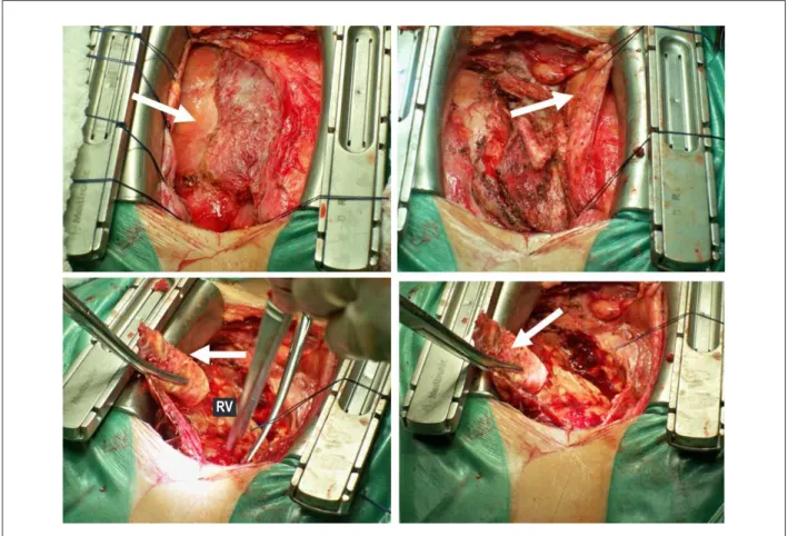
With these findings, a pericardiectomy and exploration of the cardiac mass were recommended. During the surgery, a large quantity of calcium was observed in the pericardium mostly in the right chambers, which was compressing the right ventricle and the interventricular septum (Figure 2). However, the pericardiectomy consisted only of bilateral resection of the pericardium up to the phrenic nerve. The access to the interventricular septum was not possible due to the risk of perforation of the right ventricle. Extracorporeal circulation was not used.
Patient’s rheumatic tests and negative serology ruled out the hypothesis of a rheumatic disease that could impair the
Introduction
Constrictive pericarditis is a disease that results from the thickening of the pericardium, restricting diastolic filling and evolving to reduced cardiac output. Its cause is generally unknown, but it can develop after any disease that causes acute pericarditis.
This case report is interesting because of the several differential diagnoses that have come out from the ancillary tests. The impairment of the right ventricle, and particularly that of the interventricular septum, associated with a large and indefinite mass compressing the right cavities, which resembled a mediastinal invasion on the transthoracic echocardiogram, raised doubts about the diagnosis of constrictive pericarditis. Although the thoracic CT was able to clarify the diagnosis, it also evidenced the pericardium impairment and the large quantity of calcium adhered to the structures adjacent to the heart, which could correspond to a mediastinal expansion process invading the pericardium or even a primary pericardial tumor.
This kind of impairment has not been described in medical literature. The diagnosis was confirmed during surgery.
Case Report
Lima et al Constrictive pericarditis with extensive calcification
Arq Bras Cardiol 2011; 96(1): e7-e10 pericardium, according to her history. She did not report any
epidemiology or contact with tuberculosis.
The anatomicopathologic tests evidenced a nonspecific inflammatory process whose etiology could not be established.
Discussion
Since constrictive pericarditis is a rare disease, most physicians do not make a differential diagnosis. Thus, the condition is often diagnosed lately1,2.
Figure 1 -A - The arrows indicate, on the transthoracic echocardiogram, a possible mass impairing the interventricular septum and right ventricle. B - The arrows indicate,
on the CT image, a large calciication in the pericardium and interventricultum. RV - right ventricle; LV - left ventricle; RA - right atrium; LA - left atrium.
RV LV
LA
LA
Figure 2 -Surgical aspect. The arrows indicate the calciied and thickened pericardium. RV - right ventricle. RV
Case Report
Lima et al
Constrictive pericarditis with extensive calcification
Arq Bras Cardiol 2011; 96(1): e7-e10
All heart chambers can be involved, leading to restricted diastolic filling3.
The causes of constrictive pericarditis are many, including neoplasias and disorders of the connective tissue4.
In developing countries, the primary cause of pericarditis is tuberculosis5.
The disease manifests itself mostly as right heart failure6.During cardiac auscultation, a protodiastolic
sound (pericardial knock) can be detected in patients with calcification and severe restriction. As ventricular filling is restricted, the diastolic pressure rises in right and left ventricles, increasing significantly the mean atrial pressures. Contrarily to what normally occurs during inspiration, venous return from the venae cavae to the right atrium is not accelerated in constrictive pericarditis. Consequently, the mean venous pressure does not fall during inspiration; some times it even rises (Kussmaul’s sign). Another sign that can be observed is pulsus paradoxus, which is an accentuated decrease of the systolic blood pressure during inspiration.
Ancillary tests are helpful to make the diagnosis. The chest X-ray shows pericardial calcification, mainly in profile view, besides pleural effusion without a definite etiology. The TTE confirms the presence of small ventricles with septal deviation caused by the abrupt diastolic filling; it also allows the evaluation of the pericardial thickness, presence of pericardial effusion, and signs of calcification7. The hemodynamic study
is very helpful in the diagnosis. The hemodynamic measures show diastolic pressure equalization, and the ventricular traces have the format of a square root symbol (drop and plateau), indicating a very rapid diastolic filling. Thoracic CT or MR images allow the evaluation of the myocardial thickness and are very helpful to differentiate between constrictive and restrictive pericarditis8.
Upon diagnosis, the pericardiectomy is the recommended procedure to control the disease. Releasing the restricted heart improves the cardiac function, leads to recovery, and, in most
cases, resolves the heart failure. Survival after the surgery depends on disease etiology, and patients with idiopathic constrictive pericarditis tend to have a better evolution9.
This case was worthy to be reported due to the unusual aspect of the calcification, which was very extensive and infiltrated into the interventricular septum. This extensive infiltration into the septum (Figures 1 and 2) raised a doubt about the existence of an associated disease causing septal calcification or a neoplastic disease invading the pericardium. Cases of pericardial mesothelioma, a malignant heart tumor involving the pericardium and associated with a bad prognosis, have been reported in the literature10. As TTE can not always detect this type of tumor, thoracic CT scan or MRI is required. Histochemical and anatomicopathological analyses are necessary to confirm the diagnosis, but frequently they are performed postmortem.
During the surgery, the extensive constrictive pericarditis was confirmed. The large quantity of calcium in the septal area accounted for the images obtained by echocardiography and thorax CT.
Surgical resection of the thickened pericardium and calcifications did release the ventricle, promoting patient’s recovery.
Potential Conflict of Interest
No potential conflict of interest relevant to this article was reported.
Sources of Funding
There were no external funding sources for this study.
Study Association
This study is not associated with any post-graduation program.
References
1. Asher CR, Klein AL. Diastolic heart failure: restrictive cardiomyopathy, constrictive pericarditis, and cardiac tamponade: clinical and echocardiographic evaluation. Cardiol Rev. 2002; 10 (4): 214-29. 2. Akhter MW, Nuno IN, Rahimtoola SH. Constrictive pericarditis masquerading
as chronic idiopathic pleural effusion: importance of physical examination. Am J Med. 2006; 119 (7): e1-e4.
3. Chinnaiyan KM, Leff CB, Marsalese DL. Constrictive pericarditis versus restrictive cardiomyopathy: challenges in diagnosis and management. Cardiol Rev. 2004; 12 (6): 314-20.
4. Ling LH, Oh JK, Schaff HV, Danielson GK, Mahoney DW, Seward JB, et al. Constrictive pericarditis in the modern era: evolving clinical spectrum and impact on outcome after pericardiectomy. Circulation. 1999; 100 (13): 1380-6. 5. Cooper DK, Sturridge MF. Constrictive epicarditis following Coxsackie virus
infection. Thorax. 1976; 31 (4): 472-4.
6. Tabata T, Kabbani SS, Murray RD, Thomas JD, Abdalla I, Klein AL. Difference in the respiratory variation between pulmonary venous and mitral inflow
Doppler velocities in patients with constrictive pericarditis with and without atrial fibrillation. J Am Coll Cardiol. 2001; 37 (7): 1936-42.
7. Himmelman RB, Lee E, Schiller NB. Septal bounce, vena cava plethora, and pericardial adhesion: informative two-dimensional echocardiographic signs in the diagnosis of pericardial constriction. J Am Soc Echocardiogr. 1988; 1 (5): 333-40.
8. Masui T, Finck S, Higgins CB. Constrictive pericarditis and restrictive cardiomyopathy: evaluation with MR imaging. Radiology. 1992; 182 (2): 369-73. 9. Bertog SC, Thambidorai SK, Parakh K, Schoenhagen P, Ozduran V, Houghtaling PL, et al. Constrictive pericarditis: etiology and cause-specific survival after pericardiectomy. J Am Coll Cardiol. 2004; 43 (8): 1445-52. 10. Brens RB, Diaz Guzmán D, Cabrera Peguero DC, Taveras FW, Severino
F, Llaverias R. Mesotelioma del pericardio manifiestado por cuadro de tamponada cardíaca: reporte de un caso / Pericardial mesothelioma manifested by cardiac tamponade: report of one case. Rev med domin. 1992; 53 (4): 168-70.