PONTINE AND EXTRAPONTINE OSMOTIC
MYELINOLYSIS AFTER THE SYNDROME OF
INAPPROPRIATE SECRETION OF ANTIDIURETIC
HORMONE (SIADH) ASSOCIATED WITH FLUOXETINE
Case report
Carlos Alexandre
Twardowschy
1, Cristina Buselatto
Bertolucci
2, Cleverson de Macedo
Gracia
3ABSTRACT - Osmotic demyelination syndrome (ODS) may be precipitated by aggressive correction of a hypo or hyper-osmolar states. We describe the case of a 53-year-old woman that was started on fluoxetine 20 mg/day for depression and nine days later was found to have fluoxetine-induced syndrome of inappro-priate secretion of antidiuretic hormone. After hyponatremia correction the mental status of the patient gradually improved, but subsequently she had intermittent difficulty in speaking, naming objects, mem-ory deficits and psychomotor slowness. Magnetic resonance revealed bilateral symmetric hyperintense le-sions in the basal ganglia, temporal lobe and hippocampal formation compatible with ODS. These symp-toms gradually resolved and she was discharged home without any deficits. Two months later, a new im-age showed lesion in pons and the other lesions had disappeared. Fluoxetine therapy had never been re-lated with a complication like that.
KEY WORDS: central pontine and extrapontine myelinolysis, osmotic demyelination syndrome, hypona-tremia, fluoxetine.
Mielinólise osmótica pontina e extrapontina após a síndrome da secreção inapropriada de hor-mônio antidiurético associada com fluoxetina: relato de caso
RESUMO - A síndrome de desmielinização osmótica (SDO) pode ser precipitada pela correção agressiva de um estado hiper ou hipoosmolar. Nós descrevemos o caso de mulher de 53 anos que havia iniciado o uso de fluoxetina 20 mg/dia para depressão e que nove dias depois foi diagnosticada como tendo síndrome da secreção inapropriada de hormônio antidiurético induzida por fluoxetina. Depois da correção da hipona-tremia o estado mental da paciente gradualmente melhorou, mas subsequentemente ela apresentou di-ficuldade intermitente para fala e para nomear objetos, déficits de memória recente e lentidão psicomo-tora. Ressonância magnética revelou lesões hiperintensas bilaterais e simétricas na região dos gânglios da base, lobo temporal e hipocampo compatíveis com SDO. Estes sintomas gradualmente se resolveram e a paciente foi de alta sem qualquer déficit. Dois meses mais tarde uma nova imagem cerebral mostrou lesão na ponte e ausência das lesões antigas. Até onde sabemos a terapia com fluoxetina nunca foi relacionada a uma complicação tardia como esta.
PALAVRAS-CHAVE: mielinólise central pontina e extrapontina, síndrome de desmielinização osmótica, hiponatremia, fluoxetina.
Department of Internal Medicine & Department Neurology of Nossa Senhora das Graças Hospital, Curitiba PR, Brazil: 1Internal Medi-cine Resident; 2Psychiatry Resident, Clinics Hospital, Federal University of Paraná, Curitiba PR, Brazil; 3Neurologist, Curitiba PR, Brazil. Received 8 December 2005, received in final form 23 April 2007. Accepted 11 June 2007.
Dr. Carlos AlexandreTwardowschy - Rua Dias da Rocha Filho 261 / 11 - 80040-050 Curitiba PR - Brasil. Email: [email protected] Central pontine myelinolysis (CPM) was first
de-scribed in alcoholic and malnourished patients in 19591
and subsequently has been expanded in recent years to include patients with symmetrical extrapon-tine lesions, notably in the basal ganglia, thalami and midbrain. Recently, with better understanding, these conditions were more appropriately recognized as
We describe the case of a woman, presenting with severe hyponatremia and syndrome of inappropriate secretion of antidiuretic hormone (SIADH), strongly associated with the use of fluoxetine. After correc-tion of her sodium abnormality, despite the strict ob-servance of recent therapeutics guidelines, the pa-tient developed pontine and extrapontine osmotic myelionysis. In this paper we try to relate the fluox-etine therapy as the initial cause of the ODS. As we know fluoxetine therapy had never been related with a complication like that.
CASE
A 53-year-old woman was seen in an emergency de-partment for evaluating a chest pain and dyspnea for the past two months. The patient also reported associated for-getfulness, decreasing energy, anhedonia, insomnia, anx-iety and irritability for the past four months. No psychotic symptoms were identified. She had no familiar cardiovas-cular risk. She was been treated for systemic arterial hy-pertension for the past eight years and last year was on losartan (100 mg/day) plus hydrochlorothiazide (25 mg/d). Acute myocardial infarct was ruled out and renal function
was normal. The serum sodium was 135 mmol/L. The pa-tient was discharged for a cardiologist evaluation and she was started on fluoxetine (20 mg/day).
Nine days later, the patient started with weakness and nausea in the morning. At night of the same day she be-came confused, was unable to stand or feed herself, and had one episode of vomit. Three hours later she became unresponsive and had a generalized seizure, during ap-proximately 1 minute, followed by sleepness. After the at-tack she was brought to our emergency service. On admis-sion the patient was lethargic, with impaired attention and alertness, but she was normovolemic and without focal mo-tor deficits. Her blood pressure was 210/100 mmHg. She had mild generalized muscle rigidity, Babinski`s sign bilat-erally and increased deep tendon reflex. The initial serum sodium was 105 mmol/L, the potassium 2.9 mmol/L and the complete laboratory evaluation is show in the Table. She was performed a cranium tomography and cerebrospinal fluid analysis that were normal. Thyroid stimulation hor-mone (TSH) level was within normal limits. No indications of pneumonia were found on chest X-ray. Abdomen ultra-sonography was normal.
The hyponatremia was believed to be secondary to fluoxetine-induced SIADH and it was discontinued. Her
Table. Laboratory evaluation during hyponatremia correction.
Exam/day 1st 2nd 3rd 4th 5th
Hemoglobin (g/dL) 14.4 14.6 13.5 13.1
Leucocytes (per mm3) 14.130 12.140 7.830 6.160
Band forms (%) 12 10 7 1
Platelets (per mm3) 329.000 310.000 282.000 287.000
INR 1.1
Glucose (mg/dL) 266 124 114 112
Serum Ca (mg/dL) 8.4 8.2 8.5
Serum Mg (mmol/L) 1.7 2.1 2.4 2.1
Serum Na (mmol/L) 105 114 130 130 138
Serum K (mmol/L) 2.9 3.2 2.9 3.8 3.9
Urinary Na (mmol/L) 70 27 68
Urinary K (mmol/L) 20 15
Serum osmolality (mOsm/L) 220 261 288
Urinary osmolality (mOsm/L) 400 328 224
Urea (mg/dL) 14 10 8
Creatinin (mg/dL) 0.6 0.7 0.8 0.7 0.7
AST (U/L) 29
ALT (U/L) 30
Total bilirubin (mg/dL) 1.0
pH 7.49
pCO2 33
HCO3 25
pO2 74
TSH 0.59
systolic blood pressure was controlled with oral nifedip-ine. Fluids were restricted to 1000 mL/day and because the severity of symptoms hypertonic saline solution was giv-en plus 80 mg of intravgiv-enous furosemide. The hypokale-mia was also corrected with 200 mmol K+/day. With im-provement of her hyponatremia, the patient became more alert, generalized muscle rigidity, Babinski`s sign and in-creased deep tendon reflex disappeared, but her condition subsequently worsened. She had intermittent difficulty in speaking, naming objects, memory deficits and psychomo-tor slowness. Magnetic resonance images (MRI) revealed bilateral symmetric hyperintense lesions in the basal gan-glia (Figs 1A and 2A). The serum Na+ continued to return to normal over the next few days. The patient´s speech cleared and her mentation returned to the premorbid lev-el in five days, paralllev-eling the resolution of her hyponatre-mia (Graph). The fluid restriction was discontinued
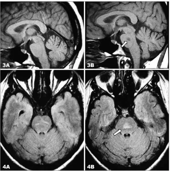
with-Column A: sixth day after hyponatremia correction. Column B: two months later.
Fig 1. (A) Coronal T2-weighted image and Fig 2. (A) Axial FLAIR image shows bilateral symmetric hyperintense lesions in the basal ganglia (Observation: the arrows are showing the lesions.). Two moths later the lesions had disappeared 1 (B) and 2 (B).
out recurrence of hyponatremia. After few days she was discharged home without any deficits. At 2-months follow-up she continued without any symptoms. Another MRI re-vealed resolution of the basal ganglia lesions (Figs 3A and 4A) and a new image in the pons (Figs 3B and 4B) compat-ible with pontine myelinolysis.
DISCUSSION
Hyponatremia is the most common electrolyte ab-normality observed in a general hospital population and it should be especially considered in patients tak-ing a selective serotonin re-uptake inhibitor2,3. First
described in 1959 by Adams et al.1
as central pontine myelinolysis and now more appropriately described as osmotic demyelination syndrome, this condition is
characterized by regions of demyelination through-out the brain most notably when hyponatremia had been corrected rapidly.
Hyponatremia causes generalized encephalopa-thy with manifestations that include malaise, nau-sea, headache, lethargy, confusion, seizures, coma, and death. Focal neurologic signs are rare. The se-verity of symptoms depends on the degree and rate of development of hyponatremia. A rapid decrease in sodium levels may lead to coma and seizures, but if hyponatremia develops slowly, patients are much less symptomatic4. The risk factors for development
of ODS after correction of chronic hyponatremia in-clude hypokalemia, malnutrition, chronic alcoholism5
, hypoxia6, burn injury, surgical removal of pituitary
Column A: sixth day after hyponatremia correction Column B: two months later
tumor and those who have undergone orthotopic liver transplant7
.
The typically histopathologic features include symmetric demyelination of the pons. The charac-teristics are a triangular to “bat-wing” lesion that spreads centrifugally from the median raphe and is located in the dorsal basis pontis with relative spar-ing of tegmentum, corticospinal and corticobulbar tracts except in the most severe cases. CPM and ex-trapontine myelinolysis (EPM) may occur alone or in conjunction8.
The cause and pathogenesis of CPM/EPM are not known. One proposed mechanism of demyelination has involved osmotic shifts with harmful metabolic consequences to oligodendrocytes. Another proposed mechanism of demyelination attributes changes to vasogenic edema developing from osmotic open-ing of the endothelial blood-brain barrier. Vascular endothelial injury due to rapid osmotic change and subsequent demyelination was also involved in the patho-etiology of EPM9. In this patient, extrapontine
abnormalities resolved in 2 months, suggesting re-versible immediate causes, but surprisingly another tardier lesion appears in the pons.
The clinical presentation of CPM typically includes progressive lethargy, quadriparesis, dysarthria, oph-thalmoplegia, dysphasia, ataxia, and reflex changes that usually occurs 2-7 days after treatment for the underlying disease or electrolyte disturbance has been initiated. Akinesis, ataxia, catatonia, choreo-athetosis, cogwheel rigidity, disorientation, dysar-thria, dystonia, extrapyramidal symptoms, emotional lability, gait disturbance, movement disorders, mut-ism, myoclonus, myokymia, parkinsonmut-ism, rigidity, and tremor have all been described in patients with EPM alone10. The Babinski´s sign, encountered in this
particular case, could be found in 21% of patients with ODS and increased deep tendon reflex in 36%11
, but they were found before correction of the hy-ponatremia, therefore, should not be attributed to EPM. However, her intermittent difficulty in speak-ing, naming objects, memory deficits and psychomo-tor slowness certainly were. She did not present any symptom of the CPM spite the MRI lesion.
Demyelination in pontine or extrapontine loca-tions does not appear exclusively in adult or alcoholic patients with hyponatremia12
but may occur when the serum sodium level is normal13,14 or elevated15,16.
The osmotic demyelination syndrome may occur even when the serum sodium abnormality is corrected within limits considered “safe”17,18. This patient
re-ported present typical lesions in MRI compatible with EPM/CPM besides carefully correction of hyponatre-mia. It is clear that rapid correction of hyponatremia is not the only factor involved.
Riggs et al.19
suggest that osmotic stress and oli-godendrocyte topography rather than rapid cor-rection of hyponatremia may be the key to the de-velopment of osmotic myelinolysis. Lateral pontine and extrapontine myelinolysis can be associated with hypernatremia and hyperosmolality20. Experimental
data shown that high and sustained levels of hyper-natremia could induce brain myelinolysis in rats21
. In both hypo and hypernatremic states, the significant event may be an increase in serum sodium or serum osmolality with sufficient rapidity and magnitude. Osmotic myelinolysis may not be exclusively an iatro-genic disorder (resulting from aggressive correction of chronic hyponatremia) but may result from other rapid changes in serum osmolality.
The distribution of cerebral demyelinating lesions in patients with hyponatremic encephalopathy could be compatible with hypoxic damage6. It appears that
ischemic hypoxia severely impairs the brain’s adap-tive mechanisms during hyponatremia by reducing cerebral oxygen availability. It may be an important comorbid factor in the morbidity associated with hy-ponatremic encephalopathy22
. Bilateral basal ganglia lesions such as these may be seen in association with hypoxia, but in this case, no respiratory disturbance, cardiac arrest, or hypotension had occurred. Hypoxic brain damage was unlikely.
Demyelination appears on CT as an area of de-creased attenuation in the central pons or in extra-pontine locations, although CT may underestimate the true extent of disease. The yield of MRI in de-termining both the number and extent of lesions in the osmotic demyelination syndrome is significantly better. Acute demyelinating lesions are symmetric and hypointense on T1 weighted images; during the subacute stage they become hyperintense on T2 weighted images probably because of the presence of endothelial injury-induced microhemorrhages. Le-sions on MRI may appear days to weeks after the onset of symptoms and may resolve completely over a period of months10. Like in this case imaging
stud-ies performed early during the illness may be un-remarkable, but still a diagnosis of central pontine myelinolysis should be suspected and a repeat imag-ing study might be required in 10-14 days to estab-lish the diagnosis23
disease. However, patients either improve clinically before the lesion regresses on MRI or they improve clinically independently of MRI findings24
.
Despite improved recognition of the classic lesions of CPM/EPM by modern neuroradiological techniques, the absence of autopsy confirmation in most recent reports raises questions about diagnostic accuracy. Over-reliance on the use of neuroimaging to assign a histologically defined diagnosis may explain why re-cent reports of clinical outcome differ from classic de-scriptions10
. In fact, some reports shown that autop-sied patients with “classic” CPM both clinically and upon neuroimaging had other histopathologies such as hemorrhagic necrosis or ischemic rarefaction of the central pons25. Thereafter, differential diagnosis with
infarct, metastasis, glioma, multiple sclerosis, enceph-alitis, and radiation or chemotherapy should be maid.
The first step in the treatment of ODS is prevent neurological injury identifying which patients are at risk of the osmotic demyelination syndrome. The risk of ODS appears to be greatest when the rate of cor-rection is greater than 10-15 mmol/L/day. In symp-tomatic acute hyponatremia, the initial rate of cor-rection can be 1-2 mmol/L/h for several hours if on any day of treatment the total daily correction is not more than 8 mmol/L/day26
.
Both normal and hypertonic saline are hypertonic to the patient’s serum osmolality in this setting. Be-cause normal saline for volume expansion may Be-cause ODS when the serum Na+
rises, half normal saline may be used to minimize the rate of rise in serum Na+.
There is some evidence in animals and in man that hypokalemia, when associated with a hyponatremic state, could predispose a patient to the development of demyelination during correction of hyponatre-mia27
. Consequently, normalizing the hypokalemia ei-ther before, or at least concomitant with, the serum Na+ correction will be a prudent and logical approach.
With the assumption that undefined myelinotoxic compounds are contributing to the demyelinating process in CPM/EPM, once symptoms have occurred, treatment and decreased morbidity could be possible with plasmapheresis. Others have reported complete recoveries after steroid administration10
. Early dexam-etasone treatment can prevent the blood-brain bar-rier disruption that is caused by the rapid correction of hyponatremia and its associative demyelinative changes and suggest that dexametasone might be effective in preventing CPM, but these are experi-mental data28
.
Complete recovery is now possible, so CPM and EPM are no longer the devastating neurological
ill-nesses they were when first described. According to the previous literature the prognosis for CPM and EPM were considered to be poor. However, milder courses and survival without substantial neurologi-cal deficit have been reported in recent years. Retro-spectively evaluation of 44 patients with CPM shows that the outcome does not depend on the severity of neurological deficits during the acute phase of the condition or on concomitant internal diseases, including the degree of hyponatremia. Of the 34 pa-tients for whom follow-up data were available, 32 survived. Of these 11 completely recovered, 11 had some deficits but were independent, and 10 were dependent24
. Earlier recognition of patients at risk of ODS, combined with a better understanding of the pathophysiology of electrolyte disturbances in diverse medical conditions, has allowed for more precise fluid and electrolyte management and bet-ter clinical outcomes.
It has been observed that ODS may occur despite strict observance of the published therapeutic guide-lines, like in this case described here. Variation in indi-vidual susceptibility to myelinolysis makes it difficult to determine a proper rate of correction Therefore, a completely safe rate of correction probably cannot be defined. Nevertheless, it seems reasonable that the rate of correction should be as low as possible, especially if the chronic hyponatremia is accompa-nied by other risk factors for ODS.
Finally, as we know, this is the first report associat-ing SIADH and hyponatremia due to fluoxetine hav-ing a tardier complication like ODS. SIADH is an im-portant, under recognized and serious complication of the selective serotonin reuptake inhibitors. Pa-tients should have their serum sodium concentration monitored, especially in the early stages of treatment.
REFERENCES
1. Adams RD, Victor M, Mancall EL. Central pontine myelinolysis: a hith-erto undescribed disease occurring in alcoholic and malnourished pa-tients. AMA Arch Neurol Psychiatry 1959;81:154-172.
2. Liu BA, Mittmann N, Knowles SR, Shear NH. Hyponatremia and the syndrome of inappropriate secretion of antidiuretic hormone associat-ed with the use of selective serotonin reuptake inhibitors: a review of spontaneous reports. CMAJ 1996;155:519-527. Erratum in: Can Med As-soc J 1996;155:1043.
3. Twardowschy CA, Bertolucci CB, Gracia CM, Brandão MAS. Severe hy-ponatremia and the syndrome of inappropriate secretion of
antidiuret-ic hormone (SIADH) associated with luoxetine. Arq Neuropsiquiatr
2006;64:142-145.
4. Laureno R, Karp BI. Myelinolysis after correction of hyponatremia. Ann Intern Med 1997;126:57-62.
5. Aguiar AC, Costa VM, Ragazzo PC, Caiado RR, Gusmao CA,
Melo-Souza SE. [Central pontine and extra-pontine myelinosis in alcoholic patient with Shoshin beriberi]. Arq Neuropsiquiatr 2004;62:733-736.
7. Singer C, Lorenzo D, Papapetropoulos S, Mesa A, Bowen B. Pontine/
extrapontine myelinolysis occurring in the setting of an eating disor
-der. Neurology 2005;64:2156-2157
8. Hadield MG, Kubal WS. Extrapontine myelinolysis of the basal ganglia
without central pontine myelinolysis. Clin Neuropathol 1996;15:96-100.
9. Waragai M, Satoh T. Serial MRI of extrapontine myelinolysis of the
basal ganglia: a case report. J Neurol Sci 1998;161:173-175. Erratum in: J Neurol Sci 1999;166:156.
10. Brown WD. Osmotic demyelination disorders: central pontine and ex
-trapontine myelinolysis. Curr Opin Neurol 2000;13:691-697.
11. Karp BI, Laureno R. Pontine and extrapontine myelinolysis: a neuro
-logic disorder following rapid correction of hyponatremia. Medicine (Baltimore) 1993;72:359-373.
12. Cadman TE, Rorke LB. Central pontine myelinolysis in childhood and adolescence. Arch Dis Child 1969;44:342-350.
13. Taneja K, Sabharwal RK, Taneja A, Mukherjee M. Central pontine my-elinolysis in a normonatremic child. Indian Pediatr 1997;34:153-156. 14. Kilinc M, Benli US, Can U. Osmotic myelinolysis in a normonatremic
patient. Acta Neurol Belg 2002;102:87-89.
15. van der Helm-van Mil AH, van Vugt JP, Lammers GJ, Harinck HI. Hy-pernatremia from a hunger strike as a cause of osmotic myelinolysis. Neurology 2005;64:574-575.
16. Brown WD, Caruso JM. Extrapontine myelinolysis with involvement of
the hippocampus in three children with severe hypernatremia. J Child Neurol 1999;14:428-433.
17. Leens C, Mukendi R, Foret F, Hacourt A, Devuyst O, Colin IM. Central
and extrapontine myelinolysis in a patient in spite of a careful correc
-tion of hyponatremia. Clin Nephrol 2001;55:248-253.
18. Kelly J, Wassif W, Mitchard J, Gardner WN. Severe hyponatraemia
sec-ondary to beer potomania complicated by central pontine myelinoly-sis. Int J Clin Pract 1998;52:585-587.
19. Riggs JE, Schochet SS Jr. Osmotic stress, osmotic myelinolysis, and ol-igodendrocyte topography. Arch Pathol Lab Med 1989;113:1386-1388. 20. McComb RD, Pfeiffer RF, Casey JH, Wolcott G, Till DJ. Lateral pontine
and extrapontine myelinolysis associated with hypernatremia and hy
-perglycemia. Clin Neuropathol 1989;8:284-288.
21. Soupart A, Penninckx R, Namias B, Stenuit A, Perier O, Decaux G. Brain myelinolysis following hypernatremia in rats. J Neuropathol Exp
Neurol 1996;55:106-113.
22. Vexler ZS, Ayus JC, Roberts TP, Fraser CL, Kucharczyk J, Arieff AI. Hypoxic and ischemic hypoxia exacerbate brain injury associated
with metabolic encephalopathy in laboratory animals. J Clin Invest 1994;93:256-264.
23. Kumar SR, Mone AP, Gray LC, Troost BT. Central pontine myelinolysis: delayed changes on neuroimaging. J Neuroimaging 2000;10:169-172.
24. Menger H, Jorg J. Outcome of central pontine and extrapontine myelin
-olysis (n=44). J Neurol 1999;246:700-705.
25. Giannetti AV, Pittella JE. Ischemic and hemorrhagic necrosis of the pons with anatomical location similar to that of central pontine myelinolysis in a chronic alcoholic patient. Clin Neuropathol 1993;12:156-159. 26. Adrogue HJ, Madias NE. Hyponatremia. N Engl J Med
2000;342:1581-1589.
27. Lin SH, Chau T, Wu CC, Yang SS. Osmotic demyelination syndrome af-ter correction of chronic hyponatremia with normal saline. Am J Med Sci 2002;323:259-262.
28. Sugimura Y, Murase T, Takefuji S, et al. Protective effect of dexameth
-asone on osmotic-induced demyelination in rats. Exp Neurol 2005;