Arquivos Brasileiros de Cardiologia - Volume 86, Nº 4, April 2006
307
Image
Image
Spontaneous Coronary Artery Dissection Causing
Acute Coronary Syndrome
Francisco Juarez C. Vasconcelos Filho, José Erirtônio F. Barreto, Raimundo Barbosa Barros,
Imara C. L. Queiroz, Rômulo Ronkaly G. Lima
Hospital de Messejana - Fortaleza, CE - Brazil
Mailing Address: Francisco Juarez C. de Vasconcelos Filho • Av. Engenheiro Leal Lima Verde, 1471 - casa 10 - 60833-520 – Fortaleza, CE - Brazil
E-mail: [email protected] Received on 09/30/05 • Accepted on 01/12/06
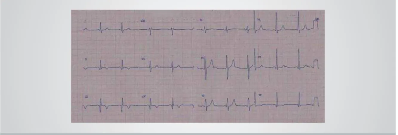
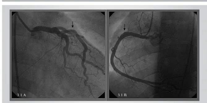
A 34-year old, healthy man, reporting very strong, oppressive precordial pain, triggered by exertional exercise (soccer game), radiating to back and left arm, and associated to cold sweating for over 1 hour and with no alleviating factor. The patient denied risk factors for CAD. The patient looked for medical assistance only 2 days after the clinical event. Physical exam showed: BP: 120x 60mmHg; HR: 60b.p.m; Cardiac auscultation: regular rhythm, S4; Pulmonary auscultation: Normal. Electrocardiogram at admission showed: sinusal rhythm with pathological Q waves in lower leads (D2, D3 and aVF) (Fig. 1). Laboratory exams at admission showed: CPK- 971 IU/L (normal < 195 IU/L); CK-MB- 59.7 IU/L (normal< 25IU/L); troponin- 2.15 ng/ml (normal<0.1 ng/ml). Elective coronary angiography identifi ed ulcerated plaque with thrombus adhered to its surface in the initial third of right coronary artery (Fig. 2.1.A) and spontaneous dissection in middle third of left anterior descending (Fig. 2.1.B); Left ventriculography showed: inferior hypokinesia (+++/+4). Atrium Flyer 3,5x16mm stent implantation was carried out in left anterior descending (Fig. 3.1.A) and infusion of IIb/IIIA inhibitor + IV heparin + clopidogrel
+ aspirin was indicated, with recommendation of a re-study 48 hours after therapeutic regimen. Control coronariography showed dissolution of intracoronary thrombus (Fig. 3.1.B).
COMMENTS
Coronary artery spontaneous dissection is a rare pathology that may lead to acute coronary syndrome with multiple clinical presentation spectra: angina pectoris, myocardium infarction or sudden death1. One
or two arteries may be injured in the same episode2. It
is more commonly found in groups of young people and it particularly focuses post-partum women3. Assumed
etiology includes: Disruption of atherosclerotic plaque, exhausting physical exercise, pregnancy related hormonal infl uence, use of contraceptives; in some cases, risk factors cannot be identifi ed.4 Treatment consists of:
clinical and drug treatment, myocardial revascularization surgery, angioplasty with stent, and at times even heart transplant.5 As for the case in question, we have presented
the use of a combination: drug therapy + stent.
Arquivos Brasileiros de Cardiologia - Volume 86, Nº 4, April 2006
308
Fig. 2.1.A – Right coronary: Showing ulcerated plaque with thrombus adhered to its surface); 2.1.B – Left anterior descending: showing spontaneous dissection in middle third) (Arrows)
Fig. 3.1.A – Left anterior descending. Angio after stent implant); 3.1.B - Right coronary control coronariography: showing dissolution of intracoronary thrombus (Arrows)
1. Pita JP, Gonzalez NV, Alvarez LP, Rodriguez JMV, Beiras AC. Spontaneous coronary artery dissection:case reports. Cath Cardiovasc Diagnosis 1994; 32: 27-32.
2. Ooi A, Lavrsen M, Monro J, Langley SM. Successful emergency surgery on triple-vessel spontaneous coronary artery dissection. Eur J Cardio Thoracic Surgery 2004; 26: 447-449.
3. Rajiv D, Gurdev S, Henry F. Spontaneous coronary artery dissection: The Clinical Spectrum. Angiology 2002;53: 89-93.
4. Hering D, Piper C, Hohmann C, Schultheiss HP, Horstkotte D. Prospective untersuchung zu haufigkeit, pathogenese und therapie spontaner, koronarangiographisch diagnostizierter koronararteriendissektionen. Z Kardiol 1988; 87: 961-970. 5. Roig S, Gómez JA, Fiol M, Guindo J, Pérez J, Carrillo A et al.
Spontaneous coronary artery dissection causing acute coronary syndrome:An early diagnosisimplies a goodprognosis. Am J Emergency Med 2003; 21: 549-551.