Bruno Carlini-Jr(a) Doglas Cecchin(b)
Gisele Damiana da Silveira Pereira(c)
Luis Alexandre Maffei Sartini Paulillo(b)
(a)Department of Restorative Dentistry, Passo
Fundo Dental School, University of Passo Fundo, Passo Fundo, RS, Brazil.
(b)Department of Restorative Dentistry,
Piracicaba Dental School, State University of Campinas, Piracicaba, SP, Brazil.
(c)Department of Restorative Dentistry, Rio de
Janeiro Dental School, Federal University of Rio de Janeiro, Rio de Janeiro, Rio de Janeiro, RJ, Brazil.
Corresponding Author:
Luiz Alexandre Maffei Sartini Paulillo E-mail: [email protected]
Received for publication on Jan 12, 2011 Accepted for publication on Apr 25, 2011
Influence of remaining coronal
structure and finish line on the fracture
strength of roots restored with metallic
posts
Abstract: The purpose of this study was to evaluate the fracture strength of roots that were prosthetically restored with metallic posts with or without any remaining coronal structure and with different inish lines. Sixty bovine incisors were sectioned below the cementoenamel junc-tion, endodontically treated, and randomly divided into six experimental groups (n = 10) containing teeth with or without any remaining coronal structure, and with a beveled shoulder, a bevel, or a shoulder inish line design. The metallic posts were luted with dual-cured resin cement. The cores were made with composite resin, and metal crowns were cemented with zinc phosphate cement. The specimens were subjected to a tangen-tial compressive load (135° angle) at a crosshead speed of 0.5 mm/min until failure, using a universal testing machine. The fracture strength data were analyzed using the ANOVA and LSMeans (least square means) tests (α = 0.05). The data indicated that the teeth with 2 mm of remain-ing coronal structure showed the highest fracture strength values when compared with the teeth without any remaining structure (p < 0.05). As to the different inish line designs, the highest fracture strength values were obtained for the beveled shoulder, followed by the bevel and then by the shoulder designs (p < 0.05). It may be concluded that, to increase fracture strength, a beveled shoulder and 2 mm of remaining coronal structure are the ideal conditions.
Descriptors: Dental Cavity Preparation; Dental Restoration Failure; Tooth Root; Post and Core Technique; Compressive Strength.
Introduction
Endodontically treated teeth frequently require indirect restorations because of extensive loss of healthy tooth structure as a result of carious lesion or fracture.1 In these cases, the use of posts is recommended to promote retention of the inal restoration.2-3
The preservation of tooth structure is an important factor in the suc-cessful restoration of endodontically treated teeth. Some authors4,5 have suggested that a tooth should have a minimum amount (2 mm) of coro-nal structure above the cementoenamel junction (CEJ) to ensure a proper shape of strength for a tooth. This coronal structure will provide a fer-rule effect with the artiicial crown that should prevent root fracture,
post fracture and dislodgement.
The strength of a prosthetic restoration depends not only on the post and remaining coronal struc-ture, but also on a suitable preparation and inish line design.4 In studies designed to investigate the frac-ture strength of metal and glass ceramic crowns, it was observed that tooth preparation with a 1.2-mm shoulder inish line and a sharp axiogingival line an-gle produced the strongest crowns, whereas crowns fabricated for preparations with a chamfer inish line produced the weakest restoration.6,7 In contrast, some authors reported that the fracture strength of crowns luted with a resin luting agent was unaf-fected by the type of inish line used.8,9 Rosen10 and Rosner11 reported that the beveled preparation de-sign promoted a reduction in casting errors, a seal against tooth fracture, appropriate adaptation of the prosthesis and the establishment of the ferrule effect.
Thus, the purpose of this study was to evaluate the fracture strength of anterior tooth roots that were prosthetically restored with metallic posts with or without 2 mm of remaining coronal structure, and with a beveled shoulder, a bevel or a shoulder inish line design. The hypothesis tested was that the fracture strength of roots might be affected by the remaining coronal structure and by the inish line design.
Methodology
Sixty recently extracted bovine lower central in-cisors stored in 0.9% thymol solution at 4 °C were selected for use in this study. The teeth were trans-versally sectioned at their cervical portion, using a diamond double-faced disk (Extec, São Paulo, Bra-zil) mounted on a low-speed handpiece, and cooled with air/water spray. Thirty roots were standardized at a length of 17 mm, whereas the other thirty were standardized at 19 mm. This difference was used to generate the 2-mm remaining coronal structure in half of the specimens.
The roots were embedded in polystyrene resin (Cromex, Piracicaba, Brazil) using a circular metal matrix (25 mm in diameter by 20 mm in height). Adhesive material and elastic-based urethane (Nex-us, Nucleus Duplex, Sweden) were used to simulate the periodontal ligament. All roots were embedded
to a depth of 15 mm, to maintain 2 mm of root length extending beyond the top of the acrylic resin in thirty roots, and 4 mm in the other thirty roots.
The root canals were prepared 1 mm short of the apex and illed with gutta-percha points (Dentsply Maillefer, Ballaigues, Switzerland) and Endometha-sone root canal sealer (Septodont, Saint-Maur-Dês-Fossés, France). After root canal illing, the gutta-percha points were all cut with size 3 Gates-Glidden burs (Dentsply Maillefer, Ballaigues, Switzerland), with a low-speed handpiece, maintaining 5 mm of illing material in the apical third of all specimens.
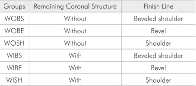
The roots without any remaining coronal ture and with 2 mm of remaining coronal struc-ture were randomly divided into 6 groups (n = 10) according to different inish line designs: beveled shoulder, bevel or shoulder (Figure 1). The groups are described in Table 1.
The post spaces were prepared to a depth of 12 mm in the roots without any remaining coronal structure and to a depth of 14 mm in the roots with 2 mm of remaining coronal structure, using a drill supplied by the post manufacturer, mounted on a low-speed handpiece. Air/water spray cooling was used during preparation, and dentin thickness was standardized.
The root dentin was etched with 37% phosphor-ic acid (FGM, Joinville, Brazil). Scotchbond MP Plus adhesive system (3M ESPE, St. Paul, USA) was then applied according to the manufacturer’s recom-mendations and light-polymerized for 40 s with a curing unit (3M, Sumaré, Brazil) using a light inten-sity of 500 mW/cm². Metallic posts (Flexi-post, EDS of Brazil, São Paulo, Brazil) were cemented with dual-cured resin cement (Enforce, Dentsply
Maille-Table 1 - Groups, remaining coronal structure and finish line designs of the roots.
Groups Remaining Coronal Structure Finish Line WOBS Without Beveled shoulder WOBE Without Bevel WOSH Without Shoulder
fer, Ballaigues, Switzerland). The cement was mixed and introduced into the post spaces with a lentulo-spiral drill using a low-speed handpiece.12 Cement was also applied on the posts before they were gen-tly seated by inger pressure and held in place for 60 s. The cement was light polymerized for 30 s on each surface (buccal, palatal, mesial and distal), for a total 2-min light polymerization cycle.
The composite resin cores (Filtek Z250; 3M ESPE) were standardized with a 0.25-mm-thick ac-etate matrix (Bio-art, São Carlos, Brazil) molded over the specimens in a vacuum-forming machine. The composite resin was placed in the acetate ma-trix in a single increment.
The inish line was prepared according to three different designs:
• beveled shoulder,
• bevel or
• shoulder.
The inish line was located at the limit between the tooth and the core for the teeth with no coro-nal remainder, and 2 mm below this limit for the specimens with remaining dental structure (Fig-ure 1). The inish line axial depth was 1 mm in all groups. All specimens were inished with a no. 3216 diamond bur (KG Sorensen, Barueri, Brazil) at high
speed and under water spray cooling.
The crown was replicated with silicone impres-sion material (Aquasil Ultra, Dentsply Maillefer, Bal-laigus, Switzerland) and poured in acrylic resin to make the other patterns. Rectangular-shaped stops with a central concavity were made on the palatine aspect of the patterns to locate and stabilize the met-al tip during the fracture strength test. Standardized acrylic resin crowns were obtained for all teeth. All crowns were cemented with zinc phosphate cement (Zinc Cement, SS White, Rio de Janeiro, Brazil). The specimens were then stored in 100% relative humid-ity, at a constant temperature of 37 (± 2) °C for a period of 48 h to ensure totally cured cement.
Next, the specimens were subjected to a com-pressive test in a universal testing machine (Emic DL 2000, Sao José dos Pinhais, Brazil). The position of the specimens was standardized using a device on the base of the apparatus, and load was applied at an angle of 135° in relation to the long axis of the roots.13,14 An increasing oblique compressive load was applied on the cingulum of the tooth’s palatal aspect (3 mm from the incisor edge), using a cylin-drical-shaped device with a round tip (2.7 mm in di-ameter). A cross-head speed of 0.5 mm/min was ap-plied until fracture. The root-post fragments set was removed from the acrylic resin after fracture and
Figure 1 - Schematic illustration of the groups under study. WOBS, WOBE and WOSH stand for the groups without any remaining coronal structure, and, respectively, with a beveled shoulder, a bevel or a shoulder design. WIBS, WIBE and WISH stand for the groups with 2 mm of remaining coronal structure, and, respectively, with a beveled shoulder, a bevel or a shoulder design. The fracture locations observed for the experimental groups were as follows: HF, horizontal fracture; CTF, transverse fracture in the cervical third of the root canal; MTF, transverse fracture in the middle third of the root canal; ATF, transverse fracture in the apical third of the root canal; LF, longitudinal fracture of the root.
WOBS
WOBE WOSH
WIBS
WIBE
WISH
observed under a stereoscopic magnifying glass at 4× magniication for fracture analysis. The fractures were classiied according to location as follows:
• horizontal fracture (HF) at the cementation line;
• transverse fracture in the cervical third of the root canal (CTF);
• transverse fracture in the middle third of the root canal (MTF);
• transverse fracture in the apical third of the root canal (ATF);
• longitudinal fracture of the root (LF) (Figure 1).
The data were analyzed by ANOVA and by LSMeans. The signiicance level was set at 5%.
Results
The compressive loads required to fracture the roots in each of the 6 groups are displayed in Table 2.
The groups with 2 mm of remaining coronal structure (WIBS, WIBE and WISH) showed high-er fracture strength values than those without any remaining coronal structure (WOBS, WOBE and WOSH) (p < 0.05), except for Groups WOBS and WISH, which did not differ statistically (p < 0.05). Regarding the preparation design, the highest frac-ture strength values were found for the beveled shoulder with or without any remaining structure (p < 0.05). However, the beveled shoulder and the bevel designs did not differ statistically (p > 0.05). Similar results were also observed when the bevel and the shoulder designs were compared within the groups with 2 mm of remaining coronal structure. The beveled shoulder design showed a fracture be-havior similar to that of the bevel and the shoulder
designs (p > 0.05). The preparation designs affected the fracture strength values in the groups without any remaining structure, considering that a statisti-cally signiicant difference was found (p < 0.05).
Groups without any remaining coronal structure (WOBS, WOBE and WOSH) typically showed an LF, whereas most fractures in the other groups were MTF and CTF (Table 3).
Discussion
This study evaluated whether the remaining cor-onal dentinal structure and inish line design could interfere with the fracture behavior of an endodonti-cally restored tooth. In general, the groups with re-maining dental structure required higher values of compressive load to promote root fracture. These results are in agreement with those of the studies by Varvara et al.15 and Ma et al.16 After prosthetic prep-aration, the remaining coronal structure becomes part of the axial wall of the core, and subsequent cementation of the crown will lead to a ferrule ef-fect that increases the fracture resistance of the root. The increase in resistance is explained by the greater preservation of dental structure and by a reduction of the lever effect of the post on the root canal walls.4
For the groups without any remaining dental structure, the beveled shoulder design presented the highest fracture resistance values, which were similar to those obtained by the group with remaining struc-ture and a shoulder design. This could be explained by the fact that the bevel extended 1 mm beyond the limit between the root and the prosthetic core, and, when embraced by a metal band, this type of inish line is capable of increasing the fracture resistance of the root/core/prosthetic crown set.4 Hence, these
Table 2 - Compressive strength values observed in the ex-perimental groups.
Group Minimum Maximum Mean ± s.d. WOBS 55.6 154.6 118.3 ± 31.4c
WOBE 49.5 154.9 98.6 ± 42.4d
WOSH 42.3 72.7 62.0 ± 10.8e
WIBS 87.5 166.4 135.8 ± 25.0a
WIBE 80.0 174.4 124.9 ± 32.8ª,b
WISH 94.0 155.0 123.2 ± 19.0b,c
Table 3 - Types of failure (number and percentage of teeth) for the experimental groups.
Groups HF CTF MTF ATF LF WOBS 2 (20%) 2 (20%) 2 (20%) 2 (20%) 2 (20%) WOBE 2 (20%) 3 (30%) 3 (30%) 1 (10%) 1 (10%) WOSH 1 (10%) 1 (10%) 3 (30%) 1 (10%) 4 (40%)
results demonstrated that, even in the presence of re-maining coronal dentin, the type of inish line design still inluences fracture resistance values.
The lowest fracture strength values were found for the shoulder design without any remaining struc-ture, a preparation coniguration which lacks a wall in dentin for the prosthetic crown to embrace. As a result, all the oblique force exerted on the crown is supported directly by the prosthetic core and trans-mitted successively to the post and root canal, gen-erating a lever effect on the root wall. The bevel de-sign was extended beyond the limit between the root and the prosthetic crown, involving about 0.5 mm of root dentinal structure. This feature allowed em-bracing only a small quantity of dental structure by the prosthetic crown, providing less ferrule effect than that achieved with the beveled shoulder, and leading to lower resistance values.
In the groups with remaining coronal dental structure, the beveled shoulder and the bevel designs showed similar behavior, a fact which could be at-tributed to the prosthetic crown having involved part of the root dentinal structure. The groups without any remaining coronal structure and with a beveled shoulder design behaved similarly to the group with remaining coronal dental structure and a shoulder design, which allows us to conclude that the ferrule effect is achieved in a similar manner by using the cervical bevel or involving the coronal structure on the axial wall of the core. Moreover, there is an in-dication of synergism, considering that the highest fracture resistance values were achieved when there was remaining coronal structure at the gingival bev-el. This combination of a remaining dental structure and the construction of a metal band can be used in cases of teeth with a short clinical crown, in which esthetics is not a preponderant factor and when the oblique forces on the prosthetic crown exceed the normal mastication forces, as is the case of teeth with ixed partial or removable dentures, adjacent to distal extensions or large prosthetic spaces.
Analysis of the fracture pattern demonstrated a greater trend towards longitudinal fractures (LF) for the groups without any remaining coronal struc-ture, whereas there was predominance of fractures in the middle and cervical thirds of the root for the
groups with remaining coronal structure, evidencing the protection exerted by the ferrule effect against longitudinal fractures.16,17 Similarly, there was a trend towards longitudinal fractures for the shoul-der design, contrasting with the beveled shoulshoul-der and bevel designs, which presented a trend towards transverse fractures in the middle third. The results of this analysis highlight the importance of preserv-ing the coronal dental structure and involvpreserv-ing it in the preparation, as well as that of the ferrule effect achieved by constructing a metal band. The beveled design showed a behavior similar to that of the bev-eled shoulder design with regard to the predominant fracture pattern. The fracture pattern has great clin-ical relevance, since it dictates the possibility of re-constructing the remaining dental structure. In this study, preservation of healthy tooth structure and its later involvement in the prosthetic preparation con-tributed to the occurrence of less catastrophic frac-tures, in comparison with those observed in teeth without any remaining coronal structure, or with-out a bevel to be embraced by the metal belt pro-vided by the metal prosthetic crown. The strength of the prosthetic restoration appears to be primarily related to the quantity of remaining coronal struc-ture involved in the preparation,16 and secondly to the inish line design.
In case of teeth without any remaining dental structure, the possibility of augmenting the clinical crown should be evaluated, or of performing root traction, in order to involve healthy dental struc-ture in the prosthetic preparation.12 In the event that these procedures are not possible, it is suggested that a beveled inish line be chosen so that a metal belt can embrace the root. The adhesive technique with resin composite and pre-fabricated iber posts is rec-ommended because it forms a single block between the root and the illing core. Nevertheless, further studies should be conducted to conirm these recom-mendations.
Conclusion
and tooth or tooth substitue core structure. J Prosthet Dent 2001 Nov;86(5):511-9
10. Rosen H. Operative procedures on mutilated endodontically treated teeth. J Prosthet Dent 1961 Sep-Oct;11:973-86. 11. Rosner D. Function, placement, and reproduction of bevels
for gold casting. J Prosthet Dent 1963;13:1160-66.
12. Fonseca TS, Alfredo E, Vansan LP, Sila RG, Sousa YT, Saquy PC, et al. Retention of radicular posts varying the application technique of the adhesive system and luting agent. Braz Oral Res. 2006 Oct; 20(4):347-52.
13. Lima AF, Spazzin AO, Galafassi D, Correr-Sobrinho L, Car-lini-Júnior B. Influence of ferrule preparation with or without glass fiber post on fracture resistance of endodontically treated teeth. J Appl Oral Sci 2010 Jul-Aug;18(4):360-3.
14. Cecchin D, Farina AP, Guerreiro CAM, Carlini-Júnior B. Fracture resistance of roots prosthetically restored with in-tra-radicular posts of different lengths. J Oral Rehabil. 2010 Fev;37(2):116-22.
15. Varvara G, Perinetti G, Di Iorio D, Murmura G, Caputi S. In vitro evaluation of fracture resistance and failure mode of internally restored endodontically treated maxillary incisors with differing heights of residual dentin. J Prosthet Dent. 2007 Nov;98(5):365-72.
16. Ma PS, Nicholls JI, Junge T, Phillips KM. Load fatigue of teeth with different ferrule lengths, restored with fiber posts, com-posite resin cores, and all-ceramic crowns. J Prosthet Dent. 2009 Oct;102(4):229-34.
17. Morgano SM, Bracket SE. Foundations in fixed prosthodon-tics: Current knowleddge and future needs. J Prosthet Dent. 1999 Dec;82(6):634-57.
References
1. Bitter K, Kielbassa AM. Post-endodontic restorations with adhesively luted iber-reinforced composite post systems: a review. Am J Dent. 2007 Dec;20(6):353-60.
2. Ottl P, Hahn L, Lauer H, Fay M. Fracture characteristics of carbon ibre, ceramic and non-palladium endodontic post systems at monotonously increasing loads. J Oral Rehabil. 2002 Feb;29(2):175-83.
3. Spazzin AO, Galafassi D, de Meira-Júnior AD, Braz R, Garbin CA. Inluence of post and resin cement on stress distribution of maxillary central incisors restored with direct resin composite. Oper Dent. 2009 Mar-Apr;34(2):223-9.
4. Sorensen JA, Engelman MJ. Ferrule design and fracture resis-tance of endodontically-treated teeth. J Prosthet Dent. 1990 May;63(5):529-36.
5. Wagnild GW, Mueller KL. Restoration of the endodontically treated tooth. In: Cohen S, Burns RC, editors. Pathways of the pulp. 8th ed. St. Louis: Elsevier; 2001. p. 765-95. 6. Doyle MG, Munoz CA, Goodacre CJ, Friedlander LD, Moore
BK. The effect of tooth preparation design on the breaking strength of Dicor crowns: II. Int J Prosthodont 1990 May-Jun;3(3):241-8.
7. Doyle MG, Goodacre CJ, Munoz CA, Andres CJ. The effect of tooth preparation design on the breaking strength of Dicor crowns: III. Int J Prosthodont 1990 Jul-Aug;3(4):327-40. 8. Bernal G, Jones RM, Brown DT, Munoz CA, Goodacre CJ.
The effect of finish line form and luting agent on the break-ing strength of Dicor crowns. Int J Prosthodont 1993 May-Jun;6(3):286-90.