*Corresponding author: F. Guan. Department of Pharmacology. College of Basic Medical Sciences. Jilin University, 130021 - 126 Xin Min Street, Changchun, Jilin, China. E-mail: [email protected]
A
vol. 52, n. 4, oct./dec., 2016 http://dx.doi.org/10.1590/S1984-82502016000400024
Effects of purine nucleotide administration on purine nucleotide
metabolism in brains of heroin-dependent rats
Kun Li
1, Ping Zhang
1, Li Chen
1, Fengying Guan
2*1School of Nursing, Jilin University, Changchun, China, 2Department of Pharmacology, School of Basic Medicine, Jilin
University, Changchun, China
Heroin is known to enhance catabolism and inhibit anabolism of purine nucleotides, leading to purine
nucleotide deiciencies in rat brains. Here, we determined the efect of exogenous purine nucleotide
administration on purine nucleotide metabolism in the brains of heroin-dependent rats. Heroin was administrated in increasing doses for 9 consecutive days to induce addiction, and the biochemical changes associated with heroin and purine nucleotide administration were compared among the treated groups. HPLC was performed to detect the absolute concentrations of purine nucleotides in the rat brain cortices.
The enzymatic activities of adenosine deaminase (ADA) and xanthine oxidase (XO) in the treated rat cortices were analyzed, and qRT-PCR was performed to determine the relative expression of ADA, XO, adenine phosphoribosyl transferase (APRT), hypoxanthine-guaninephosphoribosyl transferase (HGPRT), and adenosine kinase (AK). Heroin increased the enzymatic activity of ADA and XO, and up-regulated the transcription of ADA and XO. Alternatively, heroin decreased the transcription of AK, APRT, and HGPRT in the rat cortices. Furthermore, purine nucleotide administration alleviated the efect of heroin
on purine nucleotide content, activity of essential purine nucleotide metabolic enzymes, and transcript
levels of these genes. Our indings therefore represent a novel, putative approach to the treatment of
heroin addiction.
Uniterms: Purine nucleotide/quantitative analysis. Purine nucleotide/efects/brain rats. Heroin analysis.
Adenosine deaminase analysis. Xanthine oxidase analysis
INTRODUCTION
Opiates, such as morphine and heroin, are illegally used as recreational drugs worldwide, and can signiicantly
impair the user’s physical and mental health (Chu et al., 2009; Li et al., 2014). Opioid dependence is a signiicant
public health concern, underscoring the need for efective
treatment options (Kresina, Bruce, Mulvey, 2013; Nutt,
King, Phillips, 2010). Therefore, the exploration of the efects of repeated exposure to these compounds is
important for understanding the mechanisms of drug dependency and developing new treatment options.
Properly regulated purine nucleotide (PN) metabolism is necessary for normal brain function (Franke, 2011). Imbalances in purine nucleotide levels lead to a variety of human diseases (Burhans, Weinberger,
2007; El-Hattab, Scaglia, 2013; Kimura et al., 2003). Studies have shown that morphine and heroin can enhance
the oxidation of xanthine and hypoxanthine in the lobus
temporalis, lobus frontalis, and lobus parietalis of rats (Liu et al., 2007; Yang et al., 2006). Uric acid (UA) is the
inal oxidation product of purine metabolism in humans and higher primates, and changes in UA levels may relect
the catabolism of purine nucleotides (Liang, Clark, 2004). Furthermore, it has been reported that morphine may increase UA concentrations in the corpus striatum, as
well as in serum and extracellularly, in vitro (Enrico et al., 1997; Sumathi, Niranjali Devaraj, 2009). In patients where morphine was administered intracerebroventricularly for
cancer-related pain, the UA levels in cerebrospinal luid
samples increased significantly (Goudas et al., 1999). Morphine may also increase ATP catabolic products,
including nucleotides and oxypurines, in BALB/c mouse
strains (Di Francesco et al., 1998).
morphine (ip) over a period of 7 days in doses of 20, 30, 45, 55, 65, 85, and 95 mg/kg, respectively, to induce morphine addiction (Liu et al., 2007). Rats also received heroin (ip) over a period of 9 days in doses of 3, 3.45, 4.35, 5.7, 7.5, 9.75, 18.68, 17.16, and 21.12 mg/kg, respectively, to induce heroin addiction (Yang et al., 2006).
Signiicant increases in UA concentration in plasma were
observed in the rats following both morphine and heroin
administration. The enzymatic activities of ADA and XO were found to be enhanced signiicantly in the brain and
other organs of rats administered heroin, and morphine
was shown to increase ADA and XO gene expression in the rat brain cortex (Liu et al., 2007; Yang et al., 2006).
Our experiments using rat C6 glioma cells also revealed that the gene expression of HGPRT and AK, two
key enzymes in the purine nucleotide salvage pathway, are down-regulated by morphine, indicating that morphine inhibits purine nucleotide anabolism (Liu, Hong, Zhao,
2003). To further clarify the efects of heroin on purine
nucleotide metabolism in vivo, we measured the absolute content of purine nucleotides in rat brain cortices by HPLC,
and found that the levels of AMP and GTP are signiicantly
reduced in rats administered heroin, resulting in a purine nucleotide deficiency (Li et al., 2011). Therefore, we
hypothesize that purine nucleotide deiciency in the brain
may be one of the biochemical mechanisms of heroin
dependence. Thus, the goals of this study were to conirm
that purine nucleotide metabolism disorders are induced by heroin, and to clarify whether purine administration can counteract the biochemical changes caused by heroin.
MATERIAL AND METHODS
Animals and Drug Treatment
Adult, male Wistar rats (n = 40, 180 ± 20 g) were obtained from the Jilin University Animal Laboratory (Changchun, China), housed under controlled environmental conditions with free access to food and water, and randomly divided into four groups. Each group included 10 animals, and heroin was administered twice a day at 12-h intervals. Group I served as the control group and rats in this group received normal saline (ip) for 9 days. Rats in groups II, III, and IV were treated with heroin, heroin+PN, and PN alone, respectively. Heroin was administrated (ip) in increasing daily doses of 0.5, 0.75, 1.25, 2, 3, 4.25, 5.75, 5.75, and 5.75 mg/kg, for 9 days (Li et al., 2009). PN was administrated by gavage at a constant dose of 30 mg/kg (AMP+GMP; in equimolar concentrations), 2 h before the administration of heroin
for 9 days. On the morning of the 10th day, animals were
killed via decapitation following ether-induced narcosis. Their brains were rapidly removed and thoroughly
washed to remove the excess blood in ice-cold saline. The
cortices were removed on a pre-cooled plate, snap frozen in liquid nitrogen, and stored at -80 °C. All experimental
procedures were approved by the Experimental Animal
Ethics Committee of Jilin University.
Reagents
Heroin (99% purity) was obtained from the Public
Security Office of Jilin province, China. Adenine and
guanine nucleotides (ATP, AMP, GTP, and GMP) and the ion-pair reagent tetra butyl ammonium hydrogen sulfate (TBAHS) were purchased from Sigma. HPLC-grade acetonitrile (ACN) and methanol were purchased
from Fisher Scientiic (Pittsburgh, PA, USA). All other
chemicals used in this study were of analytical grade.
Distilled water, iltered with a Milli-Q Academic Ultrapure
Water System (Millipore, Bedford, MA, USA), was used to prepare the standard solutions and HPLC mobile phases.
ADA and XO detection kits, and Coomassie blue protein
detection kits, were purchased from Jinsite Biotechnology Co. Ltd. (Nanjing, China). PrimeScript RT Reagents Kits and SYBR PrimeScript RT-PCR kits were purchased from TaKaRa Biotechnology Co. Ltd. (Dalian, China).
Content detection of purine nucleotides
Samples were analyzed with an Agilent 1100 HPLC system equipped with an autosampler, using Agilent
Zorbsx SB-C18 (4.6×150 mm; 3.5 μm) columns. The mobile phase contained 10% methanol and 220 mmol/L KH2PO4, normalized to pH 6.5 with TBAHS. The low rate of HPLC elution was 500 μL/min. The temperature of the column was 27±1 °C. The eluate was monitored at 254 nm, with a sample size of 20 μL (Chen et al., 2007).
Cortex tissue (0.5 g) was removed from liquid
nitrogen and pulverized in 4.5 mL cold, physiologic saline with a tissue pulping machine, at 12,000 rpm in an ice-water bath. The tissue samples were sonicated
in 0.5 mL 6% HClO solution for 30 s in an ice-water
bath, adjusted with TBAHS (4 mol/L) to pH 7.0, then centrifuged. The supernatant was filtered through 0.2 pm 8-mm cellulose filters. The samples were injected directly into the HPLC column for PN measurement. By comparing retention times with pure GMP, AMP, GTP, and ATP, all compounds were chromatographically
identiied. Linearity was tested using ive known standard
area vs. concentration for GMP, AMP, GTP, and ATP. The concentrations of GMP, AMP, GTP, and ATP in the rat
cortex samples were calculated by plotting the peak area
against the known standard concentrations.
Enzyme assays
The tissue homogenate was centrifuged at 3000 rpm for 10 min at 4 °C. The sedimented fraction was discarded, leaving only the supernatant, which represented 10% of the total tissue homogenate. Protein content was estimated using Coomassie blue protein detection kits. The activity
of ADA and XO in the cortex samples were estimated
using Guisti colorimetric and colorimetric detection kits, respectively (Li et al., 2011; Yang et al., 2006).
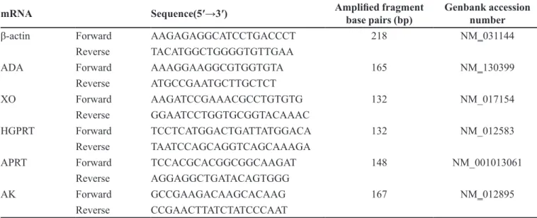
Evaluation of ADA, XO, HGPRT, APRT, and AK gene expression by qRT-PCR
Gene-speciic primers were designed and a BLAST (NCBI) search was performed to ensure their speciicity to
the target mRNA transcript. The primers were synthesized by the Jinsite Corporation (Nanjing, China). The primer
sequences for ADA, XO, HGPRT, APRT, AK, and β-actin
are summarized in Table I.
Total RNA was extracted with Trizol (Gibco BRL, Grand Island, NY, USA), normalized to a inal concentration of 500 ng/μL, and reverse transcribed using the PrimeScript
RT Reagents Kit according to the manufactures instructions. qRT-PCR analyses were performed on an ABI Prism 7000 Real Time PCR System (Applied Biosystems, Foster
City,CA,USA), in a inal volume of 20 μL, containing
10 µL SYBR Premix Ex Taq, 0.4 µL of each primer, 0.4 µL ROX Reference Dye, 2 µL RT product, and 6.8 µL dH2O, using the SYBR PrimeScript RT-PCR kit. The following qRT-PCR cycling conditions, recommended by the manufacturer, were used: 95 °C for 30 sec, followed by 40 cycles at 95 °C for 5 sec and 60 °C for 31 sec. All samples were run in 96-well optical plates in duplicate. The
cycle threshold values (Ct) were calculated and exported to Microsoft Excel for analysis. Quantiication was performed by experimental determination of Ct, deined as the cycle at which the luorescence exceeds 10 times the standard
deviation of the mean baseline emission for the early cycles (Collantes-Fernandez et al., 2002). The Pfal mathematical
model was used for qRT-PCR data analyses (Pfal, 2001). β-actin was used as a reference gene.
Statistical analyses
The data were analyzed and expressed as mean
± SD. Statistical analyses were performed using SPSS
18.0. Analysis of Variance (ANOVA) and post hoc SNK-q tests were performed to examine diferences between the groups. Signiicance was applied to values of 0.05 or 0.01 conidence.
RESULTS AND DISCUSSION
ADA and XO enzyme activity in rat cortices
The mean total protein content of the homogenates of the four groups (saline, heroin, heroin+PN, and PN)
TABLE I - Primer sequences for real-time PCR
mRNA Sequence(5′→3′) Ampliied fragment
base pairs (bp) Genbank accession number
β-actin Forward AAGAGAGGCATCCTGACCCT 218 NM_031144
Reverse TACATGGCTGGGGTGTTGAA
ADA Forward AAAGGAAGGCGTGGTGTA 165 NM_130399
Reverse ATGCCGAATGCTTGCTCT
XO Forward AAGATCCGAAACGCCTGTGTG 132 NM_017154
Reverse GGAATCCTGGTGCGGTACAAAC
HGPRT Forward TCCTCATGGACTGATTATGGACA 132 NM_012583
Reverse TAATCCAGCAGGTCAGCAAAGA
APRT Forward TCCACGCACGGCGGCAAGAT 148 NM_001013061
Reverse AGGAGGCTGATACAGTGGG
AK Forward GCCGAAGACAAGCACAAG 167 NM_012895
were 6.2 ± 0.2, 6.3 ± 0.4, 6.4 ± 0.6 and 6.2 ± 0.3 g/L,
respectively, which indicates that there was no signiicant
difference in the total tissue protein concentrations (P >0.05). The mean ADA activity of the saline, heroin, heroin+PN, and PN groups was 6.5 ± 0.8, 8.8 ± 0.9, 6.6 ± 0.8 and 6.3 ± 0.7 g/L, respectively, indicating that the
administration of heroin significantly increases cortex
ADA activity as compared with that in the saline group (P<0.05; Figure 1), and that PN administration cancels out the increased ADA activity induced by heroin (P<0.05).
The mean XO enzyme activity of each of the four
groups was 11.5 ± 1.2, 14.3 ± 1.2, 12.2 ± 1.0, and 11.7 ± 1.3
g/L, respectively. The XO activity of the heroin group was signiicantly higher than that of the saline group (P<0.05; Figure 1). The cortex XO activity of the heroin+PN group was signiicantly less than that of the heroin group
(P<0.05). PN administration alone did not result in any
signiicant diference in the examined enzyme activity as
compared to the control (P>0.05).
ADA and XO are essential enzymes for purine
catabolism. ADA irreversibly catalyzes the hydrolysis of adenosine to inosine. Inosine is then deribosylated by purine nucleoside phosphorylase, converting it to
hypoxanthine (Cristalli et al., 2001). XO catalyzes
the oxidation of hypoxanthine and xanthine to UA. Previously, our research showed that the ADA and XO
levels in many tissues of rats administered heroin in vivo
are elevated, and the efects of heroin on the brain persist
for a longer time than in other tissues (P<0.05; Yang et al., 2006). These results indicate that heroin increases the
ADA and XO levels in the cortex, and leads to increased
purine nucleotide catabolism. This study shows that the administration of purine nucleotides, in combination with
heroin, reduces ADA and XO levels dramatically (P<0.05)
as compared with the group receiving only heroin, and indicates that increased purine nucleotide catabolism resulting from heroin administration, is negated by
exogenous purine nucleotide administration.
mRNA transcript levels of key purine nucleotide metabolic enzymes
The ADA and XO mRNA levels in cortex tissue
were significantly higher in the group administered with heroin alone, as compared with those in the
saline-treated group (P<0.01). ADA and XO mRNA levels in the heroin+PN group, however, were signiicantly less
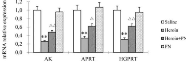
than those in the heroin-treated group (P<0.05, P<0.01; Figure 2). As shown in Figure 3, AK mRNA levels
were signiicantly lower in the group administered with
heroin alone, as compared with that in the saline-treated control group (P<0.01). APRT and HGPRT mRNA levels
were also signiicantly lower in the heroin-only group
(P<0.01). The mRNA levels of HGPRT, APRT, and AK in the heroin+PN group were higher than those in the heroin-only group (P<0.05).
Our data show significant increases in the ADA and XO mRNA levels in the cortices of rats administered
with heroin alone, as compared with those in the control
group that received only saline. Our results also show that
these increases were counteracted by purine nucleotide administration. Furthermore, we found that the tendency
of ADA and XO mRNA levels to change is correlated with
the activity of the corresponding enzyme. Therefore, we
conclude that the efect of heroin on purine nucleotide metabolism is dependent on the level of gene expression.
FIGURE 1 - ADA and XO activities in rat cortex. *P<0.05 vs.
Saline. δP<0.05 vs heroin.
FIGURE 2 - Relative mRNA level of ADA and XO in rat cortex. Relative mRNA levels are normalized against β-actin. ** P<0.01
vs. Saline. δP<0.05 vs Heroin. δδP<0.01 vs heroin.
Purine nucleotides are synthesized either from simple precursors by de novo synthesis through energetic, multistep reactions, or assembled from free purine
bases like guanine, hypoxanthine, and adenine, through
nucleotide salvage pathways (Camici et al., 2010). However, in some organs, including the brain, purine
nucleotide synthesis relies exclusively on salvage
pathways. Previously, our research showed that gene
expression levels of HGPRT, AK, and APRT, essential
enzymes in the purine nucleotide salvage pathway, are down-regulated by heroin both in vivo and in vitro (Li et al., 2011; Liu et al., 2007). HGPRT catalyzes the salvage
of the purine bases hypoxanthine and guanine to produce
nucleotides (Balasubramaniam, Duley, Christodoulou, 2014). APRT recycles adenine, produced by the polyamine pathway, to AMP. AK catalyzes the phosphorylation of adenosine to produce AMP (Camici et al., 2010; Ipata et al., 2011). As no HGPRT, APRT, or AK enzyme activity
detection kits were available, we examined the expression
levels of the HGPRT, APRT, and AK genes by qRT-PCR.
Our results reveal that heroin inhibits the transcription of
these three key enzymes involved in the purine nucleotide
synthesis pathway in the rat cortex, however, the inhibitory efects of heroin on these enzymes could be negated with the administration of exogenous purine nucleotides.
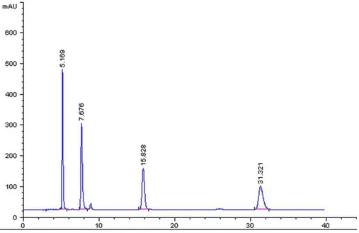
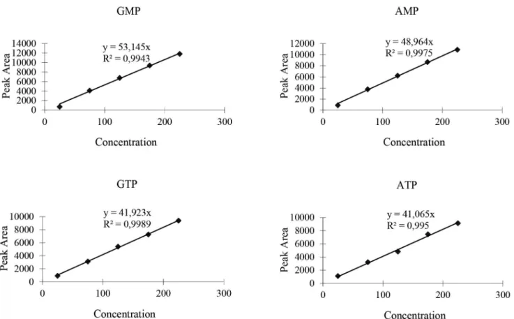
Content detection of purine nucleotides
Liquid chromatographic analyses were performed as previously described. Figure 4 shows the retention time
and area of the peak of the mixed reference solutions. Figure 5 shows the calibration curve obtained from cortex
tissue for the purine nucleotides. Figure 6 shows the HPLC
chromatogram of cortex tissue.
These results show that the proportion of AMP and
GTP in the heroin-treated group decreased signiicantly as
compared to those in the saline-treated group (Table II).
However, purine nucleotide administration signiicantly
reduced the magnitude of the AMP decrease induced by heroin (P<0.05), and alleviated the decrease in GTP,
although not signiicantly.
To verify our hypothesis that heroin dependence results in purine nucleotide deficiency, we measured the ATP, GTP, AMP, and GMP contents in the treated rat cortices. The AMP and GTP content in the group
administered heroin decreased signiicantly as compared
with those in the saline-treated group, which indicates that heroin reduces the content of some purine nucleotides in brain. Purine nucleotide administration, however,
alleviated the AMP deicit signiicantly (P<0.05), and also reduced the GTP deiciency, albeit to a lesser degree, in
heroin-dependent rats.
Purine nucleotides play an important role in DNA synthesis, energy metabolism regulation, protein synthesis and function, and enzymatic activity. Purines are also essential components of many coenzymes, including NAD and Coenzyme A, and signaling molecules like cAMP (Duval et al., 2013). However, the consequences
of AMP and GTP deiciency in the brain are not yet clear.
It has been reported that biochemical changes such as
TABLE II - The contents of GMP, AMP, GTP, ATP in rat cortex
Saline Heroin Heroin+PN PN
GMP (mg/mL) 37.35±2.59 36. 21±2.26 38.42±2.38 41.53±4.71
AMP (mg/mL) 87.78±3.92 49.04±3.11* 64.34±2.17δ 92.69±5.12
GTP (mg/mL) 15.65±1.49 4.31±0.44* 5.26±0.83 17.83±2.45
ATP (mg/mL) 4.10±0.85 4.28±1.12 3.94±0.72 4.48±0.67
*P<0.05 vs Saline; δP<0.05 vs Heroin.
FIGURE 5 - Calibration curve obtained from cortex for purine nucleotides. Diferent concentrations of purine nucleotide (25 mg/
mL, 75 mg/mL, 125 mg/mL, 175 mg/mL, 225 mg/mL) were used for the experiment.
decreased GMP concentrations are associated with opioid dependence in humans (Mannelli et al., 2009). It has also
been shown that opioid use may have toxic effects on
the central and peripheral nervous systems, leading to irreversible, pathological cellular changes. Furthermore, heroin and other opioids have been shown to inhibit the synthesis of DNA and RNA in brain tissues (Avella et al.,
2010; McLaughlin, Zagon, 2012). Our results indicate
that the effect of heroin on the metabolism of purine
nucleotides may result in purine nucleotide deiciency in
the brain, representing a putative biochemical mechanism
of heroin addiction. Our results also show that external
compensation of purine nucleotides alleviates the deficiency in purine nucleotides in rat cortices caused by heroin, although purine nucleotide administration alone does not alter purine nucleotide concentrations, essential enzyme activity, or the mRNA levels of these key enzymes. Thus, purine nucleotide administration may
represent a novel, efective approach to treating heroin
addiction.
CONCLUSIONS
Our data show that heroin promotes catabolism
and inhibits anabolism of purine nucleotides in the rat
cortex, resulting in a purine nucleotide deicit. However, the administration of exogenous purine nucleotides
can reverse these changes in nucleotide metabolism. Although our results are preliminary, they indicate that the relationship between purine nucleotide metabolism and opioid addiction deserves further study.
ACKNOWLEDGEMENTS
This study was funded by Science and Technology Department of Jilin Province (20140311066YY).
REFERENCES
AVELLA, D.M.; KIMCHI, E.T.; DONAHUE, R.N.; TAGARAM, H.R.; MCLAUGHLIN, P.J.; ZAGON, I.S.; STAVELEY-O’CARROLL, K.F. The opioid growth factor-opioid growth factor receptor axis regulates cell proliferation of human hepatocellular cancer. Am. J. Physiol. Regul. Integr. Comp. Physiol., v.298, n.2, p.R459-466, 2010.
B A L A S U B R A M A N I A M , S . ; D U L E Y, J . A . ; CHRISTODOULOU, J. Inborn errors of purine metabolism: clinical update and therapies. J. Inherit Metab. Dis., v.37, n.5, p.669-686, 2014.
BURHANS, W.C.; WEINBERGER, M. DNA replication stress, genome instability and aging. Nucleic Acids Res., v.35, n.22, p.7545-7556, 2007.
CAMICI, M.; MICHELI, V.; IPATA, P.L.; TOZZI, M.G. Pediatric neurological syndromes and inborn errors of purine metabolism. Neurochem. Int., v.56, n.3, p.367-378, 2010.
CHEN, Y.; XING, D.; WANG, W.; DING Y.; DU, L. Development of an ion-pair HPLC method for investigation of energy charge changes in cerebral ischemia of mice and hypoxia of Neuro-2a cell line. Biomed. Chromatogr., v.21, n.6, p.628-634, 2007.
CHU, F.Y.; CHIANG, S.C.; SU F.H.; CHANG, Y.Y.; CHENG, S.H. Prevalence of human immunodeficiency virus and its association with hepatitis B, C, and D virus infections among incarcerated male substance abusers in Taiwan. J. Med. Virol., v.81, n.6, p.973-978, 2009.
COLLANTES-FERNANDEZ, E.; ZABALLOS, A.; ALVAREZ-GARCIA, G.; ORTEGA-MORA, L.M. Quantitative detection of Neospora caninum in bovine aborted fetuses and experimentally infected mice by real-time PCR. J. Clin. Microbiol., v.40, n.4, p.1194-1198, 2002.
CRISTALLI, G.; COSTANZI, S.; LAMBERTUCCI, C.; LUPIDI, G.; VITTORI, S.; VOLPINI, R.; CAMAIONI, E. Adenosine deaminase: functional implications and diferent classes of inhibitors. Med. Res. Rev., v.21, n.2, p.105-128, 2001.
DI FRANCESCO, P.; TAVAZZI, B.; GAZIANO, R.; LAZZARINO, G.; CASALINUOVO, I.A.; DI PIERRO, D.; GARACI, E. Differential effects of acute morphine administrations on polymorphonuclear cell metabolism in various mouse strains. Life Sci., v.63, n.24, p.2167-2174, 1998.
DUVAL, N.; LUHRS, K.; WILKINSON, T.G. 2ND; BARESOVA, V.; SKOPOVA, V.; KMOCH, S.; VACANO, G.N.; ZIKANOVA, M.; PATTERSON, D. Genetic and metabolomic analysis of AdeD and AdeI mutants of de novo purine biosynthesis: cellular models of de novo purine biosynthesis deficiency disorders. Mol. Genet. Metab., v.108, n.3, p.178-189, 2013.
ENRICO, P.; ESPOSITO, G.; MURA. M.A.; FRESU, L.; DE NATALE, G.; MIELE, E.; DESOLE, M.S.; MIELE, M. Efect of morphine on striatal dopamine metabolism and ascorbic and uric acid release in freely moving rats. Brain Res., v.745, n.1-2, p.173-182, 1997.
FRANKE, H. Role of G protein-coupled receptors (GPCRs) for purines and pyrimidines in mediating degeneration and regeneration under neuroinlammatory processes. Purinerg. Signal, v.7, n.1, p.1-5, 2011.
GOUDAS, L.C.; LANGLADE, A.; SERRIE, A.; MATSON, W.; MILBURY, P.; THUREL, C.; SANDOUK, P.; CARR, D.B. Acute decreases in cerebrospinal luid glutathione levels after intracerebroventricular morphine for cancer pain. Anesth. Analg., v.89, n.5, p.1209-1215, 1999.
IPATA, P.L.; CAMICI, M.; MICHELI, V.; TOZZ, M.G. Metabolic network of nucleosides in the brain. Curr. Top. Med. Chem., v.11, n.8, p.909-922, 2011.
KIMURA, T.; TAKEDA, S.; SAGIYA, Y.; GOTOH, M.; NAKAMURA, Y.; ARAKAWA, H. Impaired function of p53R2 in Rrm2b-null mice causes severe renal failure through attenuation of dNTP pools. Nat. Genet., v.34, n.4, p.440-445, 2003.
KRESINA, T.F.; BRUCE, R.D.; MULVEY, K.P. Evidence-based prevention interventions for people who use illicit drugs. Adv. Prev. Med., v.2013, p.360957, 2013.
LI, K.; HE, H.; LI, H. Efect of purine nucleotides on conditioned place preference and acute withdrawal in chronic heroin dependent rats. Chin. J. Gerontol., v.29, n.22, p.2886-2888, 2009.
LI, K.; HE, H.T.; LI, H.M.; LIU, J.K.; FU, H.Y.; HONG, M. Heroin afects purine nucleotides metabolism in rat brain. Neurochem. Int., v.59, n.8, p.1104-1108, 2011.
LI, L.; ASSANANGKORNCHAI, S.; DUO, L.; MCNEIL, E.; LI, J. Risk behaviors, prevalence of HIV and hepatitis C virus infection and population size of current injection drug users in a China-Myanmar border city: results from a Respondent-Driven Sampling Survey in 2012. PLoS One, v.9, n.9, p.e106899, 2014.
LIANG, D.Y.; CLARK, J.D. Modulation of the NO/CO-cGMP signaling cascade during chronic morphine exposure in mice. Neurosci. Lett., v.365, n.1, p.73-77, 2004.
LIU, C.; LIU, J.K.; KAN, M.J.; GAO, L.; FU, H.Y.; ZHOU, H.; HONG, M. Morphine enhances purine nucleotide catabolism in vivo and in vitro. Acta Pharmacol. Sin., v.28, n.8, p.1105-1115, 2007.
LIU, J.K.; HONG, M.; ZHAO, X.D. [Efect of morphine on expression of gene of enzymes related to purine nucleotide metabolism in c6 glioma]. Zhonghua Yi Xue Za Zhi, v.83, n.1, p.46-50, 2003.
MANNELLI, P.; PATKAR, A.; ROZEN, S.; MATSON, W.; KRISHNAN, R.; KADDURAH-DAOUK, R. Opioid use affects antioxidant activity and purine metabolism: preliminary results. Hum. Psychopharmacol., v.24, n.8, p.666-675, 2009.
MCLAUGHLIN, P.J.; ZAGON, I.S. The opioid growth factor-opioid growth factor receptor axis: homeostatic regulator of cell proliferation and its implications for health and disease. Biochem. Pharmacol., v.84, n.6, p.746-755, 2012.
NUTT, D.J.; KING, L.A.; PHILLIPS, L.D. Drug harms in the UK: a multicriteria decision analysis. Lancet, v.376, n.9752, p.1558-1565, 2010.
PFAFFL, M.W. A new mathematical model for relative quantiication in real-time RT-PCR. Nucleic Acids Res., v.29, n.9, p.e45, 2001.
SUMATHI, T.; NIRANJALI DEVARAJ, S. Efect of Bacopa monniera on liver and kidney toxicity in chronic use of opioids. Phytomedicine, v.16, n.10, p.897-903, 2009.
YANG, Y.D.; ZHANG, J.Z.; SUN, C.; YU, H.M.; LI, Q.; HONG, M. Heroin afects purine nucleotides catabolism in rats in vivo. Life Sci., v.78, n.13, p.1413-1418, 2006.
Received for publication on 26th November 201