Histological Study of the Effect of Some Irrigating
Solutions on Bacterial Endotoxin in Dogs
Léa Assed Bezerra da SILVA1 Mario Roberto LEONARDO2
Sada ASSED1
Mário TANOMARU FILHO2
1Department of Clinical Pediatrics, School of Dentistry of Ribeirão Preto, USP, Ribeirão Preto, SP, Brazil 2Department of Endodontics, School of Dentistry of Araraquara, UNESP, Araraquara, SP, Brazil
The aim of this study was to evaluate, histopathologically, the effectiveness of mechanical preparation of root canals using different irrigating solutions in dog teeth filled with LPS after pulpectomy. A total of 120 root canals of 6 mongrel dogs were filled with a solution of LPS after pulpectomy. The irrigating solutions used were saline, 1, 2.5, and 5% sodium hypochlorite, and 2% chlorhexidine. No irrigation was used in the control group. The animals were sacrificed after 60 days and the teeth were fixed and demineralized. Subsequently, serial 6-µm sections were stained with hematoxylin and eosin and Mallory’s trichrome for histopathological analysis and Brown-Brenn for verification of bacterial contamination. Analysis showed that the inflammatory infiltrate was statistically less intense in the groups in which the root canals were irrigated with 5% sodium hypochlorite and 2% chlorhexidine. However, none of the irrigating solutions completely inactivated the harmful effects of LPS. Mechanical preparation associated with different irrigating solutions did not completely inactivate LPS.
Key Words: endotoxin, root canal therapy, irrigating solution.
Correspondence: Dr. Léa Assed Bezerra da Silva, Rua Hortêncio Mendonça Ribeiro, 414, 14025-590 Ribeirão Preto, SP, Brasil. Telefone: +55-16-623-3361. e-mail: [email protected]
INTRODUCTION
Endodontic microbiota (1,2) and their byproducts (3) are responsible for the accumulation of inflamma-tory and immunologic cells in the periapical region(1), as well as for biochemical changes, such as changes in pH (4), the presence of immunoglobulins (5), metabo-lites of arachdonic acid (1), enzymes (6) and peptides with inflammatory properties, such as cytokines (1). These biological events contribute to bone and cemen-tum-dentin resorption at the apical and periapical level (2).
Not only do Gram-negative microorganisms have different virulence and form products and byproducts that are toxic to apical and periapical tissues, but they also release endotoxin from their cell walls that can begin or enhance inflammatory response and bone resorption in these tissues.
Currently, the objective of treatment of root canals of both deciduous and permanent teeth with pulp necrosis and chronic periapical lesions is not only the
elimination of bacteria, but also the inactivation of endotoxins, especially the inactivation of lipid A that is the most damaging part of endotoxins. This is not accomplished when intracanal dressings that only kill residual bacteria are used after mechanical preparation. According to Dwyer and Torabinejad (7), substances that are highly alkaline, such as NaOH, detoxify LPS. However, these substances are considered highly ag-gressive to the periapical tissues.
Research has shown that irrigation with antisep-tic solutions is fundamental for the cleaning of root canals (8). However, this is temporary and limited because, even at high concentrations, sodium hypochlo-rite does not neutralize a large number of endotoxins (9). Even if sodium hypochlorite eliminated all of the microbiota in the main root canal, it does not act effectively in the entire extension of the dentin tubules, even when associated with chelating or tensoactive substances (10) or other antibacterial agents such as chlorhexidine or hydrogen peroxide (11).
histopathologically, the effectiveness of mechanical preparation of root canals using different irrigating solutions in dog teeth filled with LPS after pulpectomy.
MATERIAL AND METHODS
The 2nd, 3rd and 4th mandibular premolars and the 2nd and 3rd maxillary premolars of six mongrel dogs (age: 12-18 months) of both sexes were selected for study, obtaining a total of 120 roots that were divided into 6 groups of 20 roots each.
The animals were anesthetized intravenously with 3% sodium thiopental (30 mg/kg body weight; Abbott do Brasil Ltda., São Paulo, SP, Brazil). Stan-dard periapical radiographs were taken before treat-ment and 30 and 60 days after treattreat-ment. After isolation of the dental area with a rubber dam and disinfection with 2% digluconate chlorhexidine, crown access was made with spherical diamond points (Dentsply Maillefer, Ballaigues, Switzerland). The working length was de-termined to 2 mm short of the radiographic apex using K-files. The pulp was removed and the root canal was irrigated with 3.6 mL saline at each instrument change. The apical foramen was enlarged with K-files (#20, 25 and 30, Maillefer) to the radiographic apex. Mechani-cal preparation was performed with K-files to the work-ing length up to file #40. Irrigation with 3.6 mL saline and aspiration were done after each instrument change. A #30 K-file was used at the total length of the root to remove dentin chips and residue from the apical fora-men.
The root canals were then aspirated and dried with sterile paper points and subsequently filled with EDTA for 3 min with agitation. After washing with 3.6 mL saline, the root canals were again dried and subse-quently filled with a solution of LPS and saline, accord-ing to the method used previously by the authors (12). Briefly, under laminar air flow, 100 mg Escherichia coli endotoxin (Lipopolysaccharide B E. coli 055:B5; 9.20% Lipid A; Difco-Bacto, Detroit, MI) was sus-pended in 10 mL saline and 5 mL aliquots were kept at -70oC until use. Coronal openings were then sealed with zinc oxide/eugenol cement (Dentsply Ind. Com. Ltda., Petrópolis, RJ, Brazil) over a sterile cotton pellet placed in the pulp chamber.
No control group without LPS was used in the present study because Silva et al. (12) have already reported that surgical trauma causes only slight to
absent inflammatory reaction after 30 days.
After 10 days, the animals were again anesthe-tized and the teeth were isolated as described previ-ously and the temporary seal was removed. The 120 root canals were divided into 6 groups of 20 canals each and irrigated with 3.6 mL of the following solutions: group I = no irrigation, LPS remained in canal; group II = saline; group III = 1% sodium hypochlorite; group IV = 2.5% sodium hypochlorite; group V = 5% sodium hypochlorite; group VI = 2% digluconate chlorhexidine. All irrigating solutions were obtained from the Faculty of Pharmacy, USP, Ribeirão Preto, SP, Brazil.
Mechanical preparation of the root canals was completed with #45 to 70 K-files to the working length using 3.6 mL of the respective solution of each group for irrigation and then the crown access was filled with silver amalgam over a glass ionomer cement base. All variables were tested in the same animal and in the different quadrants.
After 60 days, the teeth were again radiographed, the animals were killed with an anesthetic overdose, and mandibles and maxillas were removed. The maxil-las and mandibles were dissected and sectioned to obtain individual roots. These specimens were washed and demineralized with buffered EDTA in a micro-wave oven (Sharp Carousel, São Paulo, SP, Brazil). The specimens were then washed in running water for 24 h, dehydrated in increasing concentrations of ethyl alcohol, cleared in xylol and embedded in paraffin blocks. Serial 6-µm sections were stained with hema-toxylin and eosin and with Mallory’s trichrome for histopathological analysis. Brown-Brenn stain, used for differential staining of positive and Gram-negative bacteria, showed that there was no contamina-tion in any experimental group.
were evaluated in all sections and the score of each root was that which occurred with the greatest frequency in the greatest number of sections. The results were ana-lyzed statistically by the Mann-Whitney nonparametric test.
RESULTS
Group I: LPS
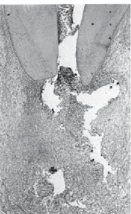
There were severe changes in the apical and periapical regions in the group filled with LPS with no irrigation. At the apical third, the cementum lacunas were enlarged and were empty or filled with necrotic tissue. There was also bleeding and inflammatory cells. The interstitial tissue invaginating from the periodon-tium had fiber dissociation, bleeding and severe mono-nuclear inflammatory infiltrate composed of mostly mononuclear cells and neutrophils. This was also seen adjacent to the foramen. There was cementum resorp-tion next to the foramen in almost all specimens. The periodontal ligament had concentrated inflammatory cells both adjacent to and at a distance from the apical foramen. There were collagen fibers parallel to the
apical surface next to the alveolar bone circumscribing the inflammatory process and active alveolar bone resportion with frequent osteoclasts (Figures 1 and 2).
Group II: Saline
A total of 19 of 20 roots were evaluated in this group due to the loss of one root during histological processing. Changes in the apical and periapical re-gions were severe in all parameters evaluated. There was a large amount of dense interstitial tissue, general-ized edema and tissue necrosis in the apical region. There was active resorption of the cementum in 17 of 19 roots, and absence of fibers and cementoblasts. In the periodontal ligament, inflammatory infiltrate was intense and predominantly mononuclear in 17 roots and moderate in 2 roots (p<0.05). This was more concen-trated at the foramen and with a considerable amount of neutrophils. There were macrophages and intense vas-cular proliferation and congestion at a distance from the apical foramen, with edema and a lack of collagen fibers circumscribing the inflammatory process. There were many osteoclasts in the alveolar bone indicating active resorption (Figures 3 and 4).
Figure 1. Group I. Histological findings showing the periodontal ligament with severe mixed inflammatory infiltrate that was more concentrated adjacent to the apical foramen. (hematoxylin and eosin: original magnification X40).
Group III: 1% Sodium Hypochlorite
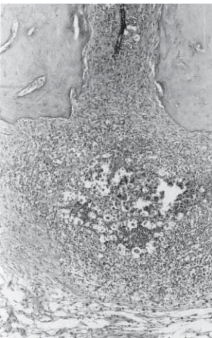
Only 19 roots were evaluated in this group be-cause one was fractured during processing. There were severe changes of the apical and periapical region similar to those of the LPS and saline groups. The mixed inflammatory infiltrate was more concentrated at the foramen with a considerable amount of neutro-phils and was severe in 12 roots and moderate in 6. There was diffuse inflammatory infiltrate in the peri-apical region and in several roots, one or more sites were adjacent to the foramen. Cementum and bone resorption was observed in 18 of 19 roots, respectively, with a large number of osteoclasts at the alveolar bone indicating active resorption (Figure 5).
Group IV: 2.5% Sodium Hypochlorite
The apical and periapical changes were similar to those of groups LPS, saline and 1% sodium hypochlo-rite with moderate/severe inflammation, cementum re-sorption, active bone rere-sorption, areas of necrosis, intense vascular proliferation and congestion and a considerable
number of osteoclasts and no osteoblasts (Figure 6).
Group V: 5% Sodium Hypochlorite
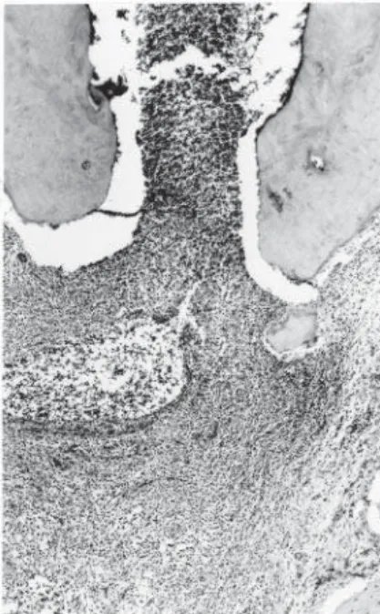
In the 20 roots irrigated with 5% sodium hy-pochlorite, there were predominantly moderate and diffuse inflamatory infiltrate at the periapex, with some neutrophils, edema and extensive bleeding in the inter-stitial tissue. There were areas of resorption of cemen-tum and an absence of cementoblasts in 9 of 19 roots (p<0.05). At a distance from the foramen, there were mononuclear inflammatory cells circumscribed by fi-bers and a discrete presence of collagen matrix. Active resorption of the alveolar bone was present in 17 of 20 roots with a large quantity of osteoclasts and an absence of osteoblasts (Figure 7).
Group VI: 2% Chlorhexidine
In the 20 canals irrigated with 2% chlorhexidine, the large lacunas in the apical cementum were empty or filled with inflammatory cells. The surface of the ce-mentum was irregular due to resorption. There were
Figure 4. Group II. Detail of Figure 3 showing areas of necrosis, severe inflammatory infiltrate, fibril dissociation and bleeding. (hematoxylin and eosin: original magnification X100).
Figure 5. Group III. Cementum resorption with a severely increased periodontal ligament containing mixed diffuse inflammatory infiltrate. Collagen fibers irregularly surround the inflammatory process. (hematoxylin and eosin: original magnification X40).
areas of necrosis and many mononuclear inflammatory cells in the interstitial tissue with tissue necrosis and fiber dissociation. There was moderate mixed inflam-matory infiltrate adjacent to the apical foramen in 10 of 20 roots, intense infiltrate in 5 roots and mild infiltrate in 5 roots. Intense vascular proliferation and congestion and generalized edema were also observed. At a dis-tance from the apical foramen, the inflammatory infil-trate was mixed with a predominance of macrophages and a discrete presence of collagen fibers circumscrib-ing the inflammatory process. The alveolar bone fre-quently had osteoclasts on its surface and there were no osteoblasts (Figure 8).
Statistical Analysis
The results of statistical analysis by the Mann-Whitney nonparametric test showed that none of the solutions used for irrigation of the root canals with pulp necrosis and a chronic periapical lesion inactivated the bacterial LPS. There was no statistical difference in dentin, cementum or bone resorption, or thickness of the periodontal ligament among the groups. However,
there was significantly less inflammatory infiltrate in both the 5% sodium hypochlorite group and the 2% chlorhexidine group than in the other groups (p<0.05).
DISCUSSION
It is well known that the predominant microor-ganisms in root canals with pulp necrosis and a chronic radiographically visible periapical lesion are anaerobic (13), especially Gram-negative (14). These Gram-nega-tive microorganisms release endotoxins (LPS) from their cell walls during bacterial multiplication or death, causing a series of important biological effects that lead to an inflammatory reaction and periapical bone resorp-tion. Thus, root canal treatment of teeth with a periapi-cal lesion must not only eliminate bacteria, but also must inactivate LPS.
During mechanical preparation, irrigating solu-tions must be selected to control infection of the root canal and if there is a periapical lesion, the ideal irrigat-ing solution must detoxify LPS as well as be biocompat-ible and have antibacterial activity.
Despite the fact that sodium hypochlorite and
Figure 6. Group IV. Areas of resorption of apical cementum at the foramen with severe inflammatory infiltrate, edema and vascular congestion and proliferation. (hematoxylin and eosin: original magnification X40).
Figure 7. Group V. Apical region with moderate predominantly mononuclear inflammatory infiltrate. There were fewer inflammatory cells at a distance from the apical foramen with a discrete presence of matrix and collagen fibers. (hematoxylin and eosin: original magnification X40).
chlorhexidine have been frequently used for irrigation in various concentrations (9,15), literature on the ef-fects of irrigating solutions on LPS is rare. Buttler and Crawford (9) studied the effect of three concentrations of sodium hypochlorite (0.58%, 2.79% and 5.20%) on LPS and reported that none of these solutions inacti-vated LPS in vivo, although in vitro this had occurred. Aibel and Stevens (16) reported that 1.2% chlorhexidine led to little or no LPS inactivation, in vitro.
In spite of the less severe inflammatory infiltrate in the groups irrigated with 5% sodium hypochlorite and 2% chlorhexidine in the present study, LPS was not totally inactivated. These results are in agreement with an in vitro study by Buck et al. (17) who reported that water, 0.12% chlorhexidine, 0.12% chlorhexidine plus 2.65% sodium hypochlorite, and 2.65% sodium hy-pochlorite had little or no effect on LPS when used for irrigation.
As reported by Nissan et al. (18), endotoxin is capable of penetrating dentin up to 0.5 mm. We believe that mechanical preparation leads to the elimination of a considerable quantity of LPS in the root canal and on the dentin. However, in the present study, none of the irrigating solutions were totally effective on the endo-toxin in the periapical region.
RESUMO
O objetivo deste estudo foi avaliar, histopatologicamente, o efeito do preparo biomecânico com diferentes soluções irrigadoras em dentes de cães preenchidos com LPS bacteriano após pulpectomia. Foram utilizados 120 canais radiculares de 6 cães, os quais foram preenchidos com solução de LPS após pulpectomia. As soluções irrigadoras usadas foram solução de hipoclorito de sódio a 1, 2.5 e 5% e solução de clorexidina a 2%. Nenhuma irrigação foi realizada no grupo controle. Os animais foram mortos após 60 dias, e os dentes foram fixados e desmineralizados. Em seguida, cortes seriados de 6 µm de espessura foram corados com hematoxilina e eosina e Tricrômico de Mallory para análise histopatológica e Brown e Brenn para verificação da contaminação bacteriana. A avaliação demonstrou que o infiltrado inflamatório foi estatisticamente menos intenso nos grupos cujos canais radiculares foram irrigados com solução de hipoclorito de sódio a 5% e de clorexidina a 2%. Contudo, nenhuma das soluções irrigadoras inativou completamente os efeitos deletérios do LPS. O preparo biomecânico coadjuvado por diferentes soluções irrigadoras não inativaram completamente a endotoxina.
ACKNOWLEDGMENTS
This research was supported by FAPESP (Fundação de Amparo à Pesquisa do Estado de São Paulo, Brazil).
REFERENCES
1. Wang CY, Stashenko P. Characterization of bone-resorbing ac-tivity in human periapical lesions. J Endod 1993;19:107-111. 2. Yamasaki M, Kumazawa M, Kohsaka T, Nakamura H,
Kameyama Y. Pulpal and periapical tissue reactions after experi-mental pulpal exposure in rats. J Endod 1994;20:13-17. 3. Ito H-O, Shuto T, Takada H, Koga T, Ainda Y, Hirata M, Koga T.
Lipopolysaccharides from Porhyromoinas gingivalis, Prevotella intermedia and Actinobacillus actinomycetemcomitans promote osteoclastic differentiation in vitro. Archs Oral Biol 1996;41:439-444.
4. McCormick JE, Weine FS, Maggio JD. Tissue pH of develop-ment periapical lesions in dogs. J Endod 1983;9:47-51. 5. Stern MH, Dreizen S, Ott TW, Levy BM. Antibody-producing
cells in human periapical granulomas and cysts. J Endod 1981;7:447-452.
6. Aqrabawi J, Schilder H, Toselli P, Franzblau C. Biochemical and histochemical analysis of the enzyme arylsulfatase in human lesions of endodontic origin. J Endod 1993;19:335-338. 7. Dwyer TG, Torabinejad M. Radiographic and histologic
evalua-tion of the effect of endotoxin on the periapical tissues of the cat. J Endod 1981;7:31-35.
8. Leonardo MR, Silva LAB, Tanomaru Filho M, Bonifácio KC, Ito IY. In vitro evaluation of the antimicrobial activity of a castor oil-based irrigant. J Endod 2001;27:717-719.
9. Buttler TK, Crawford JJ. The detoxifying effect of varying con-centrations of sodium hypochlorite on endotoxins. J Endod 1982;8:59-66.
10. Berutti E, Marini R, Angeretti A. Penetration ability of different irrigants into dentinal tubules. J Endod 1997;23:725-727. 11. Heling I, Chandler NP. Antibacterial effect of irrigant
combina-tions within dentinal tubules. Int Endod J 1998;31:8-14. 12. Silva LAB, Nelson-Filho P, Leonardo MR, Rossi MA, Pansani
CA. Effect of calcium hydroxide on bacterial endotoxin in vivo. J Endod 2002;28:94-98.
13. Sundqvist G. Ecology of the root canal flora. J Endod 1992;18:427-430.
14. Assed S, Ito IY, Leonardo MR, Silva LAB, Lopatin D. Anaerobic microorganisms in root canals of human teeth with chronic apical periodontitis detected by immunofluorescence. Endod Dent Traumatol 1996;12:66-69.
15. Leonardo MR, Tanomaru Filho M, Silva LAB, Nelson-Filho P, Bonifácio KC, Ito IY. In vivo antimicrobial activity of 2% chlorhexidine used as a root canal irrigating solution. J Endod 1999;25:167-171.
16. Aibel K, Stevens R. Effect of chlorhexidine on IL-6 induction by LPS. J Endod 1999;25:282 (abstract).
17. Buck RA, Cai J, Eleazer PD, Staat RH, Hurst HE. Detoxification of endotoxin by endodontic irrigants and calcium hydroxide. J Endod 2001;27:325-327.
18. Nissan R, Segal H, Pashley D, Stevens R, Trowbridge H. Ability of bacterial endotoxin to diffuse through human dentin. J Endod 1995;21:62-64.