Universidade do Minho
Escola de Engenharia
Sofia Rosa Evangelista Lima
Characterization of microbial populations
in a wastewater treatment plant focusing
on
Staphylococcus aureus
and
Pseudomonas aeruginosa
Dissertação de Mestrado
Mestrado em Bioengenharia
Trabalho realizado sob a orientação da
Doutora Ana Paula Rodrigues da Cunha Nicolau
e da
Nome: Sofia Rosa Evangelista Lima
Endereço Electrónico: sofia.r.evangelista.lima@gmail.com Telefone: 962568147
Número do Bilhete de Identidade: 223165050
Título da Dissertação: Characterization of microbial populations in a wastewater treatment plant focusing on Staphylococcus aureus and Pseudomonas aeruginosa
Orientador (es): Doutora Ana Paula Rodrigues da Cunha Nicolau e Doutora Maria Olívia Baptista Pereira
Ano de Conclusão: 2015
Designação do Mestrado: Mestrado em Bioengenharia
É AUTORIZADA A REPRODUÇÃO PARCIAL DESTA DISSERTAÇÃO APENAS PARA EFEITOS DE INVESTIGAÇÃO, MEDIANTE DECLARAÇÃO ESCRITA DO INTERESSADO, QUE A TAL SE COMPROMETE;
Universidade do Minho, ___ / ___ / _____
ACKNOWLEDGEMENTS
By accomplishing successfully this work, I would like to acknowledge everyone that was involved in its development and execution, either directly or indirectly.
To Dr. Ana Nicolau and Dr. Maria Olívia Pereira, I would like to express my gratitude, not only for the way I was guided throughout these months, but also for all scientific knowledge instructed.
To Ana Margarida, Diana Alves, and Susana Lopes, I specially thank, for the pleasant way they supported me, for all the patience, and for the time they made available to teach me tirelessly and thoroughly; assisting me in every doubt or concern that arose during the thesis.
To Ana Silva, I am thankful for her companionship, over those laboratory working hours, and endless support, as well as Maria João, Tânia Rodrigues and Rita Seixo.
I also would like to thank to all my Biofilm laboratory coworkers, for the availability and appreciation that they manifested since the beginning, especially to MOP Group, and NC Group.
To all my friends, but in particularly to Guilhermina Carriche, Nuno Machado, Ana Sofia Pereira, Cristina Domingues, Rui Pereira I thank for all the talks, suggestions, patience, and great affection.
Finally, I am grateful to my parents, and my sister, for always being there for me, helping me throughout this journey, cheering me up and supporting me incessantly.
ABSTRACT
Wastewater treatment plants (WWTP) have been regarded as favorable sites for the development and dissemination of antibiotic resistant bacteria (ARB). These bacteria are able to contact with human beings, either directly or indirectly, possibly leading to infections with dire consequences.
The main goal of this study was the evaluation of the efficacy of a WWTP disinfection system (through ultraviolet radiation) at reducing the bacterial load present at the final effluent. Two common pathogenic bacteria - Staphylococcus aureus and Pseudomonas aeruginosa – were used as indicators of the removal ability of the treatment system. Different colonies of both bacteria were subsequently isolated in order to inspect their antibiotic susceptibility and capacity of biofilm formation.
Four samples were collected during March and June from three different WWTP sites (raw
effluent and effluents before and after disinfection). The isolation of P. aeruginosa and S. aureus
was accomplished through the use selective growth media and biochemical confirmation tests. The susceptibility of the selected bacteria to antibiotics was determined through the modified Kirby-Bauer disk diffusion method. Lastly, the ability of the isolated bacteria to form biofilm was investigated using the crystal violet assay, and the number of viable and cultivable biofilm cells through colony counting.
The efficacy of the disinfection treatment in the reduction of bacterial load in the final effluent was effective for S. aureus, but not for the removal of P. aeruginosa. Twenty-four P. aeruginosa and
thirty-six S. aureus isolate were obtained. The majority of the colonies of both species were
susceptible to the tested antibiotics, with the exception of two: ciprofloxacine, with eight P.
aeruginosa resistant bacteria, and rifampicine, with thirteen isolates of S. aureus showing resistance. For both bacteria, the resistant isolates were found mainly in the samples collected before and after the disinfection (BD and AD). The ability of the isolated bacteria to form biofilm was variable, but generally, P. aeruginosa isolates achieved a greater biofilm mass.
In conclusion, the efficacy of the WWTP, and namely of the disinfection step, was clearly low if
one takes as indicator of its performance the removal ability of P. aeruginosa. Besides that, the
presence of resistant isolates in BD and AD samples shows that the disinfection is not effective in elimination of ARB, allowing their survival and release to the environment. Due to the great variation of formed biofilm, in both bacteria, it was not possible to establish a relationship between the amount of biofilm formation and the sites of WWTP where the isolates bacteria were collected. No relation was found between a level of resistance to antibiotics and a high capacity of biofilm formation.
RESUMO
As Estações de Tratamento de Águas Residuais (ETAR) têm sido consideradas como locais favoráveis para o desenvolvimento e disseminação de bactérias resistentes aos antibióticos (ARB) para o ambiente.Estas bactérias são capazes de entrar em contacto com o ser humano, directa ou indirectamente, podendo levar a uma infeção com consequências graves.
Este estudo teve como principal objectivo a avaliação da eficiência de uma ETAR com um sistema de desinfecção ( através da radiação ultravioleta) na redução da carga bateriana presente
no efluente final. Duas bactérias patogénicas comuns - Staphylococcus aureus e Pseudomonas
aeruginosa - foram usadas como indicadores da capacidade de remoção do sistema de tratamento. Posteriormente, diferentes colónias de ambas as bactérias foram isoladas de forma a inspecionar as suas capacidades de formação de biofilme e suceptibilidade aos antibióticos.
Quatro amostras foram recolhidas durante Março e Junho de três localizações diferentes da
ETAR (efluente bruto e os efluentes antes e depois da desinfecção). O isolamento P. aeruginosa e
S. aureus foram realizadas através do uso de meios selectivos de crescimento e de testes bioquímicos para confirmação. A susceptibilidade da bactéria aos antibióticos foi determinada através do método de difusão em disco Kirby-Bauer modificado. Por fim, a capacidade de formação de biofilme das bactérias isoladas foi analisada usando o método cristal violeta, e o número de células de biofilmes viáveis e cultiváveis através da contagem de colónias.
A eficácia do tratamento de desinfecção na redução da carga bacteriana do efluente final foi
eficaz na remoção S. aureus mas não na remoção P. aeruginosa. Foram isolados com sucesso
vinte e quatro P. aeruginosa e trinta e seis S. aureus. A maioria das colónias de ambas as
espécies foram susceptíveis aos antibióticos testados, à exceção de dois antibióticos: ciprofloxacina, com oito isolados P. aeruginosa resistentes, e rifampicina, com treze isolados de S. aureus resistentes. Em ambas as bactérias, a localização dos isolados resistentes, mostraram uma presença significativa nas amostras antes e depois da desinfecticção (AD e BD). A capacidade de formação de biofilme dos isolados de ambas as bactérias variou mas, na gobalidade, os isolados P. aeruginosa obtiveram uma maior formação de biofilme.
Em conclusão, a eficácia da ETAR, e nomeadamente a etapa da desinfecção foi claramente reduzida se se tiver em conta, como indicador do seu desempenho, a capacidade de remoção P. aeruginosa. Além disso, a presença de isolados resistentes nas amostras BD e AD mostrou que a desinfecção não é eficaz na eliminação de ARB, permitindo a sua sobrevivência e libertação para o meio ambiente. Devido à grande variação de biofilme formado, em ambas as bactérias, não foi possível estabelecer uma relação entre quantidade de biofilme formado e os locais da ETAR em que as bactérias isoladas foram recolhidas. Nenhuma relação foi observada entre o nível de resistência aos antibióticos e a elevada capacidade de formação de biofilme.
TABLE OF CONTENTS
ACKNOWLEDGEMENTS ... iii
ABSTRACT ... v
RESUMO ... vii
TABLE OF CONTENTS ... ix
LIST OF FIGURES ... xiii
LIST OF TABLES ... xv ABREVIATIONS ... xvii CHAPTER I| INTRODUCTION ... 1 1.1. Background ... 3 1.2. Main objectives ... 3 1.3. Thesis Organization ... 3
CHAPTER II| BIBLIOGRAPHIC REVIEW ... 5
2.1. Impact of antibiotic-resistant bacteria and antibiotics in human health and in environment. 7 2.2. Antibiotic resistant bacteria ... 7
2.2.1. Biofilm formation ... 8
2.3. Relationship between antimicrobial resistance and the history of antibiotics ... 11
2.4. Spread of antibiotic resistant bacteria and genes in the environment ... 12
2.5. Wastewater Treatment Plants ... 13
2.5.1. Pre-treatment ... 13
2.5.2. Primary treatment ... 14
2.5.3. Secondary treatment ... 14
2.5.4. Tertiary treatment ... 15
2.6. Wastewater treatment plants as hotspot for development and dissemination of antibiotic resistant bacteria and genes. ... 15
2.7. Mechanisms to fight the spread of antibiotic resistant bacteria using wastewater treatment plants ... 17
2.2.1. Pseudomonas aeruginosa ... 19
2.2.2. Staphylococcus aureus ... 22
CHAPTER III| MATERIALS AND METHODS ... 25
3.1. Sampling ... 27
3.2. Enumeration of cultivable bacteria ... 28
3.3. Isolation of bacteria ... 28
3.3.1. Filtration ... 28
3.3.2. Selective media and biochemistry tests ... 29
3.3.3. Cryopreservation ... 31
3.4. Biofilm formation ... 31
3.4.1. Preparation of the inoculum ... 31
3.4.2. Crystal violet staining technique ... 32
3.4.3. Colony forming units technique ... 32
3.5. Antibiotic Susceptibility Testing ... 32
3.5.1. Preparation of media ... 33
3.5.2. Preparation of antibiotic stock solution ... 33
3.5.3. Preparation of the inoculum ... 34
3.5.4. Preparation of the disks ... 34
3.5.5. Inoculation of agar plates ... 34
3.5.6. Application of antimicrobial disks and incubation of plates ... 34
3.6. Data statistical analysis ... 36
CHAPTER IV| RESULTS AND DISCUSSION ... 37
4.1. Effect of wastewater treatment plant processes on reduction of bacterial load ... 39
4.2. Isolation of bacteria ... 42
4.3. Antibiotic susceptibility testing ... 46
4.4. Biofilm formation ... 51
CHAPTER V| CONCLUSIONS AND FUTURE PRESPECTIVES ... 57
5.1. Conclusions ... 59
5.2. Future Perspectives ... 60
REFERENCES ... 61
Appendix A ... 85 Appendix B ... 88
LIST OF FIGURES
Figure 1| General overview of bacterial biofilm development. (i) reversible attachment of cells to surfaces; (ii) irreversible attachment; (iii) maturation I; (iv) maturation II; and (v) dispersion (adapted from Monroe (2007). ... 10 Figure 2| Flow diagram of the work experimental performed during the year. ... 27 Figure 3| Interpretation of the coagulase test as proposed by Turner and Schwartz, where:
Negative – no evidence of fibrin formation; 1+ Positive – small unorganized clots; 2+ Positive – small
organized clot; 3+ Positive – large organized clot; 4+ Positive – entire content of tube coagulates and
is not displaced when tube is inverted (Adapted from Turner and Schwartz (1958)). ... 30 Figure 4| The antimicrobial susceptibility disk diffusion test: MHA plate with two antibiotic disks. One of disks have inhibition zone and other not shows inhibition zone. ... 35
Figure 5| Total bacterial load (Log10 CFU/mL), of four samples, from three different locations: raw
sewage (RS); before disinfection (BD); after disinfection (AD). Growth in TSA medium at 37 ºC. ... 39
Figure 6| S. aureus bacterial load (Log10 CFU/mL) of four samples from three different locations:
raw sewage (RS); before disinfection (BD); after disinfection (AD). Growth in MSA medium at 37 ºC. ... 40
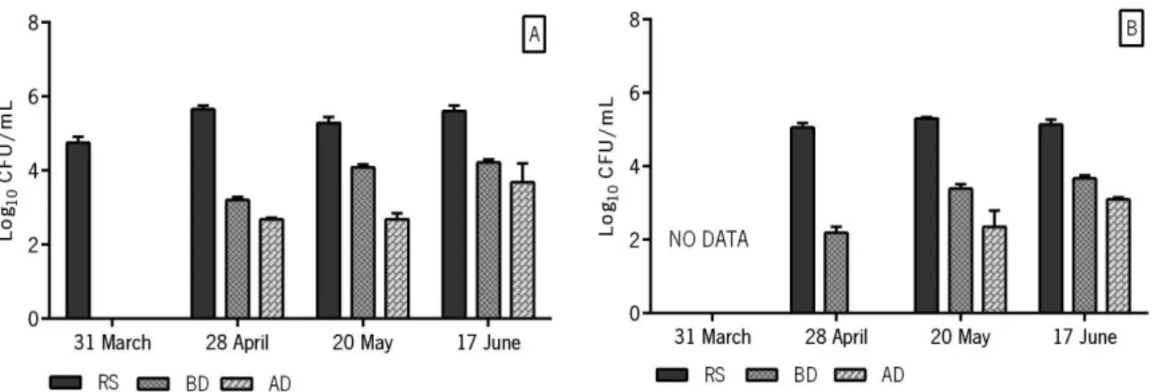
Figure 7| P. aeruginosa bacterial load (Log10 CFU/mL) of four samples taken in three different
locations: raw sewage (RS); before disinfection (BD); after disinfection (AD). Growth in CA medium at 37 ºC (A) and 42 ºC (B). ... 40 Figure 8| Isolates P. aeruginosa (PA isolates) colonies grown on CA plates, overnight at 37 °C. A) green colonies of PA isolate 10 ; B) white colonies of PA isolate 5; and C) yellow-green colonies of PA isolate 9. ... 43 Figure 9| Isolates S. aureus (SA isolates) colonies grown on MSA plates, overnight at 37 °C. A) yellow and opaque colonies SA isolate 12; and B) yellow and translucent colonies SA isolate 5. .. 45
Figure 10| Biofilm mass (OD570 nm) formed by P. aeruginosa ATCC 27853 (PA ATCC 27853) and P.
before disinfection (BD); after disinfection (AD). Growth during 24 h at 37 ºC with an agitation of 120 rpm. Bars are representative of the average biofilm biomass from three independent assays. ... 52
Figure 11| Biofilm cell (Log10 CFU/cm2) quantification of P. aeruginosa ATCC 27853 (PA ATCC
27853) and P. aeruginosa isolates (PA isolates) of four samples, from three different locations: raw sewage (RS); before disinfection (BD); after disinfection (AD). Growth during 24 h at 37 ºC with an agitation of 120 rpm. Bars are representative of the average biofilm biomass from three independent assays. ... 52
Figure 12| Biofilm mass (OD570 nm) formed by S. aureus ATCC 25923 (SA ATCC 25923) and S.
aureus isolates (SA isolates) of four samples from three different locations: raw sewage (RS); before disinfection (BD); after disinfection (AD). Growth during 24 h at 37 ºC with an agitation of 120 rpm. Bars are representative of the average biofilm biomass from three independent assays. ... 53
Figure 13|Biofilm cell (Log10 CFU/cm2) quantification of S. aureus ATCC 25923 (SA ATCC 25923)
and S. aureus isolates (SA isolates) of four samples from three different locations: raw sewage
(RS); before disinfection (BD); after disinfection (AD). Growth during 24 h at 37 ºC with an agitation of 120 rpm. Bars are representative of the average biofilm biomass from three independent assays. ... 54
Figure 14| Antibiotic susceptibility profiles of P. aeruginosa isolate 3 to amikacin (30 µg). Growth
in MHA medium for 18h, at 37 ºC. ... 88
Figure 15| Antibiotic susceptibility profiles of S. aureus ATCC 25923 to gentamicin (10 µg).
LIST OF TABLES
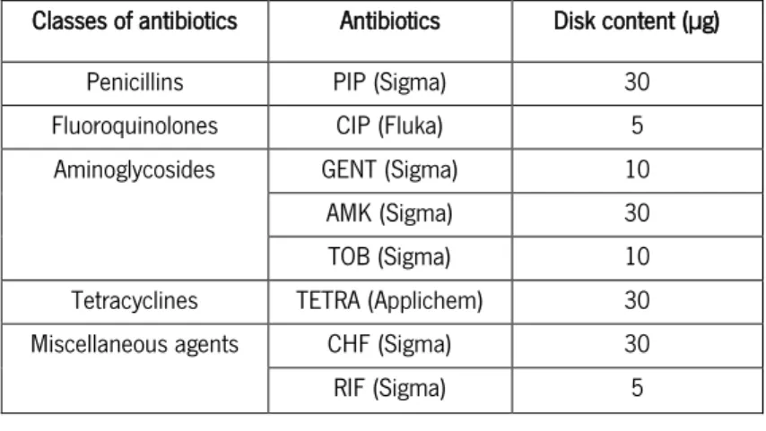
Table 1| Disk content (µg) presented in EUCAST breakpoint table (EUCAST, 2014a). The antibiotics used were: piperacillin (PIP); ciprofloxacin (CIP); gentamicin (GENT); amikacin (AMK); tobymicin (TOB); Tetracycline (TETRA); chloramphenicol (CHF); rifampicin (RIF). ... 33 Table 2| Zone diameter breakpoint presented in EUCAST breakpoint table (EUCAST, 2014a) and concentrations of antibiotics tested. The antibiotics used were: piperacillin (PIP); ciprofloxacin (CIP); gentamicin (GENT); amikacin (AMK); tobymicin (TOB); tetracycline (TETRA); chloramphenicol (CHF); rifampicin (RIF). ... 36 Table 3| Morphological features found in isolates P. aeruginosa (PA isolates) colonies observed on CA, at 42 ºC. ... 43
Table 4| Morphological features found in isolates S. aureus (SA isolates) colonies observed on
MSA, at 37 ºC. ... 44
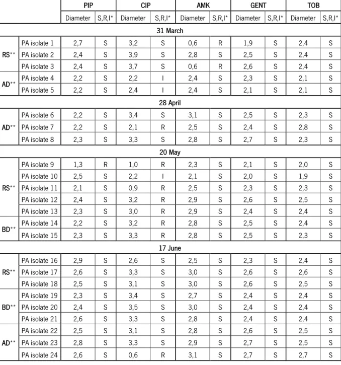
Table 5| Antimicrobial susceptibility tests (according to EUCAST) results of P. aeruginosa isolates
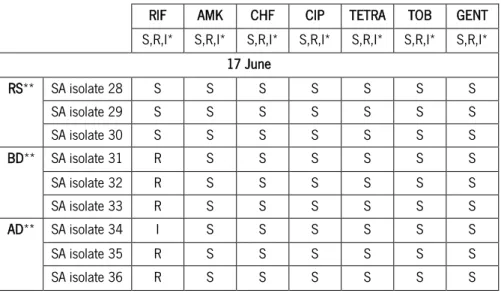
(PA isolates) for the following antibiotics: 30 µg piperacillin (PIP); 5 µg ciprofloxacin (CIP); 30 µg amikacin (AMK); 10 µg gentamicin (GENT) and 10 µg tobramycin (TOB). ... 46 Table 6| Antimicrobial susceptibility tests (according to EUCAST) results of S. aureus isolates (SA isolates) for the following antibiotics: 5 µg rifampicin (RIF); 30 µg amikacin (AMK); 30 µg chloramphenicol (CHF); 5 µg ciprofloxacin (CIP); 30 µg tetracycline (TETRA); 10 µg tobramycin (TOB); and 10 µg gentamicin (GENT). ... 48
Table 7| Antimicrobial susceptibility tests (according to EUCAST) results of P. aeruginosa isolates
for the following antibiotics: 30 µg piperacillin (PIP); 5 µg ciprofloxacin (CIP); 30 µg amikacin (AMK); 10 µg gentamicin (GENT) and 10 µg tobramycin (TOB). ... 85
Table 8| Antimicrobial susceptibility tests (according to EUCAST) results of S. aureus isolates (SA
isolates) for the following antibiotics: 5 µg rifampicin (RIF); 30 µg amikacin (AMK); 30 µg chloramphenicol (CHF); 5 µg ciprofloxacin (CIP); 30 µg tetracycline (TETRA); 10 µg tobramycin (TOB); and 10 µg gentamicin (GENT). ... 86
ABREVIATIONS
AD After Disinfection
AMK Amikacin
ARB Antibiotic Resistant Bacteria
ARG Antibiotic Resistant Genes
ATS Antimicrobial Susceptibility Testing
BD Before Disinfection
CA Cetrimide agar
CHF Chloramphenicol
CIP Ciprofloxacin
CFU Colony Forming Units
CV Crystal Violet
EUCAST European Committee on Antimicrobial Susceptibility Testing
EPS Extracellular Polymeric Substances
GENT Gentamicin
I Intermediate
MSA Mannitol Salt Agar
MHA Mueller Hinton Agar
PIP Piperacillin
RS Raw Sewage
R Resistant
RIF Rifampicin
TETRA Tetracycline
TOB Tobymicin
TSA Tryptic Soy Agar
TSB Tryptic Soy Broth
UV Ultraviolet
1.1. Background
Wastewater treatment plants (WWTP) have a crucial role in protecting public health and in the preservation of water resources. However, WWTP are a main source of release of antibiotics into the environment, being considered as one of the "hotspots" for potential evolution and spread of antibiotic resistance into the environment. The increase in bacterial resistance often drives to an increase in the duration of the treatment and expensive costs.
Unfortunately, the current WWTP are not prepared for the effective elimination of antibiotics and other chemicals as well as pathogenic microorganisms, being therefore important to focus on optimizing the WWTP and on developing new treatments for the removal of these organisms and products. Certain bacteria species known as "ESKAPE" pathogens present in hospital and care unit environments are being a source of concern in WWTP. These bacteria, major causes of nosocomial infections and known to have a high abundance of multi-resistances, are common in the sewage due to the entrance of hospital sewages in the urban WWTP. If the WWTP is not able to eliminate these microorganisms, they will be released into the environment, probably with new resistances, putting at risk the health publishes. For that, it is pivotal to inspect the ability of WWTP to remove pathogenic and antibiotics resistant bacteria in order to obtain insights about the performance of these treatment systems in this particular goal.
1.2. Main objectives
This work aimed to analyze the efficiency of a WWTP with a tertiary treatment disinfection system in reducing the bacterial load, and particularly antibiotic resistant bacteria. Staphylococcus aureus and Pseudomonas aeruginosa, two common pathogenic bacteria, were selected as indicators of the efficiency of removal through treatment with ultraviolet radiation. Moreover, it was aimed to isolate different strains of both bacteria in order to evaluate two virulence through the analysis of their susceptibility towards different antibiotics and their ability to form biofilms.
1.3. Thesis Organization
The present dissertation is organized in five chapters:
In the first chapter, it is presented and contextualized the general work subject. Likewise, the main goals of the project are defined, as well as it organization throughout the dissertation.
Characterization of microbial populations in a wastewater treatment plant focusing on pathogenic and antibiotic-resistant microorganisms
The second chapter presents an introduction about the impact of antibiotics resistant bacteria on human health and environment. The concept of antibiotics resistant bacteria and the definition of biofilm formation are also presented. A description of the relationship between antibiotics resistance and history of the antibiotics is given, and an approach of the way in which both bacteria and antibiotics resistant genes may disseminate to the environment. The concept of WWTP is introduced as well as the main steps associated with the treatment of residual water. The role of wastewater treatment plants in the development, dissemination and fight against bacteria and antibiotics resistant genes are also referred. Finally, the pathogenic microorganisms that were the target of this study are introduced.
The third chapter presents the methods and materials used throughout the execution of the experimental work. The period of sampling and the collected sites of samples in WWTP are described in this section, as well as used methods to enumeration of cultivable bacteria. All isolation of target bacteria steps are presents, since the filtration of samples to the selective media and biochemistry tests. In this part, all the isolation and confirmation work carried out, particularly in the case of P. aeruginosa, is described and justified. Lastly, the procedure for biofilm formation and antimicrobial susceptibility testing of all isolates are described.
Chapter four both presents and discusses the obtained results. During this phase, the efficiency of the WWTP was evaluated, namely on what concerns the variations of the total bacterial load, the densities of P. aeruginosa and S. aureus. At this point, the influence of temperature to distinguish P. aeruginosa from other Pseudomonas species is also discussed. The morphological characteristics of the selected isolates were described. Lastly, the antibiotic resistance profile and capacity of biofilm formation of the isolates were evaluated and discussed.
Finally, the fifth chapter presents the principal conclusions of this thesis and suggestions about future work.
2.1. Impact of antibiotic-resistant bacteria and antibiotics in human health
and in environment.
Large amounts of antibiotics for human use are released in municipal sewage (Bouki et al., 2013; Sim et al., 2010). Many antibiotics are not completely metabolized and therefore enter in the sewer through the feces (Hirsch et al., 1999). The intentional disposal of unused medicine (Kümmerer, 2003) and veterinary use also contribute to the quantities of antibiotics found in sewage, as well as the runoff from agricultural applications (Dı́az-Cruz et al., 2003; Le-Minh et al., 2010).
The biggest concern with the release of antibiotics into the environment is related to the development of antibiotic-resistant genes (ARG) and antibiotic-resistant bacteria (ARB). The increase in ARB in aquatic environments is increasing leading to the creation of selective pressures on natural bacteria species (Alpay-Karaoglu et al., 2007; Kümmerer, 2004).
The transfer of the ARB to humans may occur through several ways: water, food, plants and through manure when it is used as fertilizer (Dolliver and Gupta, 2008; Salyers, 2002). Various resistances to antibiotics are commonly found in all pathogenic and normal flora bacteria which are present in the human intestinal tract (Baquero et al., 2008; Vignesh et al., 2012).
At the environmental level, antibiotics can affect the evolution of the structure of a community, which consequently influences the ecological function of the ecosystem of the water (Aminov and Mackie, 2007; Thiele-Bruhn and Beck, 2005). Ciprofloxacin is an example of a harmful antibiotic to the environment (Kim and Aga, 2007). Its mixture with two more pharmacological products, triclosan (antisetic) and tergite NP10 (surfactant), in the aquatic environment leads to a significant impact on the rate of production of biomass and in the community structure of algae (Wilson et al., 2003).
2.2. Antibiotic resistant bacteria
As it is generally known, all living organisms struggle to adapt to the environment in order to be fit to survive (Alanis, 2005). So, it should not be a surprise for the human being the fact that bacteria have demonstrated a capacity to support and still adapt to the environment, leading to the development of different mechanisms of resistance to antibiotics (Alanis, 2005). Because of this, many strains of bacteria have become resistant and in some cases multi-resistant to antibiotics leading to their ineffectiveness in many cases (Alanis, 2005). A species is denominated
antibiotic-resistant if it is able to function, survive or persist in the presence of higher concentrations of an antimicrobial agent than others species (Smith et al., 1994).
According to Džidić et al. (2008), the increase of antibiotic resistance was due to a combination of selective pressures that enhanced the transmission of resistant organisms. Of all these factors, the most important and worrying is the continued selective pressure exerted by the routinely used antibiotics, which leads to elimination of strains sensitive and consequently a significant increase in resistant strains (Berger-Bächi, 2002; Kolář et al., 2001).
The biology of antibiotic resistance can be divided into two sections: genetic aspects and biochemical aspects (Alanis, 2005; Džidić et al., 2008). The derived-genetic resistance can be intrinsic or acquired. Intrinsic or natural resistance stems from an inherent factor structural or functional associated to an entire bacterial species (Ammor et al., 2008) where they are resistant without any additional genetic alteration (Normark and Normark, 2002). Acquired resistance to antibiotics can occur by mutations or by horizontal gene transfer (Ammor et al., 2008; Normark and Normark, 2002).
Bacteria can become resistant to antibiotics due to five biochemical mechanisms of resistance: enzymatic modification, target modification, active efflux pumps, reduced antimicrobial permeability and bypass of synthetic pathways (Bonomo and Gill, 2005; McKeegan et al., 2002; Normark and Normark, 2002; Stewart and William Costerton, 2001a). The production of biofilm through the phenotypic adaptation is another form of intrinsic resistance (Ascenzi, 2005; Kumar and Pandit, 2012).
2.2.1. Biofilm formation
More than 60 years after the first report on biofilms, they are still a concern in food, environmental and biomedical fields, among others (Flint et al., 1997; Maukonen et al., 2003; Sihorkar and Vyas, 2001; Zobell, 1943). A definition of biofilm has evolved over the last 25 years and in current days, according to Donlan and Costerton (2002), biofilm is defined as “A microbially derived sessile community characterized by cells that are irreversibly attached to a substratum or interface or to each other, are embedded in a matrix of extracellular polymeric substances that they have produced, and exhibit an altered phenotype with respect to growth rate and gene transcription”.
Biofilms can be composed by a population that developed from a unique species or a community derived from mixed microbial species (Davey and O'Toole, 2000; Mah and O'Toole,
2001). Biofilms are highly hydrated (98% of the total biofilm mass) and are composed by microorganisms (prokaryotic and eukaryotic unicellular organisms), multivalent cations, inorganic particles and extracellular polymeric substances (EPS) (Donlan and Costerton, 2002; Flemming, 1991; Wingender et al., 1999). In the majority of biofilms, the percentage of microorganisms is less than 10% of the dry mass, while the matrix can account for over 90% (Flemming and Wingender, 2010). EPS consist not only of polysaccharides, but also proteins, nucleic acids and lipids. The composition of EPS varies between species and growth conditions, and chemical communication, like quorum sensing (QS), between cells in the community stimulates its formation and secretion. QS is important in biofilm formation process and a mechanism used by cells to query extracellular environment (Renner and Weibel, 2011). EPS are responsible for the morphology and internal structure of biofilms, including surface pores and channels (Flemming and Wingender, 2010). Moreover, they can provide a matrix which allows to maintain cells position for a longer period of time in comparison to the planktonic mode (Lewandowski and Evans, 2000). The organisms associated to biofilms grow more slowly than planktonic organisms, probably because the cells are more limited by nutrients, and also because of oxygen depletion (Donlan, 2000).
Bacteria can exist in one of two types of population: planktonic or in biofilm. However in natural aquatic ecosystems more than 99.9% of the bacteria grow in biofilms on wide variety of surfaces (Donlan and Costerton, 2002). Adaptation of biofilm structure for survival in varying environments, comes from an ancient and integral component of the prokaryotic life cycle (Hall-Stoodley et al., 2004).
Bacteria that are embedded in biofilms enjoy several advantages when compared to the planktonic cells. One of the advantages is the ability of the matrix to capture and concentrate minerals and nutrients from the environment, as already mentioned (Beveridge et al., 1997; Flemming, 1991) . Multispecies biofilms can form stable microconsortia, develop physiochemical gradients and intense cell–cell communication (Flemming and Wingender, 2010). Biofilms are also an ideal place for horizontal gene transfer, since cells are maintained in close proximity to each other, and can therefore exchange genetic information (Flemming et al., 2007). Another advantage of biofilms is protection from a wide array of environmental challenges, such as UV exposure, metal toxicity, acid exposure, dehydration, salinity, predation by protozoa, bacterivorous microorganisms and bacteriophages, phagocytosis and, the most worrying for human, several antibiotics and antimicrobial agents (Espeland and Wetzel, 2001; Flemming, 1991; Huq et al.,
2008; Mah and O'Toole, 2001; McNeill and Hamilton, 2003; Shirtliff et al., 2002; Stewart and William Costerton, 2001b; Teitzel and Parsek, 2003).
Since bacteria living in biofilms have a greater resistance to antibiotics when compared to planktonic cells, and pathogenic bacteria are able to produce biofilms, this phenomena is considered a serious concern for the medical community. (Mah and O'Toole, 2001). Biofilms can form on medical devices, such as catheters or implants, and they can also cause infections on human surfaces such as teeth, skin, and the urinary tract (Hall-Stoodley et al., 2004; Hatt and Rather, 2008; Mah and O'Toole, 2001). In addition to medical equipment, various studies reveal the presence of biofilms in the food industry and in water distribution systems. These biofilms can be responsible for infections to humans, especially those belonging to risk groups (Chmielewski and Frank, 2003; Huq et al., 2008; Lee Wong, 1998; Piriou et al., 1997; Szewzyk et al., 2000; WHO, 2006).
The architecture of biofilms reveals that biofilm formation is a complex developmental process that involves several stages, each with unique characteristics (Davies, 2003; McCarty et al., 2014). The process of biofilm formation, considered as an endless cycle, is characterized by five stages (Figure 1): (i) reversible attachment of cells to surfaces; (ii) irreversible attachment; (iii) maturation I; (iv) maturation II; and (v) dispersion (Davies, 2003; Sauer et al., 2002).
Bacteria initially move to a living or non-living surface and attach to it. This corresponds to the first and critical step of biofilm formation (Sauer et al., 2002). Bacteria use a variety of extracellular organelles and proteins for sensing and attaching to the surfaces, such as flagella, pili and outer membrane proteins (Bullitt and Makowski, 1995; Thomas et al., 2004). The second stage comprises the irreversible attachment when bacteria secretes EPS that facilitate adhesion between cells and surfaces (Flemming and Wingender, 2010). The third stage corresponds to the first maturation of biofilms. Cells adsorbed on surfaces replicate and produce EPS, leading to formation
Figure 1| General overview of bacterial biofilm development. (i) reversible attachment of cells to surfaces; (ii) irreversible attachment; (iii) maturation I; (iv) maturation II; and (v) dispersion (adapted from Monroe (2007).
of microcolonies (Karatan and Watnick, 2009; Renner and Weibel, 2011). In the second maturation of biofilm, the fourth stage, the cells continue dividing and the EPS accumulates. In this stage the biofilm is capable of extending from the surface, building a three-dimensional mushroom-shaped structure (Hall-Stoodley et al., 2004). These structures are interspersed with fluid-filled channels which act as a circulatory system, allowing for the exchange of nutrients and waste products with the surrounding environment. The colonies in this mature biofilm are complex and highly differentiated. Numerous microenvironments exist within the biofilm where pH, oxygen concentration, nutrient availability and cell density differ (Kaplan, 2010). After the maturation of the biofilm, some cells detach from the biofilm and disperse (stage v) into the environment with the aim of colonizing other regions (Renner and Weibel, 2011; Stoodley et al., 2002). The dispersal process can be complex, involving numerous environmental signals, signal transduction pathways and effectors. This is important since it contributes to biological dispersion, bacterial survival, and disease transmission (Kaplan, 2010).
The transition from planktonic growth to biofilm involves multiple regulatory networks, which result in gene expression changes causing a temporal reorganization of cellular physiology (Monds and O’Toole, 2009; O'Toole et al., 2000; Prigent-Combaret et al., 2001). Thus, bacteria growing in a biofilm have phenotypical, biochemical, and morphological differences from their planktonic form (Prigent-Combaret et al., 2001).
2.3. Relationship between antimicrobial resistance and the history of
antibiotics
Throughout history, there has been an ongoing battle between the human being and the microorganisms that cause infections and diseases. From the Black Death, tuberculosis, malaria, among others, these microorganisms have affected the human population, causing morbidity and mortality.
The use of substances with antimicrobial activity for the treatment of wounds and some diseases dates back thousands of years before the "Age of Antibiotics", used by the ancient Chinese civilization, as well as by the Egyptian and Roman (Moellering, 1995). However, most of these substances were too toxic for applications in other locations. Over time, microbiology skills and techniques were developed which led to the discovery of the first natural antibiotic, in 1928, by Alexander Fleming – the penicillin.
This episode marked the beginning of the "Age of Antibiotics", but only years later, penicillin was introduced for therapeutic purposes (Moellering, 1995). During the "Age of Antibiotics", from 1940 to 1960, there was a considerable number of new antimicrobial agents used in clinical practice and it was believed that infectious diseases could be definitely controlled and prevented (Bockstael and Aerschot, 2009). However, this euphoria was of short duration, since the bacteria able to evolve, expressing various forms of resistance (Livermore, 2003; Tenover, 2006), becoming clear that the victory against bacterial pathologies was an illusion (Kim, 2013).
Throughout years, despite the efforts to control or even eliminate the bacteria, they have always managed to get around the situation in order to endure and prosper. Bacteria are survivors who inhabit the earth for more than four billion years, even in some environments where human life would be impossible.
According to Harbottle et al. (2006), resistance genes and the mechanisms of resistance existed long before the discovery and use of antibiotics. An example was the discovery in glacial samples obtained from the Canadian Arctic archipelago of bacteria resistant to antibiotics over 2000 years old (Dancer et al., 1997).
2.4. Spread of antibiotic resistant bacteria and genes in the environment
The recycling of municipal water for industrial, agricultural and urban non-potable use is an increasingly important component of the management practices of water resources in many parts of the world (Exall, 2004; Wintgens et al., 2005). However, the reuse of treated effluent increases the range of scenarios of human and environmental exposure to ARB (Le-Minh et al., 2010).
Water is not just a means of dissemination of ARB between animal and human populations, but also a route in which ARG are introduced in natural ecosystems. In these systems, non-pathogenic bacteria can serve as a reservoir of resistant genes (Baquero et al., 2008).
According to Baquero et al. (2008), there are four main genetic reactors, places where genetic evolution occurs, in which resistance to antibiotics may be developed:
First reactor: made by human and animal microbiota, with hundreds of bacterial species involved and where antibiotics exert their action.
Second reactor: involves hospitals and care centers, farms or anywhere where there are susceptible individuals exposed to bacterial exchange.
Third reactor: corresponds to sewage or other biological waste arising in the second reactor. This reactor includes lagoons and treatment plants, ideal places for bacteria to have the opportunity to mix and react genetically.
Fourth reactor: corresponds to soil and surface and ground water, where the bacteria originated from previous reactors can blend or neutralize environmental organisms.
As Baquero et al. (2008) state, the possibility of reducing the evolution of antibiotic resistance depends on the ability of humans to control the flow of active antimicrobial agents, bacterial clones biological information along these genetic reactors.
The third reactor, corresponding to the wastewater treatment plants is, at present, the subject of studies by researchers seeking to understand where antibiotics go, as well as bacteria and resistant genes (Clara et al., 2005; Gómez et al., 2007; Jones et al., 2007; Joss et al., 2005). However, the number of studies focusing exclusively on the removal of ARB in wastewater treatment systems is still limited (Michael et al., 2013).
2.5. Wastewater Treatment Plants
The wastewater treatment plants (WWTP) are facilities that promote the cleanliness of the wastewater, through the removal of pollutants coming from sewage, in order to reduce the level of suspended solids and organic and inorganic content, allowing the reuse of water (Horan, 1990). WWTP are certainly the most appropriate fate of polluted waters, contributing for the protection of public health and the preservation of water resources, as they avoid their contamination (Gray, 2010).
According to Sonune and Ghate (2004), a conventional wastewater treatment consists in the combination of physical, chemical and biological processes and operations to remove solids, organic matter and sometimes nutrients from waters. Generally, the treatment of wastewaters proceeds in four steps: (i) pre-treatment; (ii) primary treatment; (iii) secondary treatment; and (iv) tertiary treatment.
2.5.1. Pre-treatment
Pre-treatment is essentially a physical process that aims reducing the solid content of the wastewater (Horan, 1990). When the wastewater arrives at WWTP, these residual waters contain a significant amount of floating material (wood, rags, paper and fecal material), as well as heavier solids, such as grit, and suspended solids (Horan, 1990; Sonune and Ghate, 2004). Pre-treatment
prepares wastewater to the following treatments, reducing part of the organic and inorganic matter in order to prevent damage of the mechanical equipment (Horan, 1990; Vesilind, 2003). This treatment includes normally: homogenization, to obtain a liquid flow with characteristics approximately constant; pH neutralization; flotation to oils, grass and suspended solids removal (Vesilind, 2003).
2.5.2. Primary treatment
Wastewaters which were subject to preliminary treatment have already much of the solid and floating material removed. However, they still contain high concentrations of suspended particles with a size ranging between 0.05 - 10 nm, which are known as settleable solids (Horan, 1990). It is not important that these settleable solids are removed prior to the biological treatment, and in fact many biological operating units are designed to operate without a primary settling. However, a sedimentation tank may be able to remove 25-50 % of biochemical oxygen demand (BOD), 50-70 % of suspended solids, and 65 % of fat and oil (Sonune and Ghate, 2004). The reduction of the BOD loading allows the use of smaller reactors in the following step, with low energy consumption and it also results in the production of less sludge, thus enabling smaller secondary sedimentation tanks (Horan, 1990; Vesilind, 2003). There are three types of sedimentation tanks which are used in accordance with the flow: vertical flow tanks, tanks and upward flow radial flow tanks (Horan, 1990).
2.5.3. Secondary treatment
The second phase, known as secondary treatment, typically depends on biological processes to remove organic matter (approximately 90%) and/or on nutrients in aerobic, anoxic or anaerobic systems (Michael et al., 2013).
The major biological processes for wastewater treatment can be divided into two categories: fixed biomass processes and suspended biomass processes (Horan, 1990; Vesilind, 2003). The fixed biomass process (such as trickling-bed) is in the oldest form of wastewater treatment (Horan, 1990). The growth of the microorganisms, responsible for the conversion of organic matter and nutrients, occurs on the surface of a stone or plastic material, and during the passage of the waste water, its purification occurs by water infiltration into the soil (Horan, 1990; Vesilind, 2003).
In the process of suspended biomass, predominantly aerobic, high concentrations of microorganisms are obtained through the recycling of biological solids. These microorganisms convert the organic constituents and inorganic fractions into new cells and by-products can be
removed from the system by gas extraction, sedimentation or other physical means (Vesilind, 2003).
Within the biomass suspended processes used to treat wastewater, the most widely used is the activated sludge process. This process consists of a system of microorganism growth in suspension where they are provided organic matter and oxygen. The microorganisms grow into flakes, which are responsible for the transformation of organic matter into new microbial biomass, carbon dioxide and water (Horan, 1990).
The activated sludge system is colonized by a number of organisms, including bacteria (main decomposers), fungi, protozoa and rotifers. The microbial population is dominated by heterotrophic organisms, such as bacteria, fungi and protozoa, some of which require biodegradable organic matter to acquire energy and synthesize new cells. Autotrophic bacteria, which are found in varying but usually low concentrations, have the ability to use inorganic matter to gain energy as well as biomass. Finally, protozoa and rotifers are only 5 % and they are grazers or predators that feed on other bacteria or protozoa and rotifers (Vesilind, 2003).
2.5.4. Tertiary treatment
After the biological treatment, comes the third phase of the process: the tertiary treatment. The tertiary treatment may be defined as any process of treatment in which unit operations are added to the flow regime after conventional secondary treatment (Sonune and Ghate, 2004). In this step, there is the disinfection of treated wastewater and the removal of some nutrients, such as nitrogen and phosphorus that were not removed by biological processes, which contribute to eutrophication of receiving waters (Batt et al., 2007; Sonune and Ghate, 2004). In this last phase it may also be used filtration to remove suspended solids and colloidal or chemical oxidation to remove contaminants (Sonune and Ghate, 2004).
2.6. Wastewater treatment plants as hotspot for development and
dissemination of antibiotic resistant bacteria and genes.
According to Sim et al. (2010), WWTP are considered as one of the most important sources of antibiotics in the aquatic environment, because they were primarily made to remove classical contaminants (solids, nutrients and organic matter) and not emerging pollutants, such as pharmaceuticals (Clara et al., 2005; Nakada et al., 2007). Thus, according to various studies, wastewater treatment plants consisting of a variable mixture of bacteria, nutrients, and
antimicrobial agents in sub-inhibitory concentrations, become favourable locations for the proliferation of resistant bacteria, which in turn can transfer resistant genes to susceptible bacteria (Kruse and Sørum, 1994; Lindberg et al., 2004; Mach and Grimes, 1982; Poté et al., 2003).
Several studies have shown that the prevalence of ARB in the wastewater can vary significantly depending on the plant (the initial characteristics of the sewage, the type of treatment and the plant operation, among others), the target bacterial population and the antimicrobial agent being studied, as well as the methods used to determine antibiotic resistance (Guardabassi et al., 2002). As reported by Gao et al. (2012), who investigated the relation between the number of ARB and ARG and the concentration of tetracyclines and sulfonamides located in a WWTP in Michigan, there was a significant decrease of ARB and ARGs after the treatment. In a study by Iwane et al. (2001), in samples taken before and after chlorination in a WWTP, it was reported the presence of tetracycline resistant bacteria approximately 8% and 6.7%, respectively. In the same study, samples were taken near the place of discharge of the WWTP and it was found that the percentage of bacteria resistant to tetracycline was similar.
Munir et al. (2011) concludes that the WWTP are potential sources of ARB and ARG. The study consisted of the analysis of concentrations of bacteria and resistant genes to tetracycline and sulfonamide throughout the various steps of 5 WWTP in Michigam. Munir and his colleagues showed that despite a significant reduction of ARG and ARB in the raw effluent after the treatment, a final effluent with high concentrations of ARB and ARG was still discharged. Goñi-Urriza et al. (2000) evaluated the impact of a WWTP discharges into the environment by monitoring populations of ARB in the river Arga in Spain. Through the antibiotic susceptibility testing, it was found that the resistance of 21 out of 22 antibiotics tested was significantly increased among strains of Enterobacteriaceae and Aeromonas spp. collected at the local wastewater discharge.
In Portugal, Martins da Costa et al., (2006) conducted a study in 14 municipal WWTP to investigate the antimicrobial resistance of enterococci in the raw effluent, treated and in the mud. They detected the presence of multidrug resistance in 49.4% of the 983 isolates and also a high resistance to rifampicin, tetracycline, erythromycin and nitrofurantoin antibiotics. Furthermore, it was concluded that despite the decrease of enterococci in the final effluent, the concentration of enterococci was still quite high and therefore considered disturbing. In this study, it is also concluded that the worst performances in the reduction of bacterial load were in ancient WWTP and on rainy days.
2.7. Mechanisms to fight the spread of antibiotic resistant bacteria using
wastewater treatment plants
As previously stated, current WWTP are not prepared for the effective elimination of ARB and antibiotics. Thus, it is important, in the future, to invest on the optimization of the design of the WWTP and new removal treatments (Kim and Aga, 2007).
The disinfection of wastewater is currently considered as a process capable of inactivating the microorganisms and may, thus, provide an opportunity to restrict the release of ARB and ARG to the environment (Bouki et al., 2013; Rizzo et al., 2013b). Thus, disinfection is regarded as a way to reduce the risk of the spread of pathogenic resistant micro-organisms to antibiotics. Thus, at some WWTP, the effluent is disinfected before being released to the environment through chlorination or ultraviolet (UV) radiation (Michael et al., 2013).
Chlorine is a widely used disinfectant due to its strong disinfection capability, easiness of application and low cost (Anastasi et al., 2013; Ma et al., 2013). Chlorine has several purposes, among them, the oxidation of the cells, the alteration of cellular permeability, the inhibition of enzymatic activity and the damage to DNA and RNA (USEPA, 1999). This mechanism of disinfection is dependent on several factors: the microorganism, the characteristics of the wastewater and the dose of chlorine (USEPA, 1999). However, chlorine is a strong oxidant which converts organic matter dissolved in water in products which have proved to be cytotoxic and genotoxic (Hijnen et al., 2006; Zhang et al., 2012).
Disinfection of sewage waters by UV radiation is considered as an alternative to chemical disinfection since it does not produce toxic products (Hijnen et al., 2006). Thus, in order to balance public safety and environmental protection with an effective disinfection, many WWTP replaced the chemical disinfection, such as chlorination, by a UV technology (Bouki et al., 2013). The UV radiation can damage the DNA, resulting in the inhibition of cell replication and, in the case of lethal dosages, the loss of reproducibility (Rizzo et al., 2013b). The effectiveness of the UV disinfection system depends on the characteristics of the wastewater, the UV radiation intensity, the time in which the microorganisms are exposed to radiation and the configuration of the reactor (Rizzo et al., 2013b). Although disinfection with chlorine or by UV radiation can be used as weapons against ARB and ARG, studies that report their removal are few and contradictory (Bouki et al., 2013).
In the study by Huang et al. (2011), there was an increase in the proportion of various ARB after chlorination especially by chloramphenicol resistant bacteria and also reactivation of some ARB when subjected to a low dosage of chlorination. Gao et al. (2012) verified there was no apparent decrease in the concentration of tetracycline and sulfonamide resistant genes by disinfection with chlorine. The study by Li et al. (2013), which had the purpose to investigate the inactivation, reactivation and also the regrowth of pathogenic bacteria in waters disinfected with chlorine, showed that despite a reduction in the rate of reactivation and re-growth of bacteria, there was a potential risk of prevalence and bacterial re-growth. Furthermore, it was shown that disinfection with chlorine resulted in a selection of chlorine-resistant pathogenic bacteria.
In the case of UV disinfection, Conner-Kerr et al., (1998) reported an elimination of 99.9% of methicillin-resistant strain of Staphylococcus aureus or vancomycin-resistant Enterococcus faecalis in vitro at a fluence of 77 mJ/cm-2. In studies by Guo et al., (2013), there is an effective reduction
of the ARB in the final effluents after being submitted at a fluence of 5 mJ/cm-2. However, in both
studies, it was found that the UV disinfection may have selectivity for certain ARB, since there were increases and decreases of the proportion of certain ARB strains. Already in the study by Munir et al. (2011), it was found that there were no significant differences in the concentration of ARB and ARG before and after disinfection. The disinfection methods currently used are not fully effective against the spread of ARB. Thus, further studies are necessary for the application of disinfection methods effective for a complete inactivation/removal of ARB in WWTP (Bouki et al., 2013; Rizzo et al., 2013b).
Another way to reduce the spread of ARB, as well as preventing environmental changes, is the removal and transformation of antibiotics in WWTP through different processes.
According to Michael et al. (2013), the biological treatment does not remove completely the antibiotic in the treatment of wastewater and therefore alternative methods of treatment are considered as necessary to eliminate these compounds in the effluents. These processes can be biotic, i.e. biodegradation by bacteria or fungi, or abiotic, such as sorption, hydrolysis, photolysis, oxidation and reduction. Improvements of WWTP, as well as the use of technology, are considered as a possible optimization of biological treatment in what the removal of antibiotics is concerned.
The deployment of WWTP in hospitals is another way to fight the spread of pathogenic and antibiotic resistant microorganisms, since the hospital effluents are discharged in WWTP without any pre-treatment (Azar et al., 2010). Wastewaters generated in hospitals come from all medical activities (like surgery, first aid, etc.) and non-medical (such as kitchen and laundry) (Azar et al.,
2010). Besides, hospital waters have harmful pollutants such as biodegradable organic matter (protein, carbohydrate), medicinal residues and laboratory chemicals (antibiotics, phenol) and pathogenic organisms (bacteria, viruses) (Emmanuel et al., 2005; Richardson and Bowron, 1985).
The first fully-dedicated hospital wastewater treatment plant in Europe started operating in July 2011, in Marien hospital, in Gelsenkirchen, Germany. It was the first wastewater plant to focus on the elimination of micro-pollutants, as well as the purification of wastewater coming from hospital activities (MICRODYN-NADIR, 2012). In this WWTP, there is a large variety of treatment processes, such as ultrafiltration, ozonisation and activated carbon filtration.
2.8. Relevant microorganisms in hospitals that enter in the environment
As previously mentioned, the presence of bacteria in the environment has been concern to the population. However, most alarming is the presence of certain bacteria species known as "ESKAPE" pathogens in hospital and care unit environments. The "ESKAPE" pathogens group is
constituted by following bacteria: Enterococcus faecium, Staphylococcus aureus, Klebsiella
pneumoniae, Acinetobacter baumanni, Pseudomonas aeruginosa, and Enterobacter species (Rice, 2008). These microorganisms are capable of escape the effects of antimicrobial drugs and they currently cause the majority of hospital infections (Alicia I. Hidron et al., 2008; Rice, 2008). Of ESKAPE group, P. aeruginosa and S. aureus were selected for the present study because they are common microorganisms in the sewage, probably due to the entrance of hospital and care unit sewages in the urban WWTP.
2.2.1.
Pseudomonas aeruginosa
P. aeruginosa is ubiquitous in nature and therefore it is possible to find in water, soil, plant,
animals and man (Barnes et al., 2014; Giamarellou, 2000). Gessard was the first to isolate P.
aeruginosa in 1822, but only in 1890 it was acknowledged as a pathogen by Charrin (Bodey et al., 1983)
P. aeruginosa is a bacterium and belongs to the family Pseudomonadaceae. It is a non-sporing gram-negative, rod shaped, and measures 0.5 to 0.8 µm by 1.5 to 3.0 µm (Iglewski, 1996). This bacterium has one polar flagellum that confers motility (Lovewell et al., 2011) The organism is an aerobe but can grow anaerobically if nitrate, used as electron acceptor, is present (Arai, 2011;
Filiatrault et al., 2006). Several variants P. aeruginosa can be found with a range of colony
P. aeruginosa can use many environmental compounds as carbon and energy sources (Williams and Worsey, 1976). Its optimum growth temperature is 37ºC and no growth occurs at 5 ºC (Iglewski, 1996; Palleroni, 2001). However, P. aeruginosa is able to grow at temperatures as
high as 42ºC. This characteristic allows to distinguish P. aeruginosa from other Pseudomonas
species, such as Pseudomonas putida and Pseudomonas fluorescens (d'Agata, 2014; Iglewski,
1996; Oberhofer, 1979; Palleroni, 2001).
The identification of P. aeruginosa is relatively easy to make due to its ability to produce a
variety of pigments (Norberto, 2008). The most encountered pigments are pyocyanin and fluorescein. Pyocyanin is blue/green pigment that is specifically produced by this species. Fluorescein is a yellow pigment and fluoresces under ultraviolet light, but it is produced by many other Pseudomonas species too. A few strains can also produce a brown (pyomelanin) or a red (pyorubin) pigment. In some cases atypical strains are non-pigmented, normally corresponding to
environmental strains of P. aeruginosa. All strains of this species are characterized for being
catalase and oxidase positive (Norberto, 2008; Yabuuchi and Ohyama, 1972).
The ability of P. aeruginosa to survive on minimal nutritional requirements and to tolerate a
variety of physical conditions allows colonization in different sites, with a preference for moist environments (Lister et al., 2009; Schwartz et al., 2006). In the hospital, it is possible to isolate P. aeruginosa from a variety of sources, like respiratory therapy equipment, antiseptics, soap, medicines, and physiotherapy and hydrotherapy pools (Giamarellou, 2002; Pollack, 1995). This bacterium can be too found in community reservoirs like swimming pools, whirlpools, contact lens solution, home humidifiers, taps, sinks, drains, toilets, showers, vegetables, soil and rhizosphere (Halabi et al., 2001; Harris et al., 1984; Lister et al., 2009). They can even grow in distilled water, thus explaining their constant presence in the environment (Favero et al., 1971; Mena and Gerba, 2009).
They have an extensive impact at various levels, such as food spoilage and degradation of petroleum products and materials. It is also an important pathogen with a remarkable range of hosts, including mammals, insects, worms, amoeba, fungi, and other bacteria (Mena and Gerba,
2009; Siryaporn et al., 2014). Of all pseudomonads, P. aeruginosa is the most important
opportunist pathogen either for healthy or compromised humans (Mena and Gerba, 2009). In case of healthy individuals, P. aeruginosa is responsible for ear, eye and skins infections acquired from contaminated water (Mena and Gerba, 2009). These infections are caused due to extended
water-to-skin contact in damaged tissues and, in the case of eye infections, it occurs due to minor tiny scratches caused by the wearing of contact lenses (Mena and Gerba, 2009).
Most infections caused by P. aeruginosa occur in hospital environment where the colonization
rates may exceed 50% during hospitalization (Bodey et al., 1983; Pollack, 1995). This organism is responsible for approximately 10 % of nosocomial infections, and is therefore considered the major
nosocomial pathogen (Dembry et al. 1998). Nosocomial infections caused by P. aeruginosa
include pneumonia, urinary tract infection, surgical site infections, and septicemia (Spencer, 1996). Patients at the greatest risk of infection are burned patients, cystic fibrosis patients, individuals infected with HIV, elderly persons, premature infants and patients which had a disruption in the normal microbial flora as a result of antimicrobial therapy (Botzenhart and Döring,
1993; Mena and Gerba, 2009; Takesue et al., 2002). Infections caused by P. aeruginosa are not
only common, but they are also associated with high morbidity and mortality (Driscoll et al., 2007). These infections are very hard to eradicate and a major cause for therapeutic failure is due to the high number of virulence factors, as well as multiple antibacterial resistance mechanisms (Driscoll et al., 2007; Hancock and Speert, 2000; Li et al., 1994).
P. aeruginosa is intrinsically resistant to several antibiotics such as β-lactams, macrolides, tretracyclines and most fluoroquinolones (Driscoll et al., 2007). This resistance may be due to the low permeability of the outer-membrane, the constitutive expression of various efflux pumps, and
the production of antibiotic-inactivating enzymes (Hancock, 1998). Although P. aeruginosa is not
intrinsically resistant to others antibiotics, like carbapenems or polymyxins, it is able to develope resistance or to acquire new mechanisms of resistance (Mesaros et al., 2007). According to Vaisvila et al., (2001) this could be due to the large size and the versatility of its genome, and to its ubiquitous in aquatic environments, which could be also a reservoir for bacteria carrying other
resistance genes. So, the capacity of P. aeruginosa to rapidly mutate and adapt to antibacterial
threats can help to develop a multidrug-resistant phenotype and, in the worst case, a superbug (resistant to practically all antimicrobial drugs available) (Breidenstein et al., 2011; Lister et al., 2009).
The pathogenicity of this organism is due to an arsenal of virulence factors. Among them are cell-associated factors (such as lipopolysaccharide, flagella and pili, the exopolysaccharide alginate) and secreted factors (such as toxins, elastase and other proteases, phospholipases, pyocyanin) (Gooderham and Hancock, 2009; Van Delden and Iglewski, 1998). Quorum-sensing, type III secretion (pathway to inject virulence proteins into the cytosol of their eukaryotic host cells) and
many other regulatory systems are important for virulence (Gooderham and Hancock, 2009; Mota et al., 2005). These bacteria attach to the host cell surface during the infection process and this
surface act as a cue for P. aeruginosa, signaling the presence of a host. As previously mentioned
surface attachment is a critical step that enables the creation of biofilms, which are important for pathogenesis (Siryaporn et al., 2014).
2.2.2.
Staphylococcus aureus
S. aureus, a member of the Staphylococcaceae family, was discovered in 1880 by the surgeon Sir Alexander Ogston, in human pus (Foster and Geoghegan, 2015; Ogston, 1984). It is a Gram-negative, non-spore-forming and non-motile spherical organism, which presents 0.5 to 1.5 µm of
size (Foster and Geoghegan, 2015; Mathema et al., 2009). S. aureus is a facultative anaerobic,
capable to ferment a various sugars, such as mannitol, sucrose and maltose, producing acid but no gas (Charlier et al., 2009; Foster and Geoghegan, 2015). It is coagulase and catalase-positive, but oxidase-negative (Foster and Geoghegan, 2015; Sperber and Tatini, 1975) The production of coagulase by S. aureus allows to distinguish it from other staphylococcal species (Foster 1996).
The bacterium is capable to grow at a temperature range of 7–48 ºC (optimum 37 ºC)
(Kadariya et al., 2014) S. aureus originates large, circular, convex, smooth, shiny and opaque
colonies on nutrient agar and the majority produce a golden yellow pigment, which is not diffusible
into the medium. However, to identify and isolate S. aureus, it is usually used mannitol salt agar
(MSA), a selective and differential medium for Staphylococcus spp. As a result of mannitol
fermentation, normally pink medium turns yellow as well as colonies of S. aureus (Engelkirk and Duben-Engelkirk, 2008).
S. aureus is a ubiquitous organism and its predominant reservoir is the human being (Richard et al., 2005; Shirtliff et al., 2002). Although the multiple body sites where it can be found, such as axillae, intestine and perineum, it is an excellent colonizer of the moist squamous epithelium of the
anterior nares. In fact, this is the most frequent carriage site for S. aureus (Aly and Levit, 1987;
Foster and Geoghegan, 2015; Williams, 1963). According to Foster (2004) and Klutmans et al., (1997), about 20% of the population always carries one type of strain (persistent carriers); 60% carries different strains which frequently change (intermittent carriers); and only 20% do not carry
this bacterium, (noncarriers). Carriage of S. aureus appears to play an important role in the
epidemiology and pathogenesis of infections (Kluytmans et al., 1997). Carriers are presumed to be an important source spreading among individuals. Normally, the primary mode of transmission is by direct contact, skin-to-skin exchange through colonized or infected individuals (Lowy, 1998;
Miller and Diep, 2008). The contact with contaminated objects and surfaces constitute another form of transmission or even of infection, when it comes to medical equipment (Muto et al., 2003).
This organism has the ability to cause a high variety of diseases, from benign skin infections to
necrotizing pneumonia and infective endocarditis (Lowy, 1998). S. aureus is known for causing
many skin-related infections such as atopic dermatitis, carbuncles, cellulitis, follicles and furuncles (Archer, 1998; Foster and Geoghegan, 2015; Otto, 2010). It also provokes serious infections in persons debilitated by chronic illness, traumatic injury, burns or immnosuppressions (Archer, 1998; Foster 1996; van Kessel et al., 2003). In these cases the infections caused include pneumonia, deep abscesses, osteomyelitis, endocarditis, phlebitis, mastitis, meningitis, bacteremia and septic shock (Archer, 1998; Foster 1996; Otto, 2010). This pathogen is a major cause of infections associated with indwelling devices like as joint prostheses, cardiovascular devices, artificial heart valves, intravascular and intraperitoneal catheters (Foster 1996; van Kessel et al., 2003). Such as P. aeruginosa, S. aureus is an opportunistic pathogen that contributes significantly to morbidity and mortality (Sollid et al., 2014). Usually their infections are associated with hospitalized patients rather than healthy individuals in the community (Foster 1996).
Outside the hospital environment, beaches have been suggested as a potential source of
community-acquired S. aureus infection such as skin, eye and ear infections namely among
bathers (Calderon et al., 1991; Charoenca and S. Fujioka, 1995; Gabutti et al., 2000). Despite the
source of S. aureus on the beach be associated with human activity, this microorganism has
already been found in stormwater, coastal streams and wastewater which may be another source
of S. aureus dissemination on the environment (Goodwin et al., 2012; Selvakumar and Borst,
2006; Viau et al., 2011). S. aureus is also known to be a cause of food poisoning through the
release of enterotoxins (Baird and Lee, 1995; Foster and Geoghegan, 2015).
S. aureus is naturally susceptible to various antibiotics; however due to the intense selective pressure, it already acquired resistance to virtually every class of known antibiotics such as penicillin, methicillin, or vancomycin (Chambers, 2001). Since the beginning of the antibiotic era, S. aureus has responded to the introduction of new drugs by rapidly acquiring resistance through a variety of genetic mechanisms (Foster 1996; Schito, 2006). Because of that, the situation of antibiotic resistance in this organism is considered alarming and moreover this resistance is moving from the hospital to the community (Schito, 2006). According to Otto (2010), is the combination of antibiotic resistance with high virulence that makes this bacterium a dangerous pathogen.
S. aureus can express a wide number of virulence factors that allow it to adhere to surfaces, invade or avoid the immune system, and cause harmful toxic effects to the host (Foster, 2004; Holmes et al., 2005; Lowy, 1998). This pathogen expresses virulence factors such as: surface proteins that promote colonization of host tissues (proteins that bind to fibrinogen, fibronectin, collagen and laminin); factors that probably inhibit phagocytosis (capsule, immunoglobulin binding protein A); and toxins that damage host tissues ( α-toxin, β-toxin, δ-toxin, γ-toxin and leukocidin) and cause disease symptoms (enterotoxins and toxic shock syndrome toxin) (Archer, 1998; Foster 1996; Foster and Geoghegan, 2015). Biofilm formation is a virulence mechanism, which allows bacteria to persist and resist the host defenses or the antibiotics, as well as small colony variants (SCV) of S. aureus which facilitate persistent and recurrent infections (Foster, 2005; von Eiff et al., 2006).