Vol.50, Special Number : pp.117-122, September 2007
ISSN 1516-8913 Printed in Brazil BRAZILIAN ARCHIVES OF
BIOLOGY AND TECHNOLOGY
A N I N T E R N A T I O N A L J O U R N A L
Consequences
of
the
Magnetic
Field,
Sonic
and
Radiofrequency waves and Intense Pulsed Light on the
Labeling of Blood Constituents with Technetium-99m
Patricia Froes Meyer1, Sebastião David Santos-Filho1,2, Oscar Ariel Ronzio3, Ludmila Bonelli4, Adenilson de Souza da Fonseca2*, Iris do Ceu Clara Costa1, José Brandão Neto1,
Aldo da Cunha Medeiros1 and Mario Bernardo-Filho2,5
1Programa de Pós-Graduação em Ciências da Saúde; Centro de Ciências da Saúde; Universidade Federal do Rio
Grande do Norte; Av. General Gustavo Cordeiro de Farias, s/n; 59010-180; Natal - RN - Brasil. 2Laboratório de
Radiofarmácia Experimental; Departamento de Biofísica e Biometria; Instituto de Biologia Roberto Alcantara Gomes; Universidade do Estado do Rio de Janeiro; Av. 28 de setembro, 87; 20551-030; [email protected]; Rio de Janeiro - RJ - Brasil. 3Universidad de Buenos Aires; Ciudad Autónoma de Buenos Aires - Argentina. 4Universidade
Salgado de Oliveira; Belo Horizonte - MG - Brasil. 5Coordenadoria de Pesquisa; Instituto Nacional do Câncer;
Praça Cruz Vermelha, 23; 20230-130; Rio de Janeiro - RJ -Brasil
ABSTRACT
Sources of magnetic field, radiofrequency and audible sonic waves and pulsed light have been used in physiotherapy to treat different disorders. In nuclear medicine, blood constituents(Bl-Co) are labeled with technetium-99m (99mTc) are used. This study evaluated the consequences of magnetic field, radiofrequency and audible sonic waves and intense pulsed light sources on the labeling of Bl-Co with 99mTc. Blood from Wistar rats
was exposed to the cited sources. The labeling of Bl-Co with 99mTc was performed. Blood not exposed to the physical
agents was used(controls). Data showed that the exposure to the different studied sources did not alter significantly (p>0.05) the labeling of Bl-Co. Although the results were obtained with animals, the data suggest that no alteration on examinations performed with Bl-Co labeled with 99mTc after exposition to the cited agents. The biological
consequences associated with these agents would be not capable to interfere with some properties of the Bl-Co.
Key words:Blood constituents; magnetic field, sonic and radiofrequency waves, technetium-99m
*
INTRODUCTION
In physiotherapy some devices have been used to treat different disorders or to esthetical propose (Chang et al., 2007, Heinrich, 2007). These devices emit sonic and radiofrequency waves while others are capable to generate magnetic fileds (Johns et al., 2002; Heinrich, 2007). It has described positive effects of sonic waves (bioressonance) on cicatrization process in human
osteointegration suggesting osteogenesis stimulation have been described (Giordano et al., 2001).
Radionuclides have been used in investigations (clinical and basic sicences) (Saha, 2004, Joseph et al., 2006). Technetium-99m (99mTc) has been the most utilized radionuclide to label cells or molecules used as radiobiocomplexes (Bernardo-Filho et al., 2005) in the single photon emission computed tomography (SPECT) (Saha, 2004). This radionuclide has also been used in basic research (Pettersson et al., 2005; Fonseca et al., 2007).
Blood constituents labeled with 99mTc are used in nuclear medicine (Wong et al., 2004; Harel et al., 2005; Olds et al., 2005) for measurement of red cell volume detection, recognition of gastrointestinal bleeding, identification of hemangiomas, gated blood pool study and other purposes (Saha, 2004). This labeled process depends on an optimal stannous chloride concentration and can be performed using either in
vivo or in vitro methods, or by a combination of both (Saha, 2004). In the red blood cells, the transport of the 99mTc-pertechnetate ion by the band-3 system (Callahan and Rabito, 1990) and the stannous ion by the calcium channels (Gutfilen et al., 1992) to the interior of the cells have been suggested.
An experimental model based on the labeling of blood constituents with 99mTc has been used to assess some properties of synthetic and natural (Abreu et al., 2006; Fonseca et al., 2007). Moreover, no report has described the effects of physical agents used in physiotherapy on the radiolabeling of blood constituents. Thus, the aim of this work was to evaluate the effect of magnetic field, sonic and radiofrequency waves and intense pulsed light on the labeling of blood constituents with 99mTc.
MATERIALS AND METHODS
Animals
Adult male Wistar rats (3-4 months, 250-300g) were maintained in a controlled environment. The animals had free access to water and food and ambient temperature was kept at 25 ± 2ºC. Experiments were conducted in accordance with the Institutional Committee of Animal Care (Comissão de Ética para o Cuidado e Uso de
Animais Experimentais, Instituto de Biologia Roberto Alcantara Gomes, Universidade do Estado do Rio de Janeiro) with the protocol number CEA/134/2006.
Exposition of blood samples to physical agents
Heparinized blood (500µl, n=8 for each agent) was withdrawn from Wistar rats (n=8) and exposed to magnetic field (50 gauss, 30 minutes to both poles), sonic waves (3 kHz, 20 minutes), radiofrequency waves (550 kHz, 5 minutes, Vip
Eletrônica, Brazil) and intense pulsed light (2 pulses, pulse time 0.01 s, 3-7 J/cm2 to each pulse, wavelength 400-1200 nm, Radiance®, Israel).As control, blood samples no exposed to the physical agents.
Radiolabeling of blood constituents
The experiments were carried following the protocol published elsewhere (Bernardo-Filho et al., 1983). Briefly, after exposition to physical agents, 500µl of freshly prepared solution of stannous chloride (1.2 µg/ml) was added and the
incubation continued for further 1 hour. After this period of time, 100µl 99mTc (3.7MBq) as sodium pertechnetate (Na99mTcO4), recently milked from a 99Mo/99mTc generator (Instituto de Pesquisas
Energéticas e Nucleares, Comissão Nacional de Energia Nuclear, São Paulo, Brazil) were added and the incubation continued for another 10 minutes. These samples were centrifuged in a clinical centrifuge (1500rpm, 5 minutes) and aliquots (20µl) of plasma (P) and blood cells (BC) were isolated. Aliquots of 20µl of P and BC were also separated, precipitated with 1.0ml of 5% trichloroacetic acid and centrifuged (1500rpm, 5 minutes) to isolate soluble (SF) and insoluble fractions (IF). The radioactivity in P, BC, SF-P, IF-P, SF-BC and IF-BC were determined in a well counter (Packard, model C5002, Illinois, USA) and the percentage of radioactivity incorporated (%ATI) was calculated (Bernardo-Filho et al., 1983).
Statistical analysis
experimental group with the control group. InStat Graphpad software was used to perform statistical analysis (GraphPad InStat version 3.00 for Windows 95, GraphPad Software, San Diego California, USA).
RESULTS
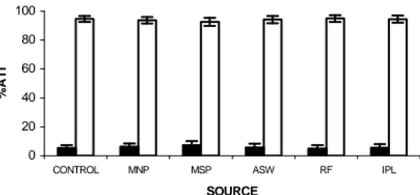
The Fig. 1 shows the ATI% in blood cells and plasma compartments from whole blood exposed to physical agents. The data indicate that, at conditions used, the magnetic field (South and North poles), sonic and radiofrequency waves and intense pulsed light did not alter significantly (p>0.05) the ATI% on the blood compartments.
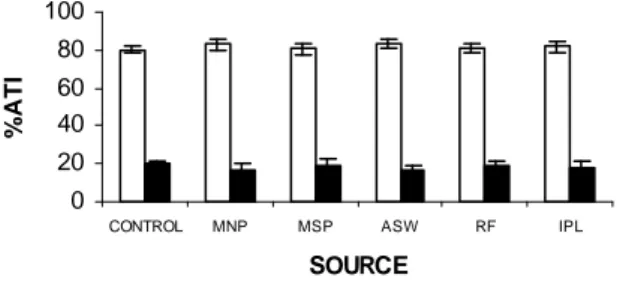
Fig. 2 shows the ATI% in insoluble and soluble fractions isolated from plasma separated from blood samples exposed to physical agents. These data indicate that magnetic field (South and North poles), sonic and radiofrequency waves and intense pulsed light have not significantly (p>0.05) modify the ATI% of fractions of plasma. The Fig. 3 shows the ATI% in insoluble and soluble fractions isolated from blood cells separated from blood samples exposed to physical agents. Similarly to the results obtained with plasma proteins, magnetic field (South and North poles), sonic and radiofrequency waves and intense pulsed light have not significantly (p>0.05) modified the ATI% of fractions of blood cells.
0 20 40 60 80 100
CONTROL MNP MSP ASW RF IPL
SOURCE
%A
T
I
Figure 1 - Effect of exposition to physical agents on the distribution of radioactivity between plasma and cellular compartments. Blood from Wistar rats was exposed to magnetic north (MNP) and south (MSP) poles, audible sonic (ASW) and radiofrequency (RF) waves and intense pulsed light (IPL). The radiolabeling procedure was performed, plasma and blood cells separated by centrifugation, the radioactivity counted and the %ATI to each fraction calculated. (□) Blood cells and (g) plasma
0 20 40 60 80 100
CONTROL MNP MSP ASW RF IPL
SOURCE
%
A
T
I
DISCUSSION
Low frequencies pulsed electromagnetic fields are one of the most athermal common therapies used in the elderly patients by physicians (Heinrich, 2007). It has suggested that the exposition to magnetic field at 15Hz is effective to increases the bone mass (Mc Leod and Rubin, 1997) increasing the local levels of PGE2 and TGF-b1 which
decrease osteoclastic bone reabsortion (Lohmann et al., 2003). Other data have suggested no effect of these electromagnetic fields on collagen synthesis (Ahmadian et al., 2006). Although reports suggest an effect of electromagnetic fields on cell function, no modifications on the distribution of radioactivity in the cellular and plasma compartments was found (Fig. 1).
0 20 40 60 80 100
CONTROL MNP MSP ASW RF IPL
SOURCE
%
A
T
I
Figure 3 - Effect ofexposition to physical agents on the fixation of radioactivity on soluble and insoluble fractions of blood cells. Blood from Wistar rats was exposed to the magnetic North (MNP) and South (MSP) poles, audible sonic (ASW) and radiofrequency (RF) waves and intense pulsed light (IPL). The radiolabeling procedure was performed; plasma and blood cells separated by centrifugation. Insoluble and soluble fractions of blood cells were obtained by precipitation and centrifugation, the radioactivity counted and the %ATI to each fraction calculated. (g) soluble fraction of blood cells and (□) insoluble fraction of blood cells
Authors have suggested that audible sonic waves could interact with proteins moving them to lymphatic system (Capponi and Ronzio, 2006). No modification on the radiolabeling of plasma and cellular proteins was induced by the source of audible sonic waves used in our experiments (Figures 2 and 3) indicating that the phenomenon reported by Capponi and Ronzio (2006) is not relevant to the studied labeled process. Thus, more studies are necessary to understand the potential applications of these mechanical waves in biomedical sciences as well their adverse effects. Radiofrequency thermal therapy of tumors is based on heating of target which induces changes in dielectric properties and protein coagulation and fat melting (Pop et al., 2003). The energy absorbed from a radiofrequency source depends strongly on the tissue dielectric properties (Strohbehn, 1983, Van de Kamer et al., 2001). As results, changes in dielectric properties during heating the tissue temperature distribution is affected and resulting thermal damage. Several numerical models for
predicting the radiofrequency thermal damage in heart muscle and liver have been proposed, but they either incorporated only temperature-dependent changes in electrical conductivity (Labont´e, 1994) or consider the conductivity to be constant (Haemmerich et al., 2001). However, no alterations on labeling of blood constituents with
99mTc were verified when blood samples were
exposed to radiofrequency waves in the conditions used in this study. In consequence, the findings described by Strohbehn, 1983, Labont´e, 1994, Van de Kamer et al., 2001, Haemmerich et al., 2001 could be not relevant to the studied labeled process with 99mTc.
2007). No alterations on the labeling of blood constituents with 99mTc after exposition to intense pulsed light could suggest a safety to this physical agent used to esthetical propose.
In conclusion, although our data have been obtained with blood from Wistar rats, the exposition to magnetic field, sonic and radiofrequency waves and intense pulsed light used in clinical physiotherapy could not alter the examinations performed in nuclear medicine based on blood constituents labeled with 99mTc. Furthermore, the biological/physical consequences associated with these physical agents would be not capable to interfere with some properties of the blood constituents.
ACKNOWLEDGEMENTS
This research was supported by Fundação de Amparo a Pesquisa do Estado do Rio de Janeiro (FAPERJ), Conselho Nacional de Desenvolvimento Científico e Tecnológico (CNPq) and Universidade do Estado do Rio de Janeiro (UERJ).
RESUMO
Fontes de campo magnético, ondas sonoras audíveis e de radiofreqüência e luz intensa pulsada são usadas para o tratamento de doenças. Constituintes sangüíneos(CS) marcados com tecnécio-99m(99mTc) são utilizados na medicina nuclear. Esse trabalho avaliou as consequências de fontes de campo magnético, ondas sonoras audíveis e de radiofreqüência e luz intensa pulsada na marcação de CS com 99mTc. Sangue de ratos Wistar foi exposto às fontes citadas. A marcação de CS com 99mTc foi realizada. Sangue não exposto foram utilizadas(controle). Resultados mostraram que os agentes físicos estudados não alteraram significativamente (p>0.05) a radiomarcação de CS. Apesar terem sido obtidos com sangue de animais, os resultados sugerem que nenhuma alteração nos exames realizados com constituintes sangüíneos com 99mTc em medicina nuclear ocorreria após a exposição às fontes avaliadas. As consequências biológicas associadas a esses agentes não seriam capazes de interferir com algumas propriedades dos CS.
REFERENCES
Abreu, P. R., Almeida, M. C., Bernardo, R. M., Bernardo, L. C., Brito, L. C., Garcia, E. A., Fonseca, A. S. and Bernardo-Filho, M. (2006), Guava extract (Psidium guajava) alters the labelling of blood constituents with technetium 99m. J. Zhejiang. Univ.
Sci. B., 7, 429-35.
Ahmadian, S., Zarchi, S. R. and Bolouri, B. (2006), Effects of extremely-low-frequency pulsed electromagnetic fields on collagen synthesis in rat skin. Biotechnol. Appl. Biochem., 43, 71-75.
Bernardo-Filho, M., Moura, I. N. S. and Boasquevisque, E. M. (1983), 99m-technetium-labeled red blood cells "in vitro". Arq. Biol. Tecnol.,
26, 455-461.
Bernardo-Filho, M.; Santos-Filho, S.D.; Moura, E.G.; Maiworm, A.I.; Orlando, M.M.C.; Penas, M.E.; Cardoso, V.N.; Bernardo, L.C. and Brito, L.C. (2005). Drug Interaction with Radiopharmaceuticals: a Review. Braz. Arch. Biol. Technol.,48, 13-28. Callahan, R. J. and Rabito, C. A. (1990), Radiolabeling
of erythrocytes with technetium-99m: role of band-3 protein in the transport of pertechnetate across the cell membrane. J. Nucl. Med., 31, 2004-2010. CapponiI, R. and Ronzio, O. (2006), Avances en
Fisioterapia. Agentes Físicos: Buenos Aires.
Chang, S. E., Ahn, S. J., Rhee, D. Y., Choi, J. H., Moon, K. C., Suh, H. S. and Soyun-Cho. Y. (2007), Treatment of facial acne papules and pustules in Korean patients using an intense pulsed light device equipped with a 530- to 750-nm filter. Dermatol.
Surg., 33, 676-679.
Dewanjee, M. K., Rao, S. A. and Penniston, J. T. (1982), Mechanism of red blood cell labeling with
99m
Technetium-pertecnetate and the role of cation pumps at RBC membrane on distribution and binding of Sn+2 and 99mTechnetium with membrane proteins and hemoglobin. J. Label. Compd. Radiopharm., 11, 1464-1466.
Fonseca, A.S.; Frydman, J.N.; Rocha, V.C. and Bernardo-Filho, M. (2007), Acetylsalicylic acid decreases the labeling of blood constituents with technetium-99m. Acta Biol. Hung. 58, 189-198. Giordano, N., Battisti, E., Cerasi, S., Fortunato, M.,
Santacroce, C. and Rigato, M. (2001), Effect of electromagnetic fields on bone mineral density and biochemical markers of bone turnover in osteoporosis: a single-blind, randomized pilot study.
Curr. Terap. Res., 62, 155-161.
Haemmerich, D., Staelin, T. S., Tungjitkusolmun, S., Lee, F. T., Mahvi, D. M. and Webster; J. G. (2001), Hepatic bipolar radio-frequency ablation between separated multiprong electrodes. IEEE Trans.
Biomed. Eng., 48, 1145–1152.
Harel, F., Dupuis, J., Benelfassi, A., Ruel, N. and Gregoire, J. (2005), Radionuclide plethysmography for non-invasive evaluation of peripheral arterial blood flow. Am. J. Physiol. Heart Circ. Physiol., 289, H258-H262.
Heinrich H. (2007), Assessment of non-sinusoidal, pulsed, or intermittent exposure to low frequency electric and magnetic fields. Health. Phys., 92, 541-546.
Johns, L. D. (2003), Nonthermal Effects of Therapeutic Ultrasound: The Frequency Resonance Hypothesis. J.
Athl. Train., 37, 293–299
Joseph, B., Kumaran, V., Berishvili, E., Bhargava, K. K., Palestro, C. J. and Gupta, S. (2006), Monocrotaline promotes transplanted cell engraftment and advances liver repopulation in rats via liver conditioning. Hepatology,44, 1411-1420. Labont´e, S. (1994), A computer simulation of
radio-frequency ablation of the endocardium IEEE Trans.
Biomed. Eng.,41, 883–890.
Lohmann, C. H., Schwartz, Z., Liu, Y., Li, Z., Simon, B. J., Sylvia, V. L., Dean, D. D., Bonewald, L. F., Donahue, H. J. and Boyan, B. D. (2003), Pulsed electromagnetic fields affect phenotype and connexin 43 protein expression in MLO-Y4 osteocyte-like cells and ROS 17/2.8 osteoblast-like cells. J. Orthop. Res.,
21, 326-334.
Mc Leod, K. J. and Rubin, C. T. (1997), The effect of low-frequency fields on osteogenesis. J. Bone Joint Surg., 74A, 920-929.
Madonna Terracina, F. S., Curinga, G., Mazzocchi, M., Onesti, M. G. and Scuderi, N. (2007), Utilization of intense pulsed light in the treatment of face and neck erythrosis. Acta Chir. Plast., 49, 51-54.
Mezzana, P. and Valeriani, M. (2007). Rejuvenation of the aging face using fractional hotothermolysis and intense pulsed light: a new technique. Acta Chir. Plast., 49, 47-50.
Olds, G. D., Cooper, G. S., Chak, A., Sivak, M. V. Jr., Chitale, A. A. and Wong, R. C. (2005), The yield of bleeding scans in acute lower gastrointestinal hemorrhage. J. Clin. Gastroenterol., 39, 273-277. Pearce, J. and Thomsen, S. (1995), Rate process
analysis of thermal damage Optical-Thermal Response of Laser-Irradiated Tissue. New York: Plenum, New York, pp. 561–605.
Pettersson, F.; Vogt, A.M.; Jonsson, C.; Mok, B.W.; Shamaei-Tousi, A., Bergstrom, S., Chen, Q. and Wahlgren M. (2005), Whole-body imaging of sequestration of Plasmodium falciparum in the rat.
Infect. Immun., 7,7736-7746.
Perez Rivera, F., Fridmanis, M., Balbi, L., Correa, A., Goñi, S. and Gaglio, P. (2002),Treating benign vascular lession cutaneous thoraxccervicofacial for intence pulsed light. Rev. Argent. Dermatol., 83, 14-22.
Pop, M., Molckovsky, A., Chin, L., Kolios, M. C., Jewett, M. A. S. and Sherar, M. D. (2003), Changes in dielectric properties at 460 kHz of kidney and fat during heating: importance for radio-frequency thermal therapy. Phys. Med. Biol.,48, 2509–2525. Raulin, C., Greve, B. and Grema, H. (2003), IPL
technology: a review. Lasers Surg. Med., 32, 78–87. Saha, G. B. (2004), Fundamentals of nuclear
pharmacy. New York: Springer-Verlag,
Strohbehn, J. (1983), Temperature distributions from interstitial RF electrode hyperthermia systems: theoretical prediction. Int. J Radiat. Oncol., 9, 1655– 1667.
Van de Kamer, J. B., Van Wieringen, N., De Leeuw, A. A. C. and Lagendijk, J. J. W. (2001), The significance of accurate dielectric tissue data for hyperthermia treatment planning. J. Int. Hyperth. 17, 123–142. Wong, K. T., Beauvais, M. M., Melchior, W. R. and
Snyder, S. P. (2004), Enhanced liver uptake of Tc-99m-labeled RBCs during gastrointestinal bleed scintigraphy using transfused RBCs compared with autologous RBCs. Clin. Nucl. Med., 29, 522-523.