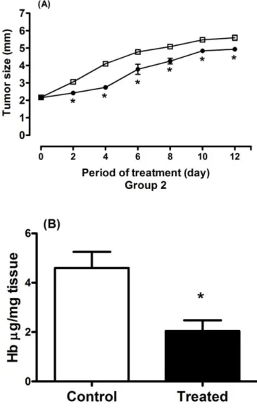
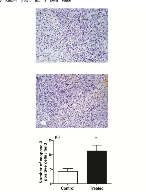
Kint3-4 promotes apoptosis and inhibition of angiogenesis in solid Ehrlich Tumor
Texto
Imagem
Documentos relacionados
didático e resolva as listas de exercícios (disponíveis no Classroom) referentes às obras de Carlos Drummond de Andrade, João Guimarães Rosa, Machado de Assis,
The probability of attending school four our group of interest in this region increased by 6.5 percentage points after the expansion of the Bolsa Família program in 2007 and
O porquê dessa classificação – Quarta e não Terceira ponto qualquer coisa – resulta de dois factores principais: o assumir que seria integrado um conjunto “substantivo”
Com o objetivo de subsidiar o processo inicial de discussão sobre a valorização dos trabalhadores em educação, na perspectiva da construção de uma política pública nacional,
Neste trabalho o objetivo central foi a ampliação e adequação do procedimento e programa computacional baseado no programa comercial MSC.PATRAN, para a geração automática de modelos
Ousasse apontar algumas hipóteses para a solução desse problema público a partir do exposto dos autores usados como base para fundamentação teórica, da análise dos dados
Peça de mão de alta rotação pneumática com sistema Push Button (botão para remoção de broca), podendo apresentar passagem dupla de ar e acoplamento para engate rápido
Assessment of electrochemotherapy effects on Ehrlich solid tumor development in this work aims to evaluate in vivo usage of the electroporator device developed by the Department
