Nutr Hosp. 2014;30(2):223-236 ISSN 0212-1611 • CODEN NUHOEQ S.V.R. 318
Revisión
Adhesion molecules and chemokines: relation to anthropometric,
body composition, biochemical and dietary variables
Renata Adrielle Lima Vieira1, Renata Nascimento de Freitas2and Ana Carolina Pinheiro Volp3
1Nutrition and Health MSc Student (Research Line: Nutrition Biochemistry and Pathophysiology). Federal University of Ouro
Preto, Ouro Preto-Minas Gerais. Brazil. 2PhD in Biochemistry and Immunology and Associate Professor at Federal University
of Ouro Preto, Ouro Preto-Minas Gerais. Brazil. 3PhD in Food Science and Technology, and Associate Professor at Federal
University of Ouro Preto, Ouro Preto-Minas Gerais. Brazil.
MOLÉCULAS DE ADHESIÓN Y QUIMIOCINAS; RELACIÓN CON VARIABLES ANTROPOMÉTRICAS,
DE COMPOSICIÓN CORPORAL, BIOQUÍMICAS Y DIETÉTICAS
Resumen
Introducción:Entre los mediadores inflamatorios invo-lucrados en la fisiopatogenia de la obesidad, se destacan las moléculas de adhesión P-selectina, E-selectina, VCAM-1, ICAM-1 y la quimiocina MCP-1. Estas desempeñan un papel crucial en la adherencia de células en las superficies endoteliales y en la integridad de la pared vascular y pue-den ser moduladas por la composición corporal y patrón alimentario.
Objetivos:Describir y discutir la relación de esas molécu-las de adhesión y quimiocina con marcadores antropométri-cos, composición corporal, bioquímicas y dietéticas.
Métodos:Se utilizaron bases científicas electrónicas para selección de artículos, sin límite de año de publicación.
Resultados: Todas las moléculas se asociaron de forma positiva con marcadores antropométricos; sin embargo, se encontraron resultados controvertidos para ICAM-1 y VCAM-1. No solamente la obesidad per si, sino también la grasa visceral está más fuertemente relacionadas con las concentraciones de E-selectina y MCP-1. La pérdida de peso influencia en la reducción de las concentraciones de esas moléculas, con excepción de la VCAM-1. La distribución de macronutrientes, el consumo excesivo de grasa saturada y trans y un patrón alimentario occidental están asociados con aumento de sus concentraciones. El inverso se pudo observar con la suplementación de ácido graso w-3 en la dieta, el patrón alimentario sano y dieta rica en calcio y productos lácteos. Ya en cuanto a los parámetros bioquímicos, las mis-mas poseen relación inversa con HDL-c y positiva con coles-terol total, triacilgliceroles, glicemia e insulinemia de ayuno y resistencia a insulina.
Conclusión:Marcadores antropométricos, composición corporal, parámetros bioquímicos y patrón alimentario ade-cuados modulan positivamente la inflamación subclínica derivada de la obesidad por medio de la reducción de las moléculas de adhesión y quimiocinas.
(Nutr Hosp. 2014;30:223-236) DOI:10.3305/nh.2014.30.2.7416
Palabras clave:Moléculas de adhesión celular. Inflama-ción. Composición corporal. Antropometria. Hábitos Ali-menticios.
Abstract
Introduction: Among the inflammatory mediators involved in the pathogenesis of obesity, the cell adhesion molecules P-selectin, E-P-selectin, VCAM-1, ICAM-1 and the chemokine MCP-1 stand out. They play a crucial role in adherence of cells to endothelial surfaces, in the integrity of the vascular wall and can be modulated by body composition and dietary pattern.
Objectives:To describe and discuss the relation of these cell adhesion molecules and chemokines to anthropometric, body composition, dietary and biochemical markers.
Methods:Papers were located using scientific databases by topic searches with no restriction on year of publication.
Results:All molecules were associated positively with anthropometric markers, but controversial results were found for ICAM-1 and VCAM-1. Not only obesity, but visceral fat is more strongly correlated with E-selectin and MCP-1 levels. Weight loss influences the reduction in the levels of these molecules, except VCAM-1. The distribution of macronutrients, excessive consumption of saturated and trans fat and a Western dietary pattern are associated with increased levels. The opposite could be observed with supplementation of w-3 fatty acid, healthy dietary pattern, high calcium diet and high dairy intake. Regarding the biochemical parameters, they have inverse relation to HDL-C and positive relation to total cholesterol, triglycerides, blood glucose, fasting insulin and insulin resistance.
Conclusion: Normal anthropometric indicators, body composition, biochemical parameters and eating pattern positively modulate the subclinical inflammation that results from obesity by reducing the cell adhesion molecules and chemokines.
(Nutr Hosp. 2014;30:223-236) DOI:10.3305/nh.2014.30.2.7416
Key words: Cell adhesion molecules. Inflammation. Body composition. Anthropometry. Dietary habits.
Correspondence: Ana Carolina Pinheiro Volp. Department of Clinical and Social Nutrition. Nutrition School. Federal University of Ouro Preto. Campus Universitário.
Morro do Cruzeiro, s/n.
CEP 35.400-000 Ouro Preto. Minas Gerais. Brazil. E-mail: anavolp@gmail.com
Abbreviations
BMI: Body mass index.
CAMs: Cell adhesion molecules. CCL2: Chemokine ligand 2. CVD: Cardiovascular disease. HDL-c: High-density lipoprotein.
HOMA-IR: Homeostatic model assessment insulin resistance.
ICAM-1: Intercellular adhesion molecule-1. IFN-γ: Interferon γ.
IL4: Interleukin 4. IL6: Interleukin 6. IL8: Interleukin 8. IL1: Interleukin 1. IR: Insulin Resistance.
LDL-c: Low-density lipoprotein.
MCP-1: Monocyte chemoattractant protein-1. MS: Metabolic syndrome.
ROS: Reactive oxygen species. T2DM: Type 2 Diabetes Melitus. TNF-α: Tumor necrosis fator-alpha. VCAM-1: Vascular adhesion molecule-1. WC: Waist circumference.
WHR: Waist-hip ratio.
Introduction
Obesity is a complex disease of multifactorial causes that is growing exponentially worldwide,1defined as excessive accumulation of body fat to such an extent that is detrimental to health.2
Studies show the relation between obesity and subclin-ical inflammation.3,4This process is related to expansion of adipocytes and infiltration of macrophages into adipose tissue, where there is an increased secretion of pro-inflammatory cytokines such as interleukin-6 (IL-6), IL-8, tumor necrosis factor (TNF-α), complement C3 and monocyte chemoattractant protein-1 (MCP-1).5-8 These pro-inflammatory cytokines can also substan-tially affect insulin sensitivity and endothelial dysfunc-tion, as well as stimulate a proliferative response in the vascular wall, which clearly promotes an increased risk for type 2 diabetes (T2DM) and cardiovascular diseases (CVD).9,10
When endothelial dysfunction occurs due to an exposure to an activation stimulus such as modified lipoproteins, pro-inflammatory cytokines or coagula-tion cascade proteases like thrombin, induccoagula-tion and expression of cell adhesion molecules (CAMs) on the surface of the endothelium increasing their interaction with circulating leukocytes can occur. Among them, we highlight the vascular cell adhesion molecule-1 (VCAM-1), the intercellular adhesion molecule-1 (ICAM-1), members of the selectin family (P-selectin and E-selectin) and MCP-1 chemokine.11,12
Studies indicate high levels of circulating CAMs in obesity, especially in obesity characterized by
accumu-lation of visceral adipose tissue.9,13,14However, studies linking these molecules and anthropometric markers of obesity are still controversial and inconclusive.15-20For some molecules, weight loss in obese individuals, whether achieved by dietary or surgical intervention, has positive relation to their levels,18,21,22but unlike it was expected, Keogh et al.23 observed a small but significant increase in VCAM-1 after 8 weeks of dietary intervention, which demonstrates that other factors may be involved in the reduction of these concentrations.
In parallel, the connection between dietary patterns and effects on inflammatory response, and conse-quently, on levels of CAMs and chemokines has been discussed.21,24,25Diet macronutrient distribution of and the amount of micronutrients can affect oxidative stress and cause inflammatory changes, which together with a model of chronic excessive intake could induce a pro-inflammatory process.25,26High calcium diets, for example, can reduce levels of MCP-1.27In addition, components of the diet such as trans fatty acids and high glycemic load are considered pro-inflammatory, while a suitable ratio of ω-3 and ω-6 polyunsaturated fatty acids is considered anti-inflammatory.21,26,28,29On the other hand, low-calorie diets have contradictory relation to levels of cell adhesion molecules. For P-selectin and ICAM-1,22,30 such diets have positive influ-ence reducing their levels, while VCAM-1 levels can increase depending on macronutrients distribution.23
Similarly, the relation of cell adhesion molecules and biochemical parameters such as fasting glucose, insulinemia, triglycerides, total cholesterol and frac-tions and presence of metabolic syndrome (MS) is also not fully elucidated and for some molecules, such rela-tions are rare.17,31,32,33
In this context, the objective of this review was to describe and discuss the role of P-selectin, E-selectin, ICAM-1, VCAM-1 and MCP-1 in inflammation and their relation to anthropometric, body composition, dietary and biochemical markers.
Methods
This review was conducted using electronic scien-tific databases, including Medline, PubMed and SciELO, using the following key words in English, Spanish and Portuguese: inflammation, obesity, cell adhesion molecules, VCAM-1, ICAM-1, E-selectin, P-selectin, CCL2 and MCP-1 chemokines, adipose tissue, anthropometry, body composition, diet(ary) pattern. The articles were selected after reading the abstract and regardless of their year of publication.
Obesity and inflammation
were overweight, of which at least 400 million were obese.34This disease results from a positive energy balance in med/long term. Excess energy is stored in adipose tissue and, if this process is prolonged, the individual develops obesity. The balance between food intake and energy expenditure is influenced by a complex interaction of genetic, environmental and social factors35 and it is associated with numerous comorbidities that affect individuals life quality, such as T2DM, CVD and some types of cancers.36
Obese individuals may develop insulin resistance (IR) and MS, and these changes may lead to T2DM. Not only excess body fat, but distribution and type of body fat exert different effects. Evidence has shown a causal relation between obesity and T2DM due to the fact that visceral adipose tissue is more metabolically active than subcutaneous and gluteal-femoral adipose tissue, causing increased production of free fatty acids, IR, hyperglycemia and consequently hyperinsu-linemia9. Moreover, infiltration of inflammation-related cytokines and white adipose tissue immune cells produces a chronic low-grade inflammation and it is, in part, responsible for the pathogenesis of insulin resistance in obese.4,37
Adipocyte hypertrophy that occurs in obesity causes an increased production of a number of pro-inflammatory cytokines and chemokines by the same cells and stromal vascular cells, such as TNF-α, IL-6, IL-1β, resistin, MCP-1, among others.5,6,7,10The increased production of these molecules triggers local effects on the endothelium, leading to an increased production of CAMs and vascular permeability, which ultimately are translated into an increase in monocyte infiltration and accumulation of macrophages.7,9Thus, the mechanism by which obesity may trigger a chronic inflammatory process is clear.
However, the inverse relation has been suggested, that is, obesity as a consequence of chronic inflamma-tion, also known as subclinical.3,38,39According to Das38 and Engström et al.,39inflammatory cytokines 6, IL-1βand TNF-αare involved in metabolism regulation and food intake by regulating insulin action in adipose tissue and modulating release of leptin by this tissue, and these effects can be enhanced by the polymor-phism of the TNF-αreceptor-2 gene that is associated with leptin resistance and obesity. Thus, it is believed to be a vicious cycle between obesity and inflammation induced by changes in adipose tissue.40
Migration of leukocytes in inflammatory response
The inflammatory response is the body’s first defense against tissue damage which aims to remove the response inducing stimulus and to initiate local tissue recovery. This response is coordinated by cellular medi-ators categorized according to their biochemical proper-ties, such as vasoactive amines, vasoactive peptides, lipid mediators, cytokines, chemokines and proteolytic enzymes.41
Initially, after tissue damage caused by injury or infection, there is a production of inflammatory media-tors such as IL-1β, TNF-αand IL-6, chemokines, as well as the expression of CAMs that produce exudate composed of large molecules as albumin and fibrinogen. After that, other plasma proteins migrate to the extravascular space together with leukocytes, which start to circulate along the endothelium through the post-capillary venules.42These changes support a key role of activated vascular endothelium, which is to promote the mobilization and recruitment of leuko-cytes to the inflammatory site.43
The migration of leukocytes into tissues depends on the binding that occurs between these cells and CAMs. Then, signals within the endothelial cells are activated, allowing the opening of narrow vascular passages that are small intercellular gaps. The movement of leukocytes through these passages is stimulated by chemokines produced by the endothelium and tissue. The majority of these cells migrate through these intercellular gaps, but in severe inflammation conditions, a small percentage of leukocytes can transcellular migrate.44
The leukocytes migration process into the extravas-cular space during inflammatory response is regulated by reactive oxygen species (ROS). This process, known as adhesion cascade, involves some steps that typically include: rolling of leukocytes on the endothe-lial cell surface followed by arrest of leukocytes on the endothelium for adhesion and high affinity and subse-quent transmigration into the inflammation tissue site (fig. 1).43,45
The rolling leukocytes express integrins in a low affinity that, after being activated by chemokines produced by activated vascular endothelium, lead to increased affinity. Subsequently, integrins stabilize selectin-mediated binding, reduce the rolling speed favoring the adhesion of leukocytes to the endothelium and promote their passage from the blood to the tissues.46
The transient and reversible interactions of leuko-cytes rolling on the endothelium are mediated by weak binding between selectins propagated on the endothe-lium and on leukocytes and by a firm adhesion inter-ceded primarily by VCAM-1 and ICAM-1, which bind to integrins 1 and 2 expressed on leukocytes.47,48
If inflammatory response persists, qualitative changes, characterized by replacement of leukocytes by macrophages and chronicity of the process, occur. The chronicity of the inflammatory state is a matter of scientific research since little is known about causes and mechanisms involved in such state and its involve-ment in developinvolve-ment of chronic diseases.49
Cell Adhesion Molecules (CAMs)
They are known to mediate cell-endothelium interac-tions of leukocytes and platelets with a critical role in many processes, including inflammation and vascular wall integrity, in addition to being essential for main-taining health and being involved in development of chronic diseases.51
Based on their structure and role, CAMs can be divided into three main families: integrins, selectins and immunoglobulin superfamily. Among them, P-selectin, E-P-selectin, ICAM-1 and VCAM-1 are rele-vant in chronic inflammatory process, and therefore have a prominent role in CVD.41,52,53The distribution and roles of these molecules in the inflammatory process are presented in table I.
Selectins
Selectins are binding protein molecules that mediate the initial low affinity interaction between leukocytes
and endothelial cells and manifest as rolling leukocytes. This transient binding, despite the low affinity, results in increased leukocyte recruitment to the periphery and subsequent firm adhesion and transendothelial migra-tion of these cells.41
In contrast to most of the other CAMs, the role of selectins is restricted to interactions between leuko-cytes and vascular endothelium, and P-selectin and E-selectin are the most important molecules in this process. Soluble forms of selectins can be detected in plasma, where high concentrations have been reported in animals and patients with inflammatory diseases.55
P-Selectin
P-selectin is identified and stored in alpha granules of platelets and in Weibel-Palade bodies of endothelial cells from where it can be rapidly mobilized to the cell
Fig. 1.—Leukocytes migra-tion process. The rolling leu-kocytes express integrins of low affinitiy that, after their activation by chemokines, acquire high affinity for en-dothelial cells. Then the se-lectin mediated binding is stabilized, the rolling speed is reduced, promoting leu-kocyte adhesion to vascular endothelium interceded by VCAM-1 and ICAM-1 and promoting the opening of in-tercellular passages, allo-wing leukocytes to pass from blood to tissues.
Integrin (low affinity)
Integrin (high affinity)
Leukocytes
Chemokines P-selectin E-selectin ICAM-1
VCAM-1
Table I
Adhesion molecules and chemokine: their cellular role and distribution
Adhesion molecules/chemokine Distribution Role
P-selectin Platelets, endothelium
Interaction between platelets and endothelium, adhesion, rolling and recruitment of leukocytes to the endothelium
E-selectin Endothelium Adhesion, rolling and recruitment
of leukocytes to the endothelium
VCAM-1 Endothelium, mild atherosclerotic Adhesion of leukocytes to injury in muscle cells endothelial cells
ICAM-1
Endothelium, leukocytes,
Adhesion and transmigration of fibroblasts, mild atherosclerotic
leukocytes injury in muscle cells
MCP-1 Epithelial, endothelial and Leukocyte recruitment
immune cells
surface in response to a variety of inflammatory agents such as cytokines and free radicals.53,56
The expression of P-selectin on the surface of endothelial cells is usually of short duration, so this makes it an ideal candidate for mediating early leuko-cyte-endothelial interactions. It has an agile kinetics that reveals its important role in recruitment of neutrophils in the early stages of inflammatory reaction.57
Few studies have reported high concentrations of P-selectin in the plasma of obese patients22,58and its rela-tion to fat distriburela-tion has not been well established. Its concentration is directly related to body mass index (BMI), waist-hip ratio (WHR) and waist circumference (WC).22,59,60 However, Pou et al.59showed that both visceral and abdominal subcutaneous fat have also been associated with P-selectin concentrations. When adjusted for BMI and WC, though, this relation did not remain significant (table II). Thus, this suggests that the relation between P-selectin and visceral adiposity is independent of BMI and WC. This is probably justified by the fact that BMI does not assess obesity itself, understood as excess adipose tissue and WC, used in clinical practice as a marker of visceral fat, also takes into consideration the abdominal subcutaneous fat. Therefore, obese patients may not have very represen-tative visceral fat, proportionally, in relation to total fat and consequently maintain homeostasis in relation to inflammatory process.
Weight loss is a method that is likely to improve concentrations of inflammatory markers and endothelial dysfunction in obese subjects. Thus, the kind of inter-vention, dietary or surgical, can influence the reduction of P-selectin concentrations. Roberts et al.61studying weight loss in obese men after 3 weeks and Ziccardi et al.22in obese women after one year, both resulting from dietary intervention and physical activity, showed decreased P-selectin concentrations (table III). This decrease was also observed after the fourth month of weight loss following surgery in morbidly obese62. This is possibly due to reduction of adiposity, which results in improved endothelial activation and is related to reduc-tion of this adhesion molecule.
However, it is unclear whether diets varying in macronutrient composition can affect inflammatory responses differently. Sharman and Volek30compared a low calorie diet with very low carbohydrate levels (< 10% carbohydrate) and low fat levels (25% fat, < 10% saturated fat and < 300 mg cholesterol) on inflam-matory markers in overweight men. After 6 weeks of intervention there was no significant reduction in absolute concentration of P-selectin for both diets. However, when inflammatory values were normalized with reduction of 1kg of body weight, there was a significant reduction of P-selectin for both treatments and it was higher for the very low carbohydrate level diet (table IV). The results of this study suggest that in a short period of time, weight loss and not diet composi-tion seems to be the underlying driving force in reducing these inflammatory markers.
Besides adiposity and macronutrient composition, biochemical parameters also influence concentrations of CAMs. Studies have shown that concentrations of P-selectin were positively associated with fasting glucose,17 triglycerides and total cholesterol and inversely associated with high-density lipoprotein (HDL-c)16,60 in apparently healthy individuals. This association of triglycerides and HDL-c was found by Miller and Cappuccio19after adjustment on age and sex (table V).
This way, it is becoming clear that P-selectin is an essential component in cardiovascular disease and therefore is a potential therapeutic target. A longitu-dinal monitoring of apparently healthy women for 3.5 years showed that concentrations of P-selectin were significantly higher in the beginning of the study among participants who subsequently developed a first cardiovascular event than those who had no event. In this study, relative risk of future events increased by 25% for each quartile increase in basal level of P-selectin.63
Therefore, concentrations of P-selectin are directly related to obesity markers and weight loss. However, as to body composition, unlike what was expected, its relation is not yet well established, since there was no association of P-selectin with visceral or subcutaneous fat. The macronutrient distribution or a healthy eating pattern also need to be elucidated, since studies indi-cate a reduction in concentrations of P-selectin resulting from weight loss induced by diet and not due to its composition. Similarly, the relation of this mole-cule with biochemical parameters should be more studied, since these are all contributing factors to cardiovascular events through its inflammatory effects on vascular endothelium.
E-Selectin
E-selectin is expressed on endothelial cells specifi-cally and strongly stimulated by inflammatory mole-cules such as TNF-α and IL-1β, and it is widely expressed in vasculature on sites of inflammation.64
Its relation to the occurrence and type of obesity, as well as to anthropometric markers is established. The concentrations of E-selectin were significantly associ-ated with BMI and WHR.16,17,19,33When compared, the concentrations of E-selectin in obese and nonobese individuals, they are significantly higher in obese, and visceral obesity is more strongly related than total fat.14,33This could be explained by production of TNF-α and IL-6 derived from visceral adipocytes on which they induce increased expression of this molecule.33
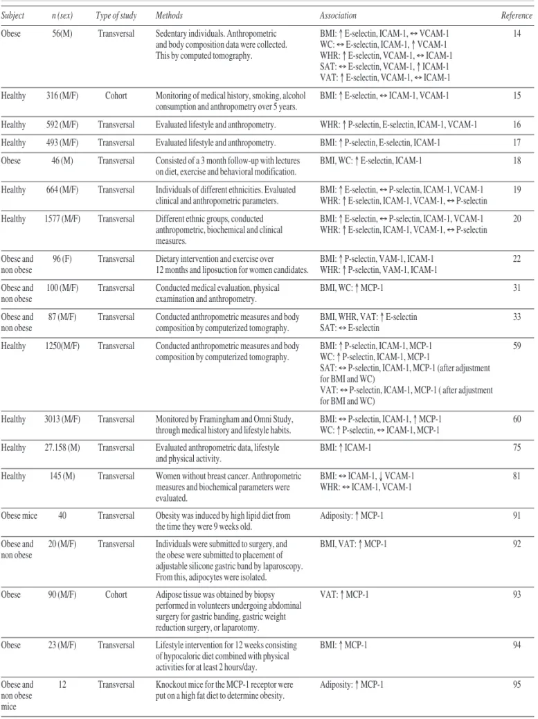
Table II
Association of adhesion molecules and chemokine with anthropometric markers and body composition
Subject n (sex) Type of study Methods Association Reference
Obese 56(M) Transversal Sedentary individuals. Anthropometric BMI: ↑ E-selectin, ICAM-1, ↔VCAM-1 14 and body composition data were collected. WC: ↔E-selectin, ICAM-1, ↑ VCAM-1
This by computed tomography. WHR: ↑ E-selectin, VCAM-1, ↔ICAM-1 SAT: ↔E-selectin, VCAM-1, ↑ ICAM-1 VAT: ↑ E-selectin, VCAM-1, ↔ICAM-1
Healthy 316 (M/F) Cohort Monitoring of medical history, smoking, alcohol BMI: ↑ E-selectin, ↔ICAM-1, VCAM-1 15 consumption and anthropometry over 5 years.
Healthy 592 (M/F) Transversal Evaluated lifestyle and anthropometry. WHR: ↑ P-selectin, E-selectin, ICAM-1, VCAM-1 16
Healthy 493 (M/F) Transversal Evaluated lifestyle and anthropometry. BMI: ↑ P-selectin, E-selectin, ICAM-1 17
Obese 46 (M) Transversal Consisted of a 3 month follow-up with lectures BMI, WC: ↑ E-selectin, ICAM-1 18 on diet, exercise and behavioral modification.
Healthy 664 (M/F) Transversal Individuals of different ethnicities. Evaluated BMI: ↑ E-selectin, ↔P-selectin, ICAM-1, VCAM-1 19 clinical and anthropometric parameters. WHR: ↑ E-selectin, ICAM-1, VCAM-1, ↔P-selectin
Healthy 1577 (M/F) Transversal Different ethnic groups, conducted BMI: ↑ E-selectin, ↔P-selectin, ICAM-1, VCAM-1 20 anthropometric, biochemical and clinical WHR: ↑ E-selectin, ICAM-1, VCAM-1, ↔P-selectin measures.
Obese and 96 (F) Transversal Dietary intervention and exercise over BMI: ↑ P-selectin, VAM-1, ICAM-1 22 non obese 12 months and liposuction for women candidates. WHR: ↑ P-selectin, VAM-1, ICAM-1
Obese and 100 (M/F) Transversal Conducted medical evaluation, physical BMI, WC: ↑ MCP-1 31 non obese examination and anthropometry.
Obese and 87 (M/F) Transversal Conducted anthropometric measures and body BMI, WHR, VAT: ↑ E-selectin 33 non obese composition by computerized tomography. SAT: ↔E-selectin
Healthy 1250(M/F) Transversal Conducted anthropometric measures and body BMI: ↑ P-selectin, ICAM-1, MCP-1 59 composition by computerized tomography. WC: ↑ P-selectin, ICAM-1, MCP-1
SAT: ↔P-selectin, ICAM-1, MCP-1 (after adjustment for BMI and WC)
VAT: ↔P-selectin, ICAM-1, MCP-1 ( after adjustment for BMI and WC)
Healthy 3013 (M/F) Transversal Monitored by Framingham and Omni Study, BMI: ↔P-selectin, ICAM-1, ↑ MCP-1 60 through medical history and lifestyle habits. WC: ↑ P-selectin, ↔ICAM-1, MCP-1
Healthy 27.158 (M) Transversal Evaluated anthropometric data, lifestyle BMI: ↑ ICAM-1 75 and physical activity.
Healthy 145 (M) Transversal Women without breast cancer. Anthropometric BMI: ↔ICAM-1, ↓VCAM-1 81 measures and biochemical parameters were WHR: ↔ICAM-1, VCAM-1
evaluated.
Obese mice 40 Transversal Obesity was induced by high lipid diet from Adiposity: ↑ MCP-1 91 the time they were 9 weeks old.
Obese and 20 (M/F) Transversal Individuals were submitted to surgery, and BMI, VAT: ↑ MCP-1 92 non obese the obese were submitted to placement of
adjustable silicone gastric band by laparoscopy. From this, adipocytes were isolated.
Obese 90 (M/F) Cohort Adipose tissue was obtained by biopsy VAT: ↑ MCP-1 93 performed in volunteers undergoing abdominal
surgery for gastric banding, gastric weight reduction surgery, or laparotomy.
Obese 23 (M/F) Transversal Lifestyle intervention for 12 weeks consisting BMI: ↑ MCP-1 94 of hypocaloric diet combined with physical
activities for at least 2 hours/day.
Obese and 12 Transversal Knockout mice for the MCP-1 receptor were Adiposity: ↑ MCP-1 95 non obese put on a high fat diet to determine obesity.
mice
al.65in surgical weight loss in individuals with morbid obesity. This suggests a reduction of endothelial acti-vation and an important role of adipose tissue in this pathophysiological mechanism.
Diet macronutrient composition can directly affect concentrations of E-selectin. When comparing different meals overloaded with glucose, fat or in combination, in healthy individuals, it was found an increase in these levels after high fat and glucose meals, and when combined, there were more pronounced effects on E-selectin.21This increase is due to hyperglycemia and postprandial hypertriglyceridemia, which have inde-pendent but cumulative effects and promote an athero-genic profile.
Moreover, a healthy dietary pattern, characterized by consumption of fruits, vegetables, fish, poultry and whole grains was associated with decreased concentra-tions of E-selectin after adjustment for age, BMI, phys-ical activity, smoking and alcohol consumption in apparently healthy women. At the same time, a Western dietary pattern characterized by higher intakes
of red meat, sweets, fries and refined grains was associ-ated with increase in these concentrations.66,67
The type of lipid in the diet may also differentially influence endothelial activation. The w-3 alpha-linolenic fatty acid consumption in overweight individ-uals with hypercholesterolemia significantly reduces E-selectin concentrations, since it activates mecha-nisms that beneficially affect both lipids/lipoproteins and CAMs on which they inhibit endothelial activa-tion68. The intake of trans fatty acids is directly associ-ated with increased concentrations of E-selectin, due to vasodilatation, and consequent increase of CAMs and reduced HDL-c, which can cause oxidation of low density lipoproteins (LDL-c). This indicates that dietary factors may influence the cardiovascular risk through modulation of endothelial function.24
The concentrations are also related to biochemical parameters. Fasting glucose, fasting insulin and triglycerides were positively associated with E-selectin,17,19,33 while HDL-c had a negative associa-tion19,33in healthy individuals. As for individuals with
Table III
Effect of weight loss on adhesion molecules and chemokine
Subject n (sex) Intervention period Methods Effect Reference
Obese 46 (F) 3 months Consisted of lectures on diet, exercise sessions ↓ E-selectin, ICAM-1 18 and behavioral modification. Emphasis on
themes of calorie restriction, increasing consumption of vegetables and grains and replacing saturated fats with unsaturated.
Obese and 96 (F) 12 months Intervention with hypocaloric diet, exercise ↓ P-selectin, VCAM-1, ICAM-1 22 non obese and liposuction for women candidates. Diet
composition: 1300 kcal, 55% carbohydrate, 22% protein, 23% fat and 25 g fibers.
Obese 99 (M/F) 8 weeks Intervention with two types of hypocaloric ↓ P-selectin, E-selectin, ICAM-1 23 diet: a low-carbohydrate and high in saturated ↑ VCAM-1
fat (4% carbohydrate, 61% lipid, 20% saturated fat) and low in saturated fat
(46% carbohydrate, 30% lipid, < 8% saturated fat).
Obese and 31 (M) 3 weeks Dietary intervention and daily exercise. Diet: ↓ P-selectin, ICAM-1 61 SM 12-15% fat, 15-20% protein and 65-70% mainly
from complex carbohydrates, > 40 g of dietary fiber.
Morbid 49 (M/F) 4 months Weight loss was due to bariatric surgery. ↓ P-selectin, E-selectin, VCAM-1, ↔ICAM-1 62 obese and
non obese
Obese and 126 (M/F) 12 months The intervention was adjustable gastric band ↓ E-selectin, ICAM-1 65 non obese or diet and physical activity. Hypocaloric diet:
1000 for men and 1100 kcal / day for women, 48% carbohydrate, 33% protein and 19% fat (olive oil).
Obese 40 18 weeks Weight loss was due to feed restriction. ↓ MCP-1 91 mice
Obese 23 (M/F) 12 weeks Intervention with low-calorie diet combined ↓ MCP-1 94 with physical activity for at least 2 hours/day.
Table IV
Qualitative and quantitative effect of diet on adhesion molecules and chemokine
Subject n (sex) Intervention period Methods Effect Reference
Type 2 50 (M/F) 4 weeks All participants ate three different diets on High fat diet: ↑ E-selectin, VCAM-1, ICAM-1 21 diabetics different days: a high fat meal (75g fat, High glucose diet: ↑ E-selectin, VCAM-1, ICAM-1 and healthy 5 g of carbohydrate and 6g of protein per High fat and glucose diet: ↑ E-selectin, VCAM-1,
m2of body surface), a meal with only 75 g ICAM-1 (more pronounced than isolated diets)
of glucose and the third meal was rich in fat and contained 75 g of glucose. Blood samples were collected at 0, 1, 2, 3, and 4 h.
Healthy 730 (F) Transversal Food intake was registered using a validated Intake of trans fatty acids: ↑ E-selectin, 24 semiquantitative FFQ. Detailed information VCAM-1, ICAM-1
about the type of fat or oil used for frying (cooking and table), brand, type and year of consumption of margarine.
αP2-agouti 30 (M) 3 weeks Mice were divided into three groups with Diet rich in calcium and dairy: ↓MCP-1 27 transgenic different diets: one group on control diet
mice (0.4% calcium carbonate), one group with a high concentration of calcium (1.2% calcium carbonate) and the third group consumed a high calcium diet derived from dairy products (1,2% nonfat dry milk).
Overweight 15 (M) 6 weeks Comparison of two hypocaloric diets: a very Low-carb diet: ↔P-selectin, ↓ICAM-1 30 low-carb diet (< 10% carbohydrate, 60% of Low-fat diet: ↔P-selectin, ↓ICAM-1
fat and 30% of protein) and a low-fat diet (20% protein, 25% fat, < 10% saturated fat and < 300 mg cholesterol, 55% carbohydrate). All volunteers consumed both diets.
Healthy 732 (F) Transversal Healthy dietary pattern was characterized Healthy dietary pattern: ↓E-selectin, 66 by higher intake of fruits, vegetables, fish, ↔VCAM-1, ICAM-1
poultry and whole grains, and Western Western dietary pattern: ↑ E-selectin, VCAM-1, dietary pattern was characterized by higher ICAM-1
intake of red and processed meats, sweets, desserts, fries and refined grains.
Healthy 486 (F) Transversal Healthy dietary pattern: rich in fruits, Healthy dietary pattern: ↓E-selectin, VCAM-1, 67 vegetables, tomatoes, poultry, tea, fruit ICAM-1
juices and whole grains. Western dietary Western dietary pattern: ↔E-selectin, ↓VCAM-1, pattern: rich in refined grains, red meat, ICAM-1
butter, processed meats, high fat dairy products, sweets and desserts, pizza, potatoes, eggs, hydrogenated fats and sodas.
Hypercho- 23 (M/F) 6 weeks Three diets: a standard diet (13% saturated, Diet Rich in ALA: ↓E-selectin, VCAM-1, ICAM-1 68 lesterolemic 13% MUFA and 9% PUFA), a diet rich in more pronounced than the other diets
individuals PUFA and ALA (8% saturated, 12% MUFA and 17% PUFA) and a diet rich in PUFA and LA (8 % saturated, 12% MUFA and 16% PUFA).
Overweight 11 (M/F) 6 hours after eating Subjects consumed three high-fat shakes: one High in saturated fat: ↑ VCAM-1, ↔ICAM-1 76 and obese rich in saturated fat, the other rich in High in monounsaturated: ↔VCAM-1, ICAM-1
monounsaturated and the third rich in w-3 High in polyunsaturated: ↔VCAM-1, ICAM-1 polyunsaturated fat. Blood samples were
collected at 0, 1, 2, 4 and 6 after eating.
Individuals 60 (M/F) 6 weeks Individuals divided into three groups fed Fish oil: ↓VCAM-1 compared with the other oils 82 with with 700 g/week of Atlantic salmon in
coronary different ways: the first with 100% fish oil, artery the second with 100% canola oil and the disease third with 50% of each oil, resulting in fillets
with high, medium and low levels of w-3 PUFAs.
Obese and 12 Transversal Knockout mice for the receptor of MCP-1 High intake of saturated fat: ↑ MCP-1 95 non obese were fed a high saturated fat diet.
mice
visceral adiposity, E-selectin is related to hyperinsu-linemia and IR,14 which was expected since this adipose tissue is highly lipolytic and has a late interfer-ence in post-insulin receptor mechanisms, resulting in inefficient glucose uptake causing such effects.
Thus, E-selectin, may play a role in atherogenesis model centered on inflammation.69The concentrations were significantly associated with coronary artery disease (OR: 1.54, 95% CI: 1.27-1.86) and occurrence of atherosclerosis of the carotid artery (OR: 1.36, 95%
CI: 1.09-1.70). The risk of coronary artery disease in individuals with high levels of E-selectin was 2.98 times higher (95% CI: 1.74-5.10).15
Therefore, E-selectin is well grounded as to the occurrence and type of obesity and visceral obesity is strongly related to these concentrations. Similarly weight loss is positively associated with its reduction. Macronutrient distribution, healthy eating pattern and type of lipids are also clearly related to E-selectin, since dietary factors can influence modulation of Table V
Association of biochemical parameters, adhesion molecules and chemokine
Subject n (sex) Type of study Association Reference
Obese 56(M) Transversal E-selectin: ↑ total cholesterol, triacylglycerols, LDL-c, fasting insulin, HOMA, ↔HDL-c, 14 fasting blood glucose
ICAM-1, VCAM-1: ↔total cholesterol, triacylglycerols, LDL-c, fasting insulin, HOMA, HDL-c, fasting blood glucose
Healthy 316 (M/F) Cohort E-selectin: ↑triacylglycerols, ↔LDL-c, ↓ HDL-c 15 ICAM-1, VCAM-1: ↔triacylglycerols, LDL-c, HDL-c
Healthy 592 (M/F) Transversal P-selectin, E-selectin, ICAM-1, VCAM-1: ↑total cholesterol, HDL-c 16
Healthy 493 (M/F) Transversal P-selectin, ICAM-1: ↑ triacylglycerols, ↔fasting blood glucose 17 E-selectin: ↔triacylglycerols, ↑ fasting blood glucose
Obese 46 (F) Transversal E-selectin: ↑ triacylglycerols, ↔total cholesterol, HOMA 18 ICAM-1: ↔triacylglycerols, total cholesterol, HOMA
Healthy 664 (M/F) Transversal P-selectin, ICAM-1, VCAM-1: ↑ triacylglycerols, ↔fasting insulin, ↓HDL-c 19 E-selectin: ↑ triacylglycerols, fasting insulin, ↓HDL-c
Healthy 1577 (M/F) Transversal E-selectin: ↑total cholesterol, triacylglycerols, fasting insulin, fasting blood glucose, ↓ HDL-c 20 P-selectin: ↔total cholesterol, triacylglycerols, fasting insulin, ↑ fasting blood glucose, ↓ HDL-c ICAM-1: ↑total cholesterol, triacylglycerols, ↔fasting insulin, fasting blood glucose, ↓ HDL-c VCAM-1: ↔total cholesterol, ↑triacylglycerols, ↔fasting insulin, fasting blood glucose, ↓ HDL-c
Obese and 100 (M/F) Transversal MCP-1: ↔total cholesterol, triacylglycerols, LDL-c, ↓ HDL-c 31 non obese
Individuals 37 (M/F) Transversal ICAM-1, VCAM-1: ↑MS, HOMA-IR 32 with MS
Obese and 87 (M/F) Transversal E-selectin: ↔total cholesterol, LDL-c, ↑triacylglycerols, ↓ HDL-c 33 non obese
Healthy 3013 (M/F) Transversal P-selectin: ↑triacylglycerols 60 ICAM-1, MCP-1: ↔triacylglycerols
Obese and 126 (M/F) Transversal E-selectin, ICAM-1: ↑insulin, HOMA 65 non obese
Healthy 30 (M) Longitudinal E-selectin: ↔fasting and postprandial blood glucose, fasting and postprandial triacylglycerol, 78 fasting and postprandial insulin
ICAM-1: ↔fasting and postprandial blood glucose, fasting and postprandial triacylglycerol, fasting insulin, ↑ postprandial insulin
VCAM-1: ↔fasting and postprandial blood glucose, fasting triacylglycerol, ↑ postprandial triacylglycerol, ↔fasting and postprandial insulin
Healthy, 40 (M/F) Transversal E-selectin: ↑total cholesterol 83 hypercholes- ICAM-1: ↑total cholesterol, triacylglycerols
terolemic and VCAM-1: ↑triacylglycerols
hypertrigly-ceridemic individuals
endothelial function. Along with these factors, total cholesterol and fractions, triglycerides, fasting insulin and fasting glucose are also associated with E-selectin. Thus, E-selectin can be considered a classic molecule for obesity and cardiovascular risk factors.
The immunoglobulin superfamily (IgSF)
The IgSF is the most abundant family of cell surface molecules, representing 50% of all leukocyte surface glycoproteins.70IgSF members are diverse, comprising soluble and membrane-bound immunoglobulin and monomeric adhesion molecules.56
These molecules are expressed on endothelial cell membranes, they are also involved in leukocyte adhe-sion and are induced by TNF-α, interferon-γ(IFN-γ) and IL-1β. The main molecules of this family are: ICAM-1 and VCAM-1, which are relevant in chronic inflammatory process, with highlights to CVD.56
Intercellular Adhesion Molecule-1 (ICAM-1)
ICAM-1 is a transmembrane glycoprotein that binds to integrins in order to facilitate transmigration of leukocytes through vascular endothelium. Intercellular adhesion function has been attributed to ICAM-1 as a result of this binding capacity. Its expression is regu-lated by agents such as TNF-αand thrombin.70, 71,72
The firm adhesion of leukocytes to the endothelium via ICAM-1 causes an increase in intracellular free Ca2 +, contractility of myosin and activation of p38 kinase. The activation of these signaling pathways results in extensive remodeling events of the cytoskeleton that alter the contractility of endothelial cells, thereby facil-itating transmigration and weakening bindings of junc-tional adhesion molecules.73,74
Its relation to anthropometric markers has been explored, but with controversial results. A study involving apparently healthy women observed a posi-tive and significant relation between BMI and ICAM-1, and showed that women in the highest BMI quintile had 14% higher concentrations than those in lower quintiles.75As to obese women, a significant positive association with BMI and WHR was found.22However, Ponthieux et al.17and Miller and Cappuccio19when studying healthy individuals of different ages and ethnicities, respectively, could not observe significant association with BMI after adjustment for age, sex, smoking status and ethnicity. This may have been observed because BMI does not measure, as it was previously mentioned, body composition, which is common among the adjustment variables.
Regarding body composition, however, both subcu-taneous and visceral abdominal fat were not associated with ICAM-1 concentrations, when adjusted for BMI and WC. Thus, the concentration of this molecule also seems to be independent of these two anthropometric
markers.59This shows that there may be alternative mechanisms to alteration of such cell adhesion mole-cule, besides type of adiposity, as possibly reduction of HDL-c and subsequent oxidation of LDL-c, which can be a risk factor for modification of the endothelium regardless body composition.
Weight loss was related to ICAM-1 concentrations. Ito et al.18and Ziccardi et al.22studied weight loss in obese women submitted to hypocaloric dietary inter-vention after 3 and 12 months, respectively, and observed a positive association with this molecule. Thus, this suggests an important role of adipose tissue in reduction of endothelial activation.
Not only the caloric value, but also qualitative charac-teristics of the diet may be associated with concentrations of this molecule and therefore with endothelial activation. Studies with women showed that a dietary pattern charac-terized by higher intakes of red and processed meats, sweets, desserts, fries and refined grains reduces ICAM-1 concentrations.66,67At the same time, consumption of a high glucose load and fat,21particularly saturated fat76and trans fatty acid24produces a significant increase in ICAM-1. Thus, it is suggested that inappropriate food intake plays an important role in endothelium functional changes caused by oxidation of LDL-c, which is the initial step in development of atherosclerosis.
Prospective studies indicate ICAM-1 concentrations as a predictor of future cardiovascular events.15,77 Biochemical parameters considered risk factors for development of CVD such as total cholesterol, triglyc-erides and HDL-c were associated with concentrations of this cell adhesion molecule, and HDL-c had an inverse association.19,20Furthermore, ICAM-1 is posi-tively related to insulin, postprandial glucose78and IR.17 Kressel et al.32used the homeostasis model assess-ment for definition of IR (HOMA-IR), and showed that individuals who had HOMA-IR ≥ 5.03 had higher concentrations of this marker when compared with those who had HOMA-IR ≤ 1.32. Therefore, these results reinforce the ideia that associations between systemic and vascular inflammation may help explain predisposition for IR in obese patients.
This way, studies on the relation of ICAM-1 with nutritional and dietary parameters present controver-sial results for anthropometric markers when adjusted for age, sex, smoking status and ethnicity, and further research is required, particularly related to body composition, in order to elucidate its relation with obesity per se and/or the distribution of body fat. However, this molecule is directly related to weight loss, with inadequate dietary pattern, high intake of saturated and trans fat and biochemical parameters, and has an inverse relation with HDL-c.
Vascular Cell Adhesion Molecule-1 (VCAM-1)
binds mainly to α4β1 integrin which is constitutively expressed on lymphocytes, monocytes and eosinophils.79
VCAM-1 can mediate both rolling and firm adhe-sion. Although it is structurally similar to ICAM-1, VCAM-1 has a unique pattern of regulation. It is not expressed in basal conditions, but is rapidly induced by pro-atherosclerotic conditions.47,79It is induced by IL-4 and high concentrations of ROS oxidized LDL-C.80
The relation of VCAM-1 concentrations with anthropometric markers is also controversial. Some researches show a positive correlation with BMI,15,16,17,22 WC and WHR,14while Miller and Cappucio19did not show any significant results between VCAM-1 and BMI after adjustment for age, sex, smoking status and ethnicity. Recently, Souza et al.81observed an inverse correlation between VCAM-1 and BMI, when studying apparently healthy women, and showed that for every increase of 1 kg/m2in BMI, there was a reduction of 1,7 ng/mL in its concentrations. Thus, there is still a gap in understanding the mechanisms that explain pathophys-iological changes that make obesity a risk factor for increased CAMs.
Results that point to a decrease in concentrations of VCAM-1 as a result of weight loss are also contradictory. Ziccardi et al.22demonstrated significant reduction of VCAM-1 after a year of nutritional intervention with low-calorie diet in obese women, while Keogh et al.23 observed a slight increase (5%) in concentrations for 8 weeks with two types of hypocaloric diet, one low in carbohydrates and high in saturated fat and the other high in carbohydrates and low in saturated fat. Although weight loss reduces adiposity and consequently reduces this molecule, this result may be due to the effect of excess fat or carbohydrate in both diets on endothelial function, since such excesses may cause lipogenesis.
Besides distribution of macronutrients, dietary patterns may also influence concentrations of VCAM-1. Individuals whose diets were based on processed foods, high glycemic index, few fruits and vegetables had significantly higher concentrations than those who had higher intake of fish, poultry, fruits, vegetables and whole grains.66,67Moreover, supplementation of w-3 polyunsaturated fatty acids significantly reduced this molecule in hypercholesterolemic individuals68with coronary artery disease.82
Biochemical parameters show little evidence of the relation with concentrations of VCAM-1 in healthy or obese individuals. Positive association of this molecule with triacylglycerols,83 IR32 and total cholesterol and nega-tive association with HDL-c16were observed. Neverthe-less, evidences indicate that VCAM-1 is one of the major CAMs involved in atherosclerotic lesions and it is the first CAM to be expressed before the development of athero-sclerotic plaques.45Its high concentration increases the risk by 2.8 times for future cardiovascular events.84
Therefore, contrary to the expected, some studies show that VCAM-1 may have an inverse relation to BMI and weight loss, which suggests that other factors may be involved in its stimulation and/or depression. However,
dietary factors such as adequate food pattern and supple-mentation of w-3 polyunsaturated fatty acids positively influence the reduction of its concentrations. Similarly, total cholesterol, triglycerides, and IR show a positive correlation and HDL-C is inversely correlated with this molecule, being this the first CAM to be altered in the development of atherosclerotic plaques.
Chemokines
Chemokines constitute a family of small chemo-tactic cytokines composed of heparin binding proteins that drive the migration of circulating leukocytes to sites of inflammation or injury.85,86These cytokines not only regulate cell chemotaxis by concentration gradi-ents, but also adhesion.87,88
According to Lezama Asencio,88chemokines are involved in metabolic disorders. The main motivation for examining chemokines is how easily their expres-sion can be documented in disorders associated with leukocyte infiltration.89 The monocyte chemotactic protein (MCP-1), also named chemokine ligand 2 (CCL2), is a key chemokine in regulation of migration and infiltration of monocytes/macrophages involved in chronic inflammatory diseases.90
Monocyte chemoattractant protein-1 (MCP-1/CCL2)
MCP-1 or CCL2 was the first human chemokine to be discovered. It is produced by many types of cell, including endothelial cells, fibroblasts, epithelial cells, smooth muscle cells, monocytes and microglial cells, constitutively or in response to extracellular stimuli such as oxidative stress, cytokines or growth factors. MCP-1/CCL2 and its receptors play a central role in develop-ment of inflammatory responses and are crucial for the recruitment of immune cells to sites of inflammation.90
In obese humans and rodents, MCP-1/CCL2 is expressed by adipose tissue and increases proportionally to adiposity.6,7,91,92A study of two independent cohorts with obese individuals showed that concentrations of MCP-1/CCL2 increased with obesity, especially with intra-abdominal visceral fat,93which is expected since this fat deposit is highly lipolytic. Thus, its concentra-tions are associated with BMI, visceral fat and WC.31,59
Moreover, its reduction is related to weight loss in individuals with severe obesity. A weight loss of 12% in obese individuals resulted in a reduction of 20% in its concentrations (p < 0,001).94Thus, it is suggested the importance of reducing adipose tissue in improve-ment of endothelial activation.
The relation of MCP-1/CCL2 with diet has been poorly described. In humans, a study by Zemel and Sun27using diets rich in calcium and dairy products resulted in significant suppression when comparing with low calcium diet. The reason for this is probably because the decrease in intracellular calcium concen-tration induced by high levels of this nutrient in the diet reduces the synthesis of fatty acid synthase and conse-quently reduces lipogenesis and increases lipolysis. This is a possible mechanism for the effect of dietary calcium in modulating adiposity. In rats, a high fat diet increases the expression of MCP-1/CCL2. Thus, it is suggested that there is an influence of diet composition on the expression of this chemokine.95
MCP-1/CCL2 also plays a key role in the pro-inflammatory pathways in vascular endothelium and development of atherosclerosis, situation in which its concentration is high. By using MCP-1/CCL2 in knockout mice to examine atherosclerosis, studies have shown that the absence of this chemokine signifi-cantly reduces deposition of lipids in arteries and formation of atherosclerotic lesions.86,90
This way, the relation of MCP-1/CCL2 with anthropo-metric markers and body composition is well established, especially with intra-abdominal visceral fat, being reduc-tion in its concentrareduc-tions influenced by weight loss. In parallel, diets rich in calcium are also related to its concentration, but more studies should be conducted in order to understand if dietary pattern and macronutrient distribution also influence positively the reduction of MCP-1/CCL2 once this chemokine is a strong link between obesity and its metabolic complications.
Conclusion/perspectives
The importance of CAMs and chemokines in subclin-ical inflammation, which can lead to development of metabolic syndrome, obesity and other chronic diseases, is well understood, being this a bidirectional process.
All molecules reviewed here relate positively with an anthropometric marker as BMI, WC, WHR, but studies also show that there is no relation of ICAM-1 with BMI after adjustment for age, sex, smoking status and ethnicity, while for VCAM-1 there may be an inverse relation, its concentrations may be higher in those with lower BMI. As for the body composition, both E-selectin and MCP-1/CCL2 are positively asso-ciated with type of fat, being the visceral one more strongly related to its concentrations. However, for P-selectin, VCAM-1 and ICAM-1, both subcutaneous and visceral abdominal fat were not associated with its concentrations.
Weight loss, whether due to hypocaloric diets with different macronutrient distribution or surgical, influ-ences positively the reduction in concentrations of all CAMs and chemokines studied, except for VCAM-1 in which distribution of macronutrients can influence more than calorie reduction, which suggests that other
factors may be involved in its stimulation and/or depression.
Besides distribution of nutrients, high consumption of trans fatty acids, saturated fat and a Western dietary pattern are associated with increased concentrations of CAMs. In contrast, individuals that supplement w-3 fatty acid in diet, have a healthy dietary pattern and a diet rich in calcium and dairy products and have lower concentrations of these molecules and MCP-1/CCL2.
Regarding biochemical parameters, all molecules and the chemokine have inverse relation of their concentrations with HDL-C and positive relation with total cholesterol and triglycerides. In addition, all molecules have positive relation with fasting glucose, fasting insulin and IR, being the ICAM-1 also related to postprandial glycemia.
Therefore, although there were significant progress in understanding the involvement of E-selectin, P-selectin, VCAM-1, ICAM-1 and the chemokine MCP-1/CCL2 in regulating subclinical inflammation, studies linking these CAMs to anthropometric markers, body composition, biochemical and dietary parameters in healthy and obese individuals are still needed to eluci-date these associations and mechanisms, since these are contributing factors for cardiovascular events due to its inflammatory effects on vascular endothelium.
Acknowledgment
We thank the Federal University of Ouro Preto by support funding which allowed the translation of this article.
References
1. WHO. Obesity: preventing and managing the global epidemic. Report of a WHO Consultation on Obesity. World Health Orga-nization Tech Report Ser2000; 894: i-xii, 1-253.
2. Pinheiro ARO, Freitas SFT, Corso ACT. Uma abordagem epidemiológica da obesidade. Rev Nutr2004; 17 (4): 523-33. 3. Prado WL, Lofrano MC, Oyama LM, Dâmaso AR. Obesidade e
adipocinas inflamatórias: implicações práticas para a prescrição de exercício. Rev Bras Med Esporte2009; 15 (5): 378-83. 4. Kalupahana NS, Moustaid-Moussa N, Claycombe KJ.
Immu-nity as a link between obesity and insulin resistance. Mol Aspects Med2012; 33 (1): 26-31.
5. Wellen KE, Hotamisligil SG. Obesity-induced inflammatory changes in adipose tissue. J Clin Invest2003; 112 (2): 1785-8. 6. Neels JG, Olefsky, JM. Inflamed fat: what starts the fire? J Clin
Invest2006; 116 (1): 33-5.
7. Gómez-Ambrosi J, Rodríguez A, Catalán V, Frühbeck G. Papel del tejido adiposo en la inflamación asociada a la obesidad. Rev Esp Obesidad2008; 6 (5): 264-79.
8. Volp ACP, Barbosa KBF, Bressan J. Triacylglycerols and body fat mass are possible independent predictors of C3 in appar-ently healthy young brazilian adults. Nutrition 2012;28 (5): 544-50.
9. Gomes F, Telo DF, Souza HP, Nicolau JC, Halpern A, Serrano Jr CV. Obesidade e doença arterial coronariana: papel da infla-mação vascular. Arq Bras Cardiol2010; 94 (2): 273-9. 10. Gustafson B. Adipose tissue, inflammation and atherosclerosis.
11. Lau DCW, Dhillon B, Yan H, Szmitko PE, Verma S. Adipokines: molecular links between obesity and atherosclerosis. Am J Physiol Heart Circ Physiol2005; 289 (5): 1807-13.
12. Zakynthinos E, Pappa N. Inflammatory biomarkers in coronary artery disease. J Cardiol2009; 53 (3): 317-33.
13. Ouchi N, Kihara S, Arita Y, Maeda K, Kuriyama H, Okamoto Y et al. Novel modulator for endothelial adhesion molecules adipocyte-derived plasma protein adiponectin. Circulation
1999; 100: 2473-6.
14. Couillard C, Ruel G, Archer WR, Pomerleau S, Bergeron J, Couture P et al.Circulating Levels of Oxidative Stress Markers and Endothelial Adhesion Molecules in Men with Abdominal Obesity. JCEM 2005; 90 (12): 6454-9.
15. Hwang SJ, Ballantyne CM, Sharrett R, Smith LC, Davis CE, Gotto Jr AM et al. Circulating adhesion molecules VCAM-1, ICAM-1, and E-selectin in carotid atherosclerosis and incident coronary heart disease cases: the Atherosclerosis Risk in Communities (ARIC) study. Circulation 1997; 96: 4219-25. 16. Demerath E, Towne B, Blangero J, Siervogel RM. The
relation-ship of soluble ICAM-1, VCAM-1, P-selectin and E-selectin to cardiovascular disease risk factors in healthy men and women.
Ann Hum Biol2001; 28 (6): 664-78.
17. Ponthieux A, Herbeth B, Droesch S, Haddy N, Lambert D, Visvikis S. Biological determinants of serum ICAM-1, E-selectin, P-selectin and L-selectin levels in healthy subjects: the Stanislas study. Atherosclerosis 2004; 172: 299-308.
18. Ito H, Ohshima A, Inoue M, Ohto N, Nakasuga K, Kaji Y, Maruyama T et al. Weight reduction decreases soluble cellular adhesion molecules in obese women. Clin Exp Pharmacol Physiol 2002; 29: 399-404.
19. Miller MA, Cappuccio FP. Cellular adhesion molecules and their relationship with measures of obesity and metabolic syndrome in a multiethnic population. Int J Obes2006; 30: 1176-82.
20. Miller MA, Sagnella GA, Kerry SM, Strazzullo P, Cook DG, Cappuccio FP. Ethnic differences in circulating soluble adhe-sion molecules. The Wandsworth heart and stroke study. Clin Sci 2003; 104: 591-8.
21. Ceriello A, Quagliaro L, Piconi L, Assaloni R, Ros RD, Maier A et al. Effect of postprandial hypertriglyceridemia and hyper-glycemia on circulating adhesion molecules and oxidative stress generation and the possible role of simvastatin treatment.
Diabetes2004; 53 (3): 701-10.
22. Ziccardi P, Nappo F, Giugliano G, Esposito K, Marfella R, Cioffi M et al. Reduction of inflammatory cytokine concentrations and improvement of endothelial functions in obese women after weight loss over one year. Circulation 2002; 105: 804-9. 23. Keogh JB, Brinkworth GD, Noakes M, Belobrajdic DP,
Buckley JD, Clifton PM. Effects of weight loss from a very-low-carbohydrate diet on endothelial function and markers of cardiovascular disease risk in subjects with abdominal obesity.
Am J Clin Nutr2008; 87 (3): 567-76.
24. Lopez-Garcia E, Schulze MB, Meigs JB, Manson JE, Rifai N, Stampfer MJ et al. Consumption of trans fatty acids is related to plasma biomarkers of inflammation and endothelial dysfunc-tion. J Nutr 2005; 135: 562-6.
25. Duncan BB, Duncan MS, Schmidt MI. Inflamação subclínica, obesidade, diabetes e doenças relacionadas. Rev Hosp Clin Porto Alegre 2005; 25 (3): 5-16.
26. Calder PC, Namanjeet Ahluwalia N, Brouns F, Buetler T, Clement K, Cunningham K et al. Dietary factors and low-grade inflammation in relation to overweight and obesity. Br J Nutr
2011; 106 (3): 5-78.
27. Zemel MB, Sun X. Dietary calcium and dairy products modu-late oxidative and inflammatory stress in mice and humans.
J Nutr 2008; 138: 1047-52.
28. Browning, L.M. n-3 Polyunsaturated fatty acids, inflammation and obesity-related disease. Proc Nutr Soc2003; 62 (2): 447-53. 29. Nanri A, Moore MA, Kono U. Impact of C-Reactive Protein on
disease risk and its relation to dietary factors: literature review.
Asian Pac J Cancer Prev2007; 8 (2): 167-77.
30. Sharman MJ, Volek JS. Weight loss leads to reductions in inflam-matory biomarkers after a very-carbohydrate diet and a low-fat diet in overweight men. Clin Sci2004; 107(4): 365-9.
31. Kim CS, Park HS, Kawada T, Kim JH, Lim D, Hubbard NE et al.Circulating levels of MCP-1 and IL-8 are elevated in human obese subjects and associated with obesity related parameters.
Int J Obes2006; 30: 1347-55.
32. Kressel G, Trunz B, Bub A, Hülsmann O, Wolters M, Lichting-hagen R et al. Markers of systemic vascular inflammation and for metabolic syndrome and insulin resistance in adults at high risk of atherosclerosis. Atherosclerosis2009; 202 (1): 263-71. 33. Zanni MV, Stanley TL, Makimura H, Chen CY, Grinspoon SK.
Effects of TNF-alpha antagonism on E-selectin in obese subjects with metabolic dysregulation. Clin Endocrinol2010; 73 (1): 48-54.
34. WHO. Obesity and overweight fact sheet. 2011.
35. Faloia E, Michetti G, De Robertis M, Luconi MP, Furlane G, Boscaro M. Inflammation as a link between obesity and meta-bolic syndrome. J Nutr Metabol 2012; 2012: 1-7.
36. Guh DP, Zhang W, Bansback, Amarsi Z, Birmingham CL, Anis AH. The incidence of co-morbidities related to obesity and overweight: A systematic review and meta-analysis. BMC Public Health2009; 9 (88): 1-20.
37. Fonseca-Alaniz MH, Takada J, Alonso-Vale MIC, Lima FB. O tecido adiposo como centro regulador do metabolismo. Arq Bras Endocrinol Metab2006; 50 (2): 216-29.
38. Das UN. Is obesity an inflammatory condition? Nutrition2001; 17: 953-66.
39. Engström G, Hedblad B, Stavenow L, Lind P, Janzon L, Lindgärde F. Inflammation-sensitive plasma proteins are asso-ciated with future weight gain. Diabetes2003; 52 (8): 2097-2101.
40. Oliveira JS, Bressan J. Tecido adiposo como regulador da infla-mação e da obesidade, Buenos Aires. Rev Digital, 2010; 15 (150).
41. Cruvinel WM, Júnior DM, Araújo JAP, Catelan TTT, Souza AWS, Silva NP et al. Fundamentos da imunidade inata com ênfase nos mecanismos moleculares e celulares da resposta inflamatória. Rev Bras Reumatol2010; 50 (4): 434-61. 42. Fujiwara N, Kobayashi K. Macrophages in inflammation. Curr
Drug Targets: Inflammation Allergy2005; 4 (3): 281-6. 43. Dimasi D, Sun WY, Bonder CS. Neutrophil interactions with the
vascular endothelium. Int. Imunophamacol2013; 13: 1567-76. 44. Mamdouh Z, Mikhailov A, Muller WA. Transcellular
migra-tion of leukocytes is mediated by the endothelial lateral border recycling compartment. J Exp Med 2009; 206 (12): 2795-808. 45. Cook-Mills JM, Marchese ME, Abdala-Valencia H. Vascular
cell adhesion molecule-1 expression and signaling during disease: regulation by reactive oxygen species and antioxi-dants. Antioxid Redox Signal 2011; 15 (6): 1607-38.
46. Langer H, Chavakis T. Leukocyte-endothelial interactions in inflammation. J Cell Mol Med2009; 13: 1211-20.
47. Zhang J, Alcaide P, Liu L, Sun J, He A, Luscinskas FW et al. Regulation of endothelial cell adhesion molecule expression by mast cells, macrophages, and neutrophils. PLoS ONE2011; 6 (1). 48. Su, Y, Lei X, Wu L, Liu L. The role of endothelial cell adhesion molecules P-selectin, E-selectin and intercellular adhesion molecule-1 in leukocyte recruitment induced by exogenous methylglyoxal. Immunology2012; 137 (1): 65-79.
49. Medzhitov R. Origin and physiological roles of inflammation.
Nature2008; 454 (7203): 436-44.
50. Hope SA, Meredith IT. Cellular adhesion molecules and cardiovascular disease. Part I. Their expression and role in atherogenesis. Intern Med J2003; 33 (8): 380-6.
51. Jaitovich A, Etcheverry GJ. Moléculas de adhesión: su papel en la fisiopatologia cardiovascular. Medicina (B Aires)2004; 64 (5): 455-62.
52. Steeber DA, Tedder TF. Adhesion molecule cascades direct lymphocyte recirculation and leukocyte migration during inflammation. Immunol Res 2000; 22 (20): 299-317.
53. Golias CH, Tsoutsi E, Matziridis A, Makridis P, Batistatou A, Charalabopoulos K. Leukocyte and endothelial cell adhesion molecules in inflammation focusing on inflammatory heart disease. In vivo2007; 21: 757-70.
evaluation of inflammation in human nutritional studies. Br J Nutr2013; 109 (1): 3-25.
55. Ley K, Laudanna C, Cybulsky MI, Nourshargh S. Getting to the site of inflammation: the leukocyte adhesion cascade updated.
Nat Rev Immunol 2007; 7: 678-89.
56. Carlos TM, Harlan JM. Leukocyte-endothelial adhesion mole-cules. Blood1994; 84 (7): 2068-101.
57. Blann AD, Nadar SK, Lip GYH. The adhesion molecule P-selectin and cardiovascular disease. Eur Heart J 2003; 24 (24): 2166-79.
58. Maggio ABR, Wacker J, Montecucco F, Galan K, Pelli G, Mach F et al. Serum resistin and inflammatory and endotelial activation markers in obese adolescentes.J Pediatr2012; 161: 1022-7.
59. Pou KM, Massaro JM, Hoffmann U, Vasan RS, Maurovich-Horvat P, Larson MG et al. Visceral and subcutaneous adipose tissue volumes are cross-sectionally related to markers of inflammation and oxidative stress: The Framingham Heart Study. Circulation 2007; 116: 1234-41.
60. Fontes JD, Yamamoto JF, Larson MG, Wang N, Dallmeier D, Rienstra M et al. Clinical correlates of change in inflammatory biomarkers: The Framingham Heart Study. Atherosclerosis
2013; 228: 217-23.
61. Roberts CK, Won D, Pruthi S, Kurtovic S, Sindhu RK, Vaziri ND et al. Effect of a short-term diet and exercise intervention on oxidative stress, inflammation, MMP-9, and monocyte chemo-tactic activity in men with metabolic syndrome factors. J Appl Physiol2006; 100: 1657-65.
62. Vázquez LA, Pazos F, Berrazueta JR, Fernández-Escalante C, García-Unzueta MT, Freijanes J et al. Effects of changes in body weight and insulin resistance on inflammation and endothelial function in morbid obesity after bariatric surgery.
JCEM2005; 90 (1): 316-22.
63. Ridker PM, Buring JE, Rifai N. Soluble P-Selectin and the risk of future cardiovascular events.Circulation 2001; 103: 491-5.
64. Antoine M, Tag CG, Gressner AM, Hellerbrand C, Kiefer P. Expression of E-selectin ligand-1 (CFR/ESL-1) on hepatic stel-late cells: Implications for leukocyte extravasation and liver metastasis. Oncol Rep 2009; 21 (2): 357-62.
65. Pontiroli, AE, Pizzocri P, Koprivec D, Vedani P, Marchi M, Arcelloni C et al.Body weight and glucose metabolism have a different effect on circulating levels of ICAM-1, E-selectin, and endothelin-1 in humans. Eur J Endocrinol 2004; 150 (2): 195-200.
66. Lopez-Garcia E. Major dietary patterns are related to plasma concentrations of markers of inflammation and endothelial dysfunction. Am J Clin Nutr2004; 80 (4): 1029-35.
67. Esmaillzadeh A, Kimiagar M, Mehrabi Y, Azadbakht L, Hu FB, Willett WC. Dietary patterns and markers of systemic inflammation among Iranian women. J Nutr2007; 137 (4): 992-8.
68. Zhao G, Etherton TD, Martin KR, West SG, Gillies PJ, Kris-Etherton PM. Dietary alpha-linolenic acid reduces inflamma-tory and lipid cardiovascular risk factors in hypercholes-terolemic men and women. J Nutr 2004; 134 (11): 2991-7. 69. Wenzel K, Felix S, Kleber FX, Brachold R, Menke T, Schattke
S et al. E-selectin polymorphism and atherosclerosis: an associ-ation study. Hum Mol Genet 1994; 3 (11): 1935-7.
70. Konstantopoulos K, Kukreti S, McIntire LV. Biomechanics of cell interactions in shear fields. Adv Drug Deliv Rev 1998; 33 (1): 141-64.
71. Bella J, Kolatkar PR, Marlor CW, Greve JM, Rossmann MG. The structure of the two amino-terminal domains of human ICAM-1 suggests how it functions as a rhinovirus receptor and as an LFA-1 integrin ligand. Proc Nat Acad Sci USA 1998; 95: 4140-5.
72. Petruzzelli L, Takami M, Humes D. Structure and function of cell adhesion molecules. Am J Med 1999; 106: 467-76. 73. Yang L, Froio RM, Sciuto TE, Dvorak AM, Alon R,
Luscin-skas FW. ICAM-1 regulates neutrophil adhesion and transcel-lular migration of TNF-α-activated vascular endothelium under flow. Blood 2005; 106 (2): 584-92.
74. Lawson C, Wolf S. ICAM-1 signaling in endothelial cells.
Pharmacol Rep 2009; 61: 22-32.
75. Mora S, Lee IM, Buring JE, Ridker PM. Association of physical activity and body mass index with novel and traditional cardiovas-cular biomarkers in women. JAMA 2006; 295: 1412-9.
76. Peairs AD, Rankin JW, Lee YW. Effects of acute ingestion of different fats on oxidative stress and inflammation in over-weight and obese adults. Nutr J2011; 10: 122.
77. Ridker PM, Hennekens CH, Roitman-Johnson B, Stampfer MJ, Allen J. Plasma concentration of soluble intercellular adhesion molecule 1 and risks of future myocardial infarction in appar-ently healthy men. Lancet1998; 351: 88-92.
78. Rubin D, Claas S, Pfeuffer M, Nothnage M, Foelsch UR, Schrezenmeir J. s-ICAM-1 and s-VCAM-1 in healthy men are strongly associated with traits of the metabolic syndrome, becoming evident in the postprandial response to a lipid-rich meal. Lipids in Health and Disease2008, 7: 32.
79. Ley K, Huo Y. VCAM-1 is critical in atherosclerosis. J Clin Invest 2001; 107 (10): 1209-10.
80. Lee YW, Kühn H, Hennig B, Neish AS, Toborek M. IL-4-induced oxidative stress upregulates VCAM-1 gene expression in human endothelial cells. J Mol Cell Cardiol2001; 33 (1): 83-94.
81. Souza CI, Rosa DD, Ettrich B, Cibeira GH, Giacomazzi J, Tusset P et al. Association of adipokines and adhesion mole-cules with indicators of obesity in women undergoing mammography screening. Nutr Metab 2012; 50: 1529-35. 82. Seierstad SL, Seljeflot I, Johansen O, Hansen R, Haugen M,
Rosenlund G et al. Dietary intake of differently fed salmon: the influence on markers of human atherosclerosis. Eur J Clin Invest2005; 35 (1): 52-9.
83. Hackman A, Abe Y, Insull Jr W, Pownall H, Smith L, Dunn K et al. Levels of soluble adhesion molecules in patients with dyslipidemia. Circulation 1996; 93: 1334-8.
84. Blankenberg S, Rupprecht HJ, Bickel C, Peetz D, Hafner G, Tiret L et al. Circulating cell adhesion molecules and death in patients with coronary artery disease. Circulation2001; 104: 1336-42.
85. D’ambrosio D, Bordignon PP, Sinigaglia F. Chemokine recep-tors in inflammation: an overview. J Immunol Methods 2003; 273 (1-2): 3-13.
86. Charo IF, Taubman MB. Chemokines in the pathogenesis of vascular disease. Circ Res 2004; 95: 858-66.
87. Fernandez EJ, Lolis E. Structure, function, and inhibition of chemokines. Ann Rev Pharmacol2002; 42: 469-99.
88. Lezama Asencio P. Rol de quimiocinas y sus receptores em la inflamación. Ver Méd Vallejiana 2006; 3 (2): 133-9.
89. Gerard C, Rollins BJ. Chemokines and disease. Nat Immunol
2001; 2 (2):108-15.
90. Deshmane SL, Kremlev S, Amini S, Sawaya BE. Monocyte Chemoattractant Protein-1 (MCP-1): An Overview. J Inter-feron Cytokine Res2006; 29 (6): 313-26.
91. Takahashi K, Mizuarai S, Araki H, Mashiko S, Ishihara A, Kanatani A et al. Adiposity Elevates Plasma MCP-1 Levels Leading to the Increased CD11b-positive Monocytes in Mice.
J Biol Chem2003; 278 (47): 46654-60.
92. Bruun JM, Lihn AS, Pedersen SB, Richelsen B. Monocyte chemoattractant protein-1 release is higher in visceral than subcutaneous human adipose tissue (AT): implication of macrophages resident in the AT. J Clin Endocrinol Metab
2005; 90 (4): 2282-9.
93. Harman-Boehm I, Blüher M, Redel H, Sion-Vardy N, Ovadia S, Avinoach E et al.Macrophage infiltration into omental versus subcutaneous fat across different populations: effect of regional adiposity and the comorbidities of obesity. J Clin Endocrinol Metab 2007; 92 (6): 2240-7.
94. Christiansen T, Richelsen B, Bruun JM. Monocyte chemoat-tractant protein-1 is produced in isolated adipocytes, associated with adiposity and reduced after weight loss in morbid obese subjects. Int J Obes2005; 29 (1): 146-50.