Recebido 16 de julho de 2018. Aceito 13 de agosto de 2019.
Mast cell quantification in the skin of dogs with hormonal dermatosis
[Quantificação de mastócitos em cães com dermatoses hormonais]
“Artigo Científico/Scientific Article”
Glícia Meneses Costa
1, Steffi Lima Araujo
1*, Francisco Antônio Félix Xavier Júnior
1,
Glayciane
Bezerra de Morais
2, João Alison de Moraes Silveira
3; Daniel de Araujo Viana
4,
Janaina Serra Azul Monteiro Evangelista
2.
1Programa de Pós-graduação em Ciências Veterinárias, Faculdade de Veterinária, Universidade Estadual do Ceará, Fortaleza-CE, Brasil.
2Faculdade de Veterinária, Universidade Estadual do Ceará, Fortaleza-CE, Brasil.
3 Departamento de Fisiologia e Farmacologia, Universidade Federal do Ceará, Fortaleza-CE, Brasil. 4 PATHOVET, Anatomia Patológica e Patologia Clínica Veterinária, Fortaleza-CE, Brasil.
*Corresponding author/Autor para correspondência: E-mail: [email protected]
Abstract
The mast cells are important in physiological and pathological skin events. They play an important role in the homeostatic regulatory mechanisms in the skin and thyroid gland. Mast cells present a barrier to difference external environmental stimuli and play a mediating role in the presence of infectious agents under the epidermis. This study aimed to quantify the number of mast cells in histological sections of the skin of healthy dogs and dogs with hypothyroidism and hyperadrenocorticism and to determine the distribution of mast cell numbers in the superficial dermis and deep dermis. When we compared the total mast cell count per high power field in dogs with hypothyroidism, hyperadrenocorticism and healthy dogs, only dogs with hypothyroidism had a significant difference in the quantification of mast cells per high power field, (p < 0.05). After analyzing our results, it was possible to conclude that animals with hypothyroidism produce greater amount of mast cells in the superficial dermis than patients with hyperadrenocorticism and healthy animals.
Keywords: hypothyroidism; hyperadrenocorticism; endocrinopathies. Resumo
Os mastócitos são importantes em eventos fisiológicos e patológicos da pele. Eles desempenham um papel importante nos mecanismos reguladores homeostáticos da pele e da glândula tireoide. Os mastócitos apresentam uma barreira para diferenciar estímulos ambientais externos e desempenham um papel mediador na presença de agentes infecciosos sob a epiderme. Este estudo teve como objetivo quantificar o número de mastócitos em cortes histológicos na pele de cães saudáveis e com hipotireoidismo e hiperadrenocorticismo e também determinar a distribuição dos números de mastócitos na derme superficial e na derme profunda. Quando comparamos a contagem total de mastócitos por campo de alta potência em cães com hipotireoidismo, hiperadrenocorticismo e cães saudáveis, apenas os cães com hipotireoidismo tiveram uma diferença significativa na quantificação de mastócitos por campo de alta potência (p <0,05). Após a análise dos nossos resultados, foi possível concluir que animais com hipotireoidismo produzem maior quantidade de mastócitos na derme superficial do que pacientes com hiperadrenocorticismo e animais saudáveis.
Palavras-chave: hipotireoidismo; hiperadrenocorticismo; endocrinopatias.
Introduction
The mast cells are important in physiological and pathological skin events. It has been reported
that they release biologically active substances like histamine, heparin, serotonin, neutrophil
chemotactic factors, platelet activating factors, proteolytic enzymes, leukotrienes and prostaglandins in response to mechanical, chemical and immunological stimuli (Warton et al., 1986).
Those cells are involved in the maintenance of homeostasis in the whole of biological organism (Galli, 2000; Gurish and Austen, 2001). They play an important role in the homeostatic regulatory mechanisms in the skin and in the thyroid gland. At region of the dermis and hypodermis reside the mast cells in healthy rats. Mast cells present a barrier to different external environmental stimuli and play a mediating role in the presence of infectious agents under the epidermis (Senol and Fireman, 1997). The papillary dermis is normally rich in cells such as fibroblasts, macrophages, mast cells and other inflammatory cells under certain conditions (Harper and Grove, 1979; Azzarone and Macieira-Coelho, 1982;).
The first line of defense in the skin protection against antigens is the dermal mast cells mediators that take part in events preceding inflammation presenting ability to respond quickly to allergens (Benyon, 1989).
The superficial dermis has showed a higher count of mast cells than the deeper dermis in dog (Persinger et al., 1983; Becker et al., 1985), cats (Foster, 1994), chicken and quails (Kurtdede, 1995), and humans skin (Cowen et al., 1979; Eady et al., 1979; Walton and DeSouza, 1983; Marshall et al., 1987). Meanwhile, rats have had more mast cells in the deeper dermis than in the superficial dermis (Persinger et al., 1983; El Sayed and Dyson, 1993).
Skin located in the ear of healthy dogs contains the highest numbers of mast cells and they are associated with superficial blood vessels and the adnexa (Emerson and Cross, 1965).
A current research by Müntener et al. (2012) demonstrated that dogs with hypothyroidism had dermal inflammation otherwise there was no significant difference when compared to healthy animals. In other study, 98% (22/23) dogs with hypothyroidism had no evidence of inflammation, only one dog had a mild mast cell infiltrate. In the group with hypothyroidism and signs of seborrhea mast cell infiltrate in the dermis had been observed (Rojko et al., 1978). Animals with hyperadrenocorticism showed histopathological signs of inflammation, but only when associated with dermal collagen mineralization (Rojko et al., 1978; Doerr et al., 2013).
Therefore, this study aimed to quantify the number of mast cell in histological sections at skin of healthy dogs and dogs with hypothyroidism and hyperadrenocorticism and to determine the distribution of mast cells numbers in the superficial dermis and deep dermis.
Material and Methods
Experimental animals
The experimental group consisted of 16 dogs of both sexes, being 6 males and 10 females, aged between 2 and 14 years (7±2,5 years). They were evaluated in the Veterinary Hospital from Ceará State University and private veterinary clinics, in Ceará, Brazil from 2016 to 2017. The animal inclusion criteria were history of hypothyroidism or hyperadrenocorticism through clinical and laboratory evaluations. TSH, free T4 (fT4), and total T4 (TT4) measurements were used to confirm hypothyroidism and low-dose dexamethasone suppression test to confirm hyperadrenocorticism. The control group was compound of 8 dogs, 2 males and 6 females healthy.
Sample preparation
Skin biopsies were collected with a 3 mm biopsy punch. Samples were obtained from the dorsal trunk area of eight healthy dogs, eight
hypothyroidism dogs and eight
hyperadrenocorticism dogs, those used as the study material. The samples were collected, fixed in 10% buffered formalin and processed by standard histological processing techniques. Biopsy specimens were dehydrated in ethanol (70- l00%), cleared in xylene, and embedded in paraffin. Sections (4 µm) were stained by Hematoxylin and Eosin (HE) and Toluidine Blue (TB).
Mast cells in the dorsal trunk area from all 24 dogs were counted evenly. Twenty high power fields (magnification x400) from superficial dermis (fields1-10) and deep dermis and adnexal tissues (fields 11-20) were examined. All mast cells with a nucleus were counted for each field. The slides were analyzed under light microscopy (Nikon® Eclipse Ni with an attached Nikon® DS RI1camera).
Statistical analysis
For mast cell quantification assays, data were expressed as animal status versus amount of mast cells in the superficial dermis and deep dermis. These data were compared using the Two-Way ANOVA, followed by Tukey’s multiple
comparisons post-test. For compare, the total mast cell count per high power field in dogs of all 3 groups we used One-Way ANOVA followed by Dunnet´s comparisons post-test. We used the statistical software GraphPad PRISM® v7.04 (GraphPad Software, CA, USA). Results were reported as mean ± SEM. Differences for all analyses were considered significant at p < 0.05.
Results
The epidermal thickness in the control dogs was normal (Figures 1 and Figura 4). Areas of epidermal hyperkeratosis and mild epidermal pigmentation were observed in some animals in that group. Most of hair follicles were in telogen phase.
Figure 1. Clinical and histopathological signs observed within the Control group. A dog with no skin alterations. B – Photomicrograph of normal skin with no histological alterations within the epidermis and dermis, or the arrector pili muscles as seen in detail in C (HE, 100x).
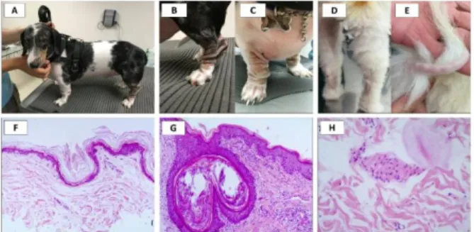
Hyperpigmentation and caudal alopecia were presented in hypothyroidism dogs (7 dogs, 87.5%). Epidermal scale was observed in all dogs. Clinical evidence of seborrhea (scales, crutis, rancid odor without pruritus) were founded in this group. The histopathological sections of the skin showed irregular acanthosis with areas of orthokeratosis and parakeratosis that ranges light of hair follicles forming keratin plugs (comedones). Vacuolization of the pilo-erector muscle was detected in 6 (75%) dogs of this group. Inflammatory infiltrate was distributed in the dermis, mainly in the superficial dermis, consisting mostly of mast cells (Figures 2, 4B and 4E).
Different degree of alopecia and hyperpigmentation were showed by hyperadrenocorticism dogs. Others clinical signs observed in this group were abdominal distention and striae. All dogs exhibited epidermal atrophy and hyperkeratois. In this animals an excessive trichilemmal keratinization, follicular dystrophy or follicular atrophy and sebaceous glands atrophy were increased. Calcinosis cuts (mineralization of dermal collagen) were presented in 4 dogs (50%) and inflammatory infiltrate was detected around it, consisting largely of mast cells (Figure 3, 4C and 4F).
Figure 2. Clinical and histopathological signs observed within the Hypothyroidism group. A – A dog with symmetric alopecia and poor haircoat quality. B and C – Same dog seen in A. Alopecia in the chest, lichenification with mild hyperpigmentation within the limbs. D and E – Another dog with symmetric alopecia of the distal limbs and tail. F – Photomicrograph of the skin showing normal epidermal thickness with orthokeratosis (HE, 100X). G – Photomicrograph of the skin showing mild irregular acanthosis and keratin plugs (comedones). Also, there is infiltration of inflammatory cells within the dermis and in this group sometimes showed arrector pili muscle degeneration, as seen in detail in H. (HE, 100X).
Figure 3. Clinical and histopathological signs observed within the Hyperadrenocorticism group. A and B – A dog with dorsal and ventral symmetric alopecia and lichenification with mild hyperpigmentation. C and D – Abdominal distension with evident striae and erythematous irregular plaques and crusts in the same dog. E and F – Dorsal poor hair coat quality with alopecia and crusts in all dorsal area. G – Photomicrograph of the skin showing atrophic epidermis with keratin plugs (comedones) and atrophic sebaceous glands, showed in detail in H (HE, 100 and 200X). I - Photomicrograph of the skin showing mild irregular acanthosis and diffuse mineralization with mild infiltration of inflammatory cells within (HE, 100X).
Mast cells were readily identified as fusiform, round, oval or pear-shaped cells with round or oval nuclei. When we compared the total mast cell count per high power field in dogs of all three groups there was no significant difference between the control group (4.04 ± 0.288) and the hypothyroidism (26.63 ± 10.780) and hyperadrenocorticism (23.38 ± 14.670) groups.
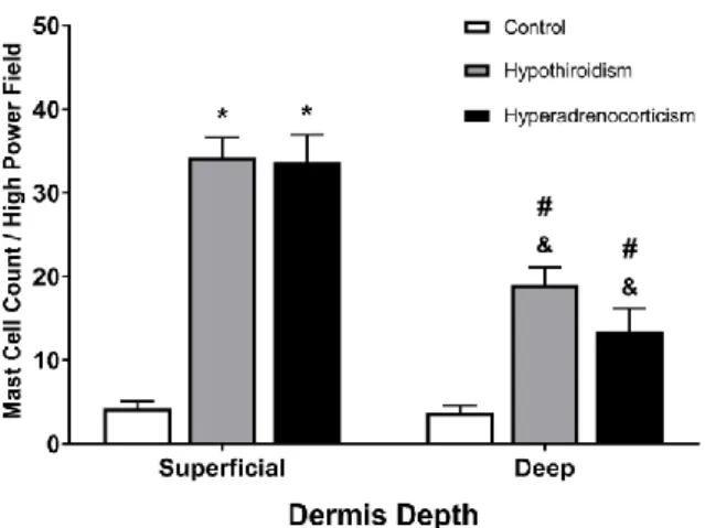
There was statistical difference in the mast cells mean count per high power field between the control group in superficial dermis (34.25 ± 2.390)
and hyperadrenocorticism group in superficial dermis (33.75 ± 3.220) as well as there was found in the control group in deep dermis (3.75 ± 0.840) and hypothyroidism group in deep dermis (13.50 ± 2.688). In dogs with hypothyroidism the superficial dermis had more mast cells than the deeper dermis with a mean mast cells count per high power field of (34 ± 10.39 and 18.75± 4.14), respectively, and this difference was significant (p < 0.05) (Figure 5). No mast cells were observed in the epidermis of any section.
Figure 4. Photomicrographs of lateral trunk skin from control dogs and dogs with hypothyroidism and hyperadrenocorticism, stained by Hematoxylin and Eosin and Toluidine Blue. Source: author's collection. Lateral trunk skin from control dogs (A) and dogs with hypothyroidism (B) and hyperadrenocorticism (C) stained by HE (100x). Letters D, E and F lateral trunk skin from control dogs and dogs with hypothyroidism and hyperadrenocorticism respectively stained by TB (400x). Nikon® Eclipse Ni with an attached Nikon® DS RI1camera.
Figure 5. Mast cell count per high power field in dermis depth (superficial and deep). * Significant mean difference between control group and hypothyroidism and hyperadrenocorticism group. & Difference between control group in deep dermis and hypothyroidism and hyperadrenocorticism in deep dermis group. # Difference between hypothyroidism in superficial dermis and deep dermis groups and the difference between hyperadrenocorticism in superficial dermis and deep dermis groups.
Discussion
The histopathological findings for describe hormonal dermatosis were compatible with findings in hypothyroidism and hyperadrenocorticism dogs of this study (Rojko et al., 1978; Müntener et al., 2012). These authors described sebaceous gland atrophy, epidermal melanosis, excessive tricolemal keratinization, orthokeratotic hyperkeratosis, follicular dilatation and atrophy and predominantly telogenic hair follicles.
To our knowledge, the present study was the first which mast cell have been visualized and quantified in skin of dogs with hypothyroidism and hyperadrenocorticism and the results comparing with the skin of healthy dogs.
Mast cells are well-characterized cells that house the majority of histamine and inflammatory factors causing allergic disease (Beaven, 2009) and contribute to a variety of physiological and pathological processes involving innate and adaptive immunity (Galli et al., 2011). Mast cells reside in many parts of the body, especially where foreign environmental agents try to invade the host, such as skin, airway, and gastrointestinal tract. Mast cells in the skin are predominantly localized in the upper dermis and close to blood vessels, skin appendages, and neurons (Sanchez-Pathan et al., 2008).
The elevated infiltration of mast cells in hypothyroid animals therefore agrees with an ongoing inflammatory process. There is evidence that mast cells can synthesize and store hormones among which are thyroid stimulating hormone (TSH) and the thyroid hormone (T3) (Csaba and Pállinger, 2009a, b; Thangam et al., 2018). Furthermore, evidence also indicates that mast cells express T3 receptors and that tissue mast cells population increased in hypothyroidism (Siebler et al., 2002), thus suggesting that the health or sick thyroid could condition mast cell hormone levels and/or that mast cells may represent an alternative source of packaged T3 locally deliverable (Thangam et al., 2018). The hypothalamus, throughout the release of the TSH, stimulates mast cells increasing the T3 content. T3 is co-stored with histamine in mast cell granules or is degraded to 3-iodothyronamine (T1AM) and/or 3-iodothyroacetic acid (TA1). T1AM and TA1 derived from circulation or produced inside mast cells trigger mast cell degranulation releasing T3 and histamine which mediates pain, itch and central
effects including neuroprotection/neuroinflamma-tion (Landucci et al., 2019).
A study realized by Rojko (1978) in hypothyroid dogs with seborrheic lesions (five of 28 dogs), acanthosis, parakeratosis and mild inflammatory, mast cells infiltration in the dermis had been present however changes in hair follicles were similar to those described for the classical form. The relationship between thyroid dysfunction and the pathogenesis of the seborrheic histologic lesion is still not known. In the present study we also observed a mast cells infiltration in the dermis of dogs with hypothyroidism.
In our work, we noted that mast cells were especially found in the superficial dermis of hypothyroidism dogs. Kurtdede et al. (2005) observed that the average of mast cell count per mm² in the superficial dermis and deep dermis are similar in the cheek area. Meanwhile, in areas such as pinna, thorax and thigh the mast cell count was found to be higher in the superficial dermis than in the deeper dermis. Other authors had obtained similar results in a diversity of different diseases and situations. In cats with diseases that do not affecting the skin the superficial dermis had more mast cells than the deeper dermis (Foster, 1994). Auxilia and Hill (2000) have studied the localization of the mast cells in the skin of dogs without skin disease and they showed the same results. Superficial dermis usually showed a higher count of mast cells than the deeper dermis in the skin of dogs. However, in rats they were more abundant in the deeper dermis than in the superficial dermis (Persinger et al., 1983; El Sayed and Dyson, 1993).
In dogs with hyperadrenocorticism the inflammation was present only in association with mineralization of dermal collagen (calcinosis cutis) (four dogs). In the present study, there was no
statistical difference between
hyperadrenocorticism and healthy dogs. Perhaps that happened because only half of the animals with hyperadrenocorticism enrolled had mineralization of dermal collagen (calcinosis cutis). This symptom has had an uncommon occurrence but has been a dermatological characteristic in dogs with hyperadrenocorticism (Frank, 2006; Doerr et al., 2013).
Conclusion
After analyzing our results, it was possible to conclude that animals with hypothyroidism produce greater amount of mast cells in the
superficial dermis than patients with hyperadrenocorticism and healthy animals.
The increase in the amount of these cells is justified by their role in the inflammation process, and in the production/ secretion of thyroid hormone. Thus, a large number of mast cells lead to the production of thyroid hormones in the dermis of dogs with hypothyroidism.
Conflicts of Interest
The authors declare that there is no conflict of interest
The Ethics Committee
The research Project was approved by the ethics committee of the Animal Use of Ceará State University, under number 11585871-7/10 – CEUA 1534880/2016.
Acknowledgments
We thank Comissão de Aperfeiçoamento de Pessoal do Nível Superior (CAPES), Conselho Nacional de Desenvolvimento Científico e Tecnológico (CNPq), and Fundação Cearense de Apoio ao Desenvolvimento Científico e Tecnológico (FUNCAP) for providing all resources necessary for this study development.
References
Auxilia, S.T.; Hill, P.B. Mast cell distribution, epidermal thickness and hair follicle density in normal ca-nine skin: possible explanations for the predilection sites of atopic dermatitis?
Veterinary Dermatology, 11: 247-254, 2000.
Azzarone, B.; Macieira-Coelho, A. Heterogeneity of the kinetics of proliferation within human skin fibroblastic cell populations. Journal of
Cell Science, 57: 177-187, 1982.
Beaven, M.A. Our perception of the mast cell from Paul Ehrlich to now. European Journal of
Immunology, 39:11–25, 2009.
Becker, A.B.; Chung, K.F.; McDonald, D.M.; Lazarus, S.C.; Frick, O.L.; Gold, W.M. Mast cell heterogeneity in dog skin. The Anatomical
Record, 213: 477-480, 1985.
Benyon, R.C. The human skin mast cell. Clinical
& Experimental Allergy, 19: 375-387, 1989.
Cowen, T.; Trigg, P.; Eady, R.A.J. Distribution of mast cells in human dermis: development of a mapping technique. British Journal of
Dermatology, 100: 635-641, 1979.
Csaba, G.; Pállinger, É. Is there a possibility of intrasystem regulation by hormones produced
by the immune cells? Experiments with extremely low concentrations of histamine.
Acta Physiologica Hungarica, 96: 369–374,
2009a.
Csaba, G.; Pállinger, É. Thyrotropic hormone (TSH) regulation of triiodothyronine (T3) concentration in immune cells. Inflammation
Research, 58: 151–154, 2009b.
Doerr, K.A.; Outerbridge, C.A.; White, S.D.; Kass, P.H.; Shiraki, R.; Lam, A.T.; Affolter, V.K. Calcinosis cutis in dogs: histopathological and clinical analysis of 46 cases. Veterinary
Dermatology, 24: 355–79, 2013.
Eady, R.A.J; Cowen, T.; Marshall, T.F.; Plummer, V.; Greaves, M.W. Mast cell population density, blood vessel density and histamine content in normal human skin. British Journal
of Dermatology, 100: 623-633, 1979.
El Sayed, S.; Dyson, M. Histochemical heterogeneity of mast cells in rat dermis.
Biotechnic & Histochemistry, 68: 326-332,
1993.
Emerson, J. L., Cross, R. F. The distribution of mast cells in normal canine skin. American
Journal of Veterinary Research, 26: 1379-82,
1965.
Foster, A.P. A Study of the number and distribution of cutaneous mast cells in cats with disease not affecting the skin. Veterinary Dermatology, 5(1): 17-20, 1994.
Frank, L.A. Comparative dermatology-canine endocrine dermatoses. Clinics in Dermatology, 24: 317-325, 2006.
Galli, S.J. Mast cells and basophils. Current
Opinion in Hematology, 7: 32-39, 2000.
Galli, S.J.; Borregaard, N.; Wynn, T.A. Phenotypic and functional plasticity of cells of innate immunity: macrophages, mast cells and neutrophils. Nature Immunology, 12: 1035– 44, 2011.
Gurish, M.F.; Austen, K.F. The diverse roles of mast cells. The Journal of Experimental
Medicine, 194:1-5, 2001.
Harper, R.A.; Grove, G. Human skin fibroblasts derived from papillary and reticular dermis: Differences in growth potential in vitro.
Science, 204: 526-528, 1979.
Kurtdede, N.; Yörük, M. Tavuk ve bıldırcın deri-sinde mast hücrelerinin morfolojik ve histometrik incelen-mesi. Veterinary Journal
of Ankara University, 42: 77-83, 1995.
Landucci, E.; Laurino, A.; Cinci, L.; Gencarelli, M.; Raimondi, L. Thyroid Hormone, Thyroid
Hormone Metabolites and Mast Cells: A Less Explored Issue. Frontiers in Cellular Neuroscience, 13: 1-7, 2019.
Marshall, J.S.; Ford, G.P.; Bell, E.B. Formalin sensitivity and differential staining of mast cells in human dermis. British Journal of
Dermatology, 117: 29-36, 1987.
Müntener, T.; Schuepbach-Regula, G.; Frank, L.; Rüfenacht, S.; Welle, M.M. Canine noninflammatory alopecia: a comprehensive evaluation of common and distinguishing histological characteristics. Veterinary Dermatology, 23(3): 206-244, 2012.
Persinger, M.A.; Lapage, P.; Simard, J.P.; Barker, G.H. Mast cell number in incisional wounds in rat skin as a function of distance, time and treatment. British Journal of Dermatology, 108: 179-187, 1983.
Rojko, J.L.; Hoover, E.A.; Martin, S.L. Histologic interpretation of cutaneous biopsies from dogs with dermatologic disorders. Veterinary
Pathology, 15: 579-589, 1978.
Sanchez-Patan, F.; Anchuelo, R.; Vara, E.; Garcia, C.; Saavedra, Y.; Vergara, P. Prophylaxis with ketotifen in rats with portal hypertension: involvement of mast cell and eicosanoids.
Hepatobiliary & Pancreatic Diseases International, 7:383–94, 2008.
Senol, M.; Fireman, P. Human skin mast cell: Current concepts. Turkish Journal Dermatology, 6(1-2): 56-62, 1997.
Siebler, T.; Robson, H.; Bromley, M.; Stevens, D.A.; Shalet, S.M.; Williams, G.R. Thyroid status affects number and localization of thyroid hormone receptor expressing mast cells in bone marrow. Bone, 30: 259–266, 2002.
Thangam, E.B.; Jemima, E.A.; Singh, H.; Baig, M.S.; Khan, M.; Mathias, C.B. The role of histamine and histamine receptors in mast cell-mediated allergy and inflammation: the hunt for new therapeutic targets. Frontiers in Immunology, 9:1873, 2018.
Walton, S.; DeSouza, E.J. Variation in mast cell numbers in psoriasis and lichen planus: comparisons with normal skin. Dermatologica, 166: 236-239, 1983.
Warton, A.; Papadimitriou, J.M.; Goldie, R.G.; Paterson, J.W. An ultrastructural study of mast cells in the alveolar wall of normal and asthmatic lung. The Australian Journal of
Experimental Biology and Medical Science,