Ana Georgina Gomes Alves
Paracoccidioides brasiliensis: Study of the sexual cycle
Julho de 2012 U M in ho | 2 01 2 An a G eo rg in a G om es A lv es P a ra c o c c id io id e s b ra si li e n si s: S tu d y o f th e s e x u a l c yc le
Ana Georgina Gomes Alves
Paracoccidioides brasiliensis: Study of the sexual cycle
Paracoccidioides brasiliensis: Estudo do ciclo sexual.
Dissertação de Mestrado
Mestrado em Genética Molecular
Trabalho efectuado sob a orientação do
Doutor Fernando José dos Santos Rodrigues
co-orientação da
Doutora Paula Gonçalves
e da
Doutora Maria João Sousa
ii Nome: Ana Georgina Gomes Alves
Endereço electrónico: ana_g_alves@hotmail.com Número do Bilhete de Identidade: 12981961
Título da dissertação:
Paracoccidioides brasiliensis: Study of the sexual cycle Paracoccidioides brasiliensis: Estudo do ciclo sexual.
Orientador:
Fernando José dos Santos Rodrigues Co-Orientadora:
Paula Gonçalves Co-Orientadora: Maria João Sousa Ano de conclusão: 2012
Designação do Ramo de Conhecimento do Mestrado: Genética Molecular
DE ACORDO COM A LEGISLAÇÃO EM VIGOR, NÃO É PERMITIDA A REPRODUÇÃO DE QUALQUER PARTE DESTA TESE/TRABALHO
Universidade do Minho, 31 de Julho de 2012
iii
The work presented in this thesis was done in the Laboratory of Microbiology, Microbiology and Infection Research Domain of the Life and Health Sciences Research Institute (ICVS), School of Health Sciences, University of Minho, Braga, Portugal (ICVS/3B’s – PT Government Associate Laboratory, Braga/Guimarães, Portugal). The financial support was given by the Fundação para a Ciência e a Tecnologia (PTDC/BIA-MIC/108309/2008).
v Agradecimentos
Após três longos anos, chega ao fim mais uma caminhada!! Vejo-o como o começo de um novo futuro…
E esse começo deve-se a todos os que me rodearam nesta caminhada, por vezes nada fácil.
Consegui terminar, mas confesso que não apenas graças a mim, mas graças a Eles, que estiveram sempre presentes.
A todos Eles, que me apoiaram e seguiram, tenho apenas duas palavras: Muito obrigada!! Ao Prof. Fernando Rodrigues, sem o qual não teria sido possível a realização desta tese e deste mestrado. Pelo incentivo que me deu para iniciar esta etapa e por me ter permitido realizá-la. Muito obrigada por todo o know-how científico, pela ajuda, pelo tempo e todos os conselhos pessoais e profissionais.
À Prof. Paula Gonçalves, por ter aceitado ser minha co-orientadora e por todo o apoio científico dedicado durante este percurso. Obrigada.
À Prof. Maria João Sousa, por ter aceitado ser minha co-orientadora, e pelo apoio demonstrado não só durante o mestrado, mas durante todo o percurso académico. Obrigada.
Aos meus AMIGOS de laboratório. Sim, passados quatro anos desde o meu primeiro dia no ICVS, é isso que sinto, que fiz muitas e boas amizades, com as quais sei que posso contar sempre.
Ao Mark… O que seria de mim sem ele durante este percurso? Muito obrigada por TUDO. Sem o seu apoio, ajuda e amizade, nada disto seria possível. Deu-me muitas vezes força para continuar. Apesar de eu ser uma vervelend, esteve sempre presente e ajudou-me imenso.
À Belém, à Xana, à Júlia, ao Agostinho, ao Nuno, à Ana Mesquita que sempre me apoiaram em tudo. Estiveram sempre lá para ajudar, fosse de manhã, de tarde ou à noite. À Jéssica e ao Pedro, por todas as conversas proibidas e por todas as pizzas encomendadas fora de horas. Muito obrigada pelo apoio durante esta etapa. Às vezes não é fácil, mas sempre souberam o que dizer e quando o dizer… Sim Pedro, é desta!!
Ao João, que se tornou um dos meus melhores amigos. Como costumo dizer, o meu irmão mais novo. Tantas noites, tantas horas de trabalho… Mas chega a um ponto que
vi
para sempre. E porque um percurso como este não é apenas trabalho… Obrigada por todo o apoio, em TUDO. Estarei sempre aqui…
Às minhas meninas, Dina, Carina, Sofia, Maria e Ana que apesar de fisicamente longe estão sempre bem pertinho de mim. Muito obrigada.
Aos Bomboémia. Pois é, muitas vezes o meu ponto de refúgio, o local de libertação de todo o stress acumulado!! Obrigada por todas as vezes que tocávamos a Zumba quando percebiam que não tinha sido um dia fácil. Obrigada por todas as festas, carinho, amizade e companheirismo. Não vos posso agradecer um a um, mas fica aqui registado que foram muito importantes neste percurso.
À Mariana, à Natália, à Nita, à Márcia, à Noni, à Espanhola, ao Alex, à Ju, à Raquel, à Ana Real, à Rute, que são, sem dúvida, a minha segunda família, e que foram os que mais sentiram este meu percurso. Desculpem cada café, viagem, cinema, jantar desmarcados. É também graças ao vosso apoio, amizade e companheirismo que esta caminhada terminou. Obrigada por fazerem parte da minha vida e por me deixarem fazer parte da vossa.
Ao Chico, que me deu o maior impulso final. Que tem estado sempre presente e que me apoia incondicionalmente com carinho, compreensão e muita paciência. Obrigada por teres aparecido na minha vida.
Last but not least, À minha família.
Modéstia à parte eu posso dizer que tenho “a melhor família do mundo”. Obrigada por estarem presentes em todos os momentos da minha vida.
Em particular aos meus pais e ao meu irmão que fizeram de mim o que sou hoje. Por todos os ensinamentos, por todo o apoio, compreensão, paciência que tiveram comigo ao longo destes anos. Terminei mais uma etapa da minha vida académica, graças a eles. Não há palavras que possam descrever o quão grata estou por fazerem parte de mim. Muito obrigada.
vii
Paracoccidioides brasiliensis: Study of the sexual cycle.
The thermodimorphic fungal pathogen Paracoccidioides brasiliensis is the etiological agent of paracoccidioidomycosis, one of the most prevalent systemic mycoses endemic in Latin America, occurring mainly in Brazil, Colombia and Venezuela. The morphological transformation of P. brasiliensis is characterized by the existence of two different morphological forms: a mycelium/conidial form that is present at environmental temperatures (below 25°C); and a multiple budding yeast form, present at temperatures of the mammalian host (37°C).
The sexual cycle in P. brasiliensis has not been observed in nature or laboratory conditions. Nevertheless, in the present study, we detected low expression levels of mating-related genes, such as α-pheromone, α and a-pheromone receptors (PREB and
PREA), and heterothallic mating loci (MAT1-1 and MAT1-2), in yeast and mycelial forms,
and verified that heterothallic strains of opposite mating-types are able to express α-pheromone, and both pheromone receptors. In order to further evaluate the functional activity of mating-related genes, particularly α-pheromone and its cognate receptor (PreB), we took advantage of the heterologous expression of these P. brasiliensis genes in the corresponding Saccharomyces cerevisiae null mutants. Through several functional tests, including cell cycle arrest and shmoo formation, we showed that S. cerevisiae strains heterologously expressing PREB respond to synthetic α-pheromone of P.
brasiliensis. In addition, mating ability of S. cerevisiae non-fertile strains was restored by
the expression of PREB or α-pheromone in the corresponding null mutants. In general, this study demonstrates novel evidences for the existence of a functional mating signaling system in P. brasiliensis.
ix
Paracoccidioides brasiliensis: Estudo do ciclo sexual.
O fungo patogénico termodimórfico Paracoccidioides brasiliensis é o agente etiológico da paracoccidioidomicose, uma das mais prevalentes micoses sistémicas, endémica da América Latina, ocorrendo principalmente no Brasil, Colômbia e Venezuela. A transformação morfológica de P. brasiliensis é caracterizada pela existência de duas formas distintas: a temperaturas ambientais (25°C) existe sob a forma de micélio/conídeo; e a temperaturas do hospedeiro (37°C) sob forma de levedura.
Apesar de ainda não ter sido observado a existência de ciclo sexual em P. brasiliensis, no presente estudo detetámos níveis de expressão de genes relacionados com a reprodução sexuada em diversos fungo, tais como, feromona-α, recetor da feromona α e a (PREB e
PREA) e ainda o MAT locus (MAT1-1 e MAT1-2). Verificámos que estirpes heterotálicas,
de tipos de acasalamento opostos, têm a capacidade de expressar a feromona-α e ambos os recetores. De forma a avaliar a atividade funcional de genes relacionados com a reprodução sexuada, particularmente a feromona-α e respetivo recetor (PreB), procedemos à expressão destes genes de P. brasiliensis em estirpes mutantes de
Saccharomyces cerevisiae. Através de vários testes funcionais, incluindo paragem de
ciclo celular e formação de shmoos, mostrámos que a estirpe de S. cerevisiae que expressa PREB tem a capacidade de responder à feromona-α sintética de P. brasiliensis. Para além disso, a capacidade de acasalamento de estirpes de S. cerevisiae não férteis foi restabelecida pela expressão heteróloga da feromona-α e PREB. Este estudo demonstra novas evidências para a existência de um sistema de sinalização de acasalamento funcional em P. brasiliensis.
xi
Table of contents
CHAPTER I - INTRODUCTION ... 1
1.1.Paracoccidioides brasiliensis 3 1.1.1. Fungal pathogens and thermal dimorphism ... 3
1.1.2. P. brasiliensis and paracoccidioidomycosis ... 3
1.1.3. Ecological habitats of P. brasiliensis isolates ... 5
1.1.4. Phylogeny, cryptic speciation and genome ploidy of P. brasiliensis ... 6
1.2.Sexual reproduction in fungi 8 1.2.1. General aspects of sexual reproduction in fungi ... 8
1.2.1.1. Genetics of the mating in Saccharomyces cerevisiae ... 9
1.2.1.2. Mating regulation in Candida albicans... 12
1.2.1.3. Mating in Aspergillus fumigatus ... 14
1.2.1.4. Mating in Histoplasma capsulatum ... 15
1.2.1.5. Mating in Cryptococcus neoformans ... 17
1.2.2. Biogenesis of the mating pheromones ... 19
1.2.3. Intracellular mating signaling pathway ... 21
1.2.4. Mating in Paracoccidioides brasiliensis ... 25
1.3.Aims 26 CHAPTER II - MATERIAL AND METHODS ... 27
2.1.Strains and culture conditions 29 2.2.Identification of MAT loci in P. brasiliensis strains 30 2.3.In silico identification of P. brasiliensis mating-related genes 30 2.4.Real-time-PCR analysis of mating gene expression in P. brasiliensis strains 31 2.5.Heterologous expression of P. brasiliensis mating-related genes in S. cerevisiae 32 2.5.1. Construction of a MF(α)1/2 double mutant strain of S. cerevisiae ... 32
2.5.2. Construction of heterologous expression plasmids ... 32
2.5.3. S. cerevisiae transformation ... 33
2.5.4. Genomic DNA extraction from S. cerevisiae ... 34
2.5.5. Halo assay ... 35
2.5.6. Shmoo assay ... 35
2.5.7. Cell cycle analysis by flow cytometry ... 35
2.5.8. Quantitative mating assays ... 36
2.6.Statistical analysis 37 CHAPTER III – RESULTS AND DISCUSSION ... 39
3.1.Mating-type analysis of P. brasiliensis strains 41 3.2.Pheromone response pathway in P. brasiliensis 42 3.2.1. Identification and characterization of P. brasiliensis α-pheromone ... 44
xii
mycelium 50
3.4.Heterologous expression of P. brasiliensis mating-related genes in S. cerevisiae 53
3.4.1. Halo assay ... 53
3.4.2. Shmoo assay... 55
3.4.3. Cell cycle arrest assay... 57
3.4.4. Mating assay ... 62
CHAPTER IV – CONCLUSIONS AND FUTURE PERSPECTIVES ... 65
REFERENCES ... 69
1
3
1.1. Paracoccidioides brasiliensis
1.1.1.
Fungal pathogens and thermal dimorphismWorldwide over a hundred thousand fungal species are known, though only a limited number are related to human disease [1]. This is thought to be due to the high temperature of the mammalian body, which is considered a barrier against infections by fungi, as most of them have a higher growth rate at environmental temperatures [2]. For several human pathogenic fungi, the abrupt temperature change between the environment (below 25°C) and human body (37°C) is characterized by a morphological transition between yeast and mycelium form, called thermodimorphism, being this ability considered an important virulence factor [1].
The morphological form that the fungus displays at environmental or host temperature can vary according to the fungal species (Figure 1). Important fungal pathogens that show such dimorphic behavior include Candida albicans, Sporothrix schenkii, Penicillium
marnefii, Coccidioides immitis and the closely related Histoplasma capsulatum, Blastomyces dermatitidis and Paracoccidioides brasiliensis [3-5].
1.1.2.
P. brasiliensis and paracoccidioidomycosisThe thermodimorphic fungal pathogen P. brasiliensis is the etiological agent of paracoccidioidomycosis (PCM), one of the most prevalent systemic mycoses endemic in Latin America, occurring mainly in Brazil, Colombia and Venezuela [6]. It is estimated that about 10 million people may be infected, though only 2% develop the disease [6, 7]. The morphological transformation of P. brasiliensis is reflected by the existence of two different morphological forms: a mycelium/conidial form that is present at environmental temperatures (below 25°C) ; and a multiple budding yeast form, present at temperatures of the mammalian host (37°C) (Figure 1) [1].
4
Figure 1 – Dimorphic fungi – Morphological forms at different temperatures of different fungus species [8].
It is believed that the infection starts by inhalation of airborne conidia derived from the mycelial form. When the conidia reach the epithelial cells of the lung, the morphological switch to the yeast form occurs and yeast cells start to spread into the blood system, leading to disseminated infection (Figure 2) [6, 9]. After the inhalation of conidia, the infected individuals can present two main clinical forms of PCM, the acute or sub-acute form (juvenile type) and the chronic form (adult type). The juvenile type of PCM develops within weeks or months after contact with the fungus and is more severe, leading to higher rates of mortality. In contrast, the adult type of PCM can be in latency for several years and accounts to more than 90% of the cases [10].
PCM occurs mainly in rural populations and is more common in males. The mechanisms underlying this gender susceptibility is thought to be related with hormonal regulation of
P. brasiliensis form switch. In vitro studies have shown that female hormones such as
oestrogens block the conidia- or mycelium-to-yeast transition, probably via a cytosolic steroid-binding protein [11, 12]. In vivo studies with mice corroborate these results, and revealed that 96h after conidia inoculation, the transition to yeast is accomplished in lungs of the male mice, but not in female mice [13]. On the other hand, Pinzan et al. have recently shown that the interference exerted by sexual hormones in the immune response against P. brasiliensis impact the differences in the clinical incidence and progression of PCM between males and females [13].
5
Figure 2 – Hypothetical biological cycle of P. brasiliensis (adapted from [14]). In this picture are highlighted some selective forces that may be involved in the supposed ecological niches in hosts and in saprobic environments.
1.1.3.
Ecological habitats of P. brasiliensis isolatesFor a long time, studies involving P. brasiliensis were made using clinical isolates, since the isolation of this pathogen from the environment is difficult to achieve [15]. In fact, PCM has a prolonged latency period, which together with the absence of outbreaks makes it difficult to determine the exact habitat of P. brasiliensis [14, 16]. The fungus has been repeatedly isolated from nine-banded armadillo (Dasypus novemcinctus) in Brazil, being present in 75-100% of animals captured in endemic PCM areas [15-17]. Despite this high incidence in the species D. novemcinctus, independent of age or gender, the animals do not show visible signs of PCM disease [17]. The fungus has been identified, by PCR in soil samples collected from armadillos burrow and faeces of naturally infected armadillos. However, until now it has not been possible to isolate P. brasiliensis from its saprobic form [17]. The maintenance of a parasitic and saprobic form could contribute to sexual reproduction, since P. brasiliensis was found in restricted and well defined areas, which increases the probability to encounter individuals of the same pathogenic fungal species [17]. Bagagli et al. [17] collected some data suggesting that P. brasiliensis strains, isolated from armadillos, exhibit a wider spectrum of genotypes when compared to clinical isolates, in part because the same animal can acquire multiple infections with distinct strains belonging to different genotypes.
6
1.1.4.
Phylogeny, cryptic speciation and genome ploidy of P.brasiliensis
In classic systematics, P. brasiliensis was included in the anamorphic phylum Deuteromycota and in Hyphomycetes class, due to the absence of a traceable sexual phase [1]. However, morphological and phylogenetic studies together with the development of molecular tools made it possible to place P. brasiliensis in the phylum Ascomycota, inside the order Onygenales, family Onygenaceae sensu lato [1, 17]. More recently, a new family was proposed (Ajellomycetaceae), distinct from Onygenaceae
sensu lato, which includes the genera Blastomyces, Histoplasma, Emmonsia, and Paracoccidioides [14, 17, 18].
Furthermore, Matute et. al [19], performed a study where through the analysis of eight regions from five nuclear coding genes, they found that P. brasiliensis is stratified in at least three distinct species: S1 (species 1 from Brazil, Argentina, Paraguay, Peru and Venezuela), PS2 (phylogenetic species 2 from Brazil and Venezuela) and PS3 (phylogenetic species from Colombia). Recently, Teixeira et al. [20] showed that 17 genotypically similar isolates, including strain Pb01, were distinct from the three cryptic species previously described and proposed the new “Pb01-like-cluster” as a new species (Paracoccicioides lutzii).
Both mycelia and yeast form of P. brasiliensis are characterized by a multinucleate nature, while conidia, the supposedly infectious form, comprise only a single nucleus [21]. The knowledge on genetic composition as well as on the mechanisms involved in dimorphism and virulence of P. brasiliensis is limited, which is partly due to the lack of a known teleomorphic (sexual) phase and few (cyto)genetic tools for this fungus [7, 21]. The genome characterization and chromosomal mapping of P. brasiliensis were previously accessed by distinct approaches [7, 22, 23]. Pulse field gel electrophoresis (PFGE), allowed to determine the size of the fungus genome, initially estimated to be approximately 23-31 Mb. PFGE analysis also revealed the presence of 4-5 chromosomes (2-10 Mb) [22, 23]. However, when these results were compared to the ones obtained by microfluorometry (45.7 to 60.9 Mb), suggested the possibility of the existence of haploid and diploid (or aneuploid) isolates of the fungus [21, 23]. Later on, Almeida et al. [21] evaluated ploidy and genome size of P. brasiliensis, by a flow cytometry (FCM) protocol [24]. They reported a genome size ranging from 26.3 ± 0.1 to 35.5 ± 0.2 Mb per uninucleated yeast cell. Concerning the ploidy of P. brasiliensis, they showed a ploidy
7
ratio between 1.0 and 1.1, which means that the analyzed isolates present a haploid, or at least aneuploid, DNA content. No association was detected between genome size/ploidy and the clinical-epidemiological features of the studied isolates; however, additional studies of a higher number of isolates are needed [21].
Total genome sequences of three P. brasiliensis strains (Pb01, Pb03, and Pb18) that were recently published by the Broad Institute of Harvard and MIT (http://www.broadinstitute.org/annotation/genome/paracoccidioides_brasiliensis/MultiHo me.html) indicated that the two P. brasiliensis genomes of Pb18 and Pb03 are similar in size (30.0 Mb and 29.1 Mb respectively), while the P. lutzii (Pb01-like) genome is nearly 3 Mb larger at 32.9 Mb [25].
8
1.2. Sexual reproduction in fungi
1.2.1.
General aspects of sexual reproduction in fungiIn eukaryotes it’s established that sexual reproduction allows for genetic recombination. The maintenance of the sexual cycle is essential to purge the genome of deleterious mutations, and for the generation of genetic variation, where the combination of beneficial genes in sexual offspring from two parents increases fitness in novel or changing ecological niches [26-28].
Although in some eukaryotes, including many pathogenic fungi, a sexual cycle was not yet established, many fungi are known to undergo sexual reproduction, and diverse patterns of sexual recombination occur throughout the five fungal phyla Ascomycota, Basidiomycota, Chytridiomycota, Mucoromycotina and Microsporidia [28]. Moreover, with the increasing number of completed fungal genome sequences it becomes clear that many of these fungi once thought asexual, actually have retained the genetic machinery for sexual reproduction [29].
It is well established that fungal sexual development is orchestrated by transcription factors encoded by a genetic locus called the mating-type or MAT locus, which determines the sex of the fungus [30]. Mating communication between two haploid cells is achieved by the production of small peptide pheromones from one cell, that are sensed by specific receptors on the surface of the opposite cell [31]. Fungi exhibit either of two mating patterns: self-fertile (homothallic) fungi in which sexual reproduction can occur between genetically identical cells without the need of a mating partner, or cells of one mating-type have the ability to undergo mating-type switch; or self-sterile (heterothallic) fungi in which mating occurs only when a cell encounters an opposite mating-type partner [30, 32, 33].
The fungal subkingdom Dikarya, encompassing the phyla Ascomycota and Basidiomycota, includes the most common human fungal pathogens, such as Candida
albicans, Aspergillus fumigatus and Cryptococcus neoformans [34]. An important
difference between these two phyla lies in the mating-type composition, as fungi belonging to the phylum Ascomycota have just two mating-types, while the ones that belong to the phylum Basidiomycota can have multiple mating-types [31]. Another important difference is the type of sexual reproduction structures produced by both phyla. The sexual progeny from Ascomycetes is characterized by the formation of spores
9
(ascospores) enclosed within an ascus. In the subphylum Pezizomycotina the ascocarp can be defined as cleistothecium, perithecium, apothecium and ascostroma, depending on the structure [28, 35]. In contrast to the ascomycetes, the sexual spores of basidiomycetes are exposed to the air on the surface of basidia [28]. The genetic details concerning sexual reproduction in these two phyla will be further discussed in the next sections.
1.2.1.1.Genetics of the mating in Saccharomyces cerevisiae
S. cerevisiae grows mostly as yeast and can exist either as a haploid or diploid cell,
though the predominant ploidy isolated from nature is diploid [28]. In the sexual cycle of
S. cerevisiae, there are haploid cells with two different mating-types, MATa and MATα,
which can mate to form diploid a/α cells (Figure 3) [28, 36]. Each haploid mating-type cell secretes a small mating peptide pheromone that signals the opposite cell for mating: a cells produce a-factor and α cells produce α-factor. Each type of pheromone is sensed by specific receptors on the opposite mating-type cell: a cells encode the α-receptor (STE2) and α cells encode the a-receptor (STE3). After pheromone recognition, the cells develop projections called shmoos, characterized by a polarized growth toward a mating partner, ultimately leading to cell fusion, followed by nuclear fusion (Figure 3). The efficiency of the cell fusion requires cell synchronization, which is achieved by a cell cycle arrest in the G1 phase [28, 37, 38].
After the mating process the resulting diploid cell loses the capacity to mate, as expression of haploid-specific genes necessary for sexual fusion, are repressed. These diploid cells however are capable to undergo meiosis and sporulation (Figure 3) [33, 39]. The expression of mating-specific genes in S. cerevisiae is regulated by three transcription factors encoded at the MAT locus. The MATa locus encodes a1, an HD2 class homeodomain transcription factor, while the MATα locus encodes α1, an alpha box transcription factor, and α2, an HD1 class homeodomain transcription factor [28, 39, 40]. The expression of a cell-specific genes is regulated only by MCM1, a transcription factor that belongs to the MADS box family. MCM1 alone binds to the P box of a-specific upstream regulatory sequences (URSs) and promotes the transcription of a-cell specific genes. In contrast, for the transcription of α cell-specific genes, the presence of α1 is necessary, which together with MCM1 binds to the PQ box of α-specific URSs. The α2 transcription factor binds to two α2 binding sites that flank the P box of a-specific URSs, repressing the transcription of a cell-specific genes (Figure 4) [39, 40].
10
Figure 3 – Mating in S. cerevisiae. Two haploid cells secrete pheromone, sensed by the opposite mating-type. The cells develop shmoos and the cell/nuclear fusion occurs, resulting in the production of diploid cells. While in the presence of nutrients the diploid cell divides by mitosis, in the absence of nutrients, cells will undergo meiosis, forming four haploid cells [41].
The diploid a/α cells formed during mating do not produce any kind of receptor or pheromone, since α2 in combination with a1 block the α1 expression and consequently
11
the expression of α cell-specific genes (Figure 4). The heterodimer a1-α2 also represses haploid-specific genes, which includes, among others, genes necessary for mating (such as STE4, STE5 and STE12), and RME1, a protein responsible for the inhibition of meiosis [31, 39, 40].
Figure 4 – Regulation of cell-type specific genes. Transcription factors encoded by each MAT locus act with MCM1 transcription factor to regulate the expression of asg (a specific genes/mRNAs), αsg (α specific genes/mRNAa), and hsg (haploid specific genes/mRNAss) [41].
Some strains of S. cerevisiae are homothallic, which means that a single haploid cell can switch mating-type and undergo a self-fertile sexual cycle. As referred above, the yeast cell type is determined by the expression of specific genes at the MAT locus; however in
S. cerevisiae there are also two silenced loci called HMLα and HMRa that contain
non-transcribed copies of the a or α genes, respectively. Recombination between MATa and
HMLα or MATα and HMRa results in a mating-type switch [28, 39, 40]. The homothallic
switching endonuclease (HO) is the enzyme responsible for this process, and is expressed only in haploid cells, particularly in mother cells [28, 36]. The gene conversion process is
12
initiated by the cleavage of a double-stranded DNA, promoted by the HO endonuclease [39, 42]. The repair of the DNA break allows the copy of information from HML or HMR to MAT, allowing mating between two strains initially of the same mating-type [39].
S. cerevisiae has been used as a model for the study of mating in other fungi, but recent
studies on human fungal pathogens showed novel mating paradigms that differ substantially from the S. cerevisiae model [29].
1.2.1.2. Mating regulation in Candida albicans
Candida species are the most common human fungal pathogens, with C. albicans being
responsible for about half of all Candida infections. C. albicans is a commensal organism in the gastrointestinal tract, oral and vaginal mucosa, and is present in about 70% of healthy individuals [43]. However, it is an opportunistic fungus, that has the ability to cause both local mucosa infections and systemic infections, in particular in immunocompromised individuals [44].
C. albicans belongs to the phylum Ascomycota, Saccharomycetaceae family [43], and in
this sense is closely related to S. cerevisiae. C. albicans exists naturally as a diploid yeast and for more than 100 years was considered an asexual organism [45]. Hull and Johnson [46], describe mating-type like (MTL) loci, homologous to the MAT loci present in the yeast S. cerevisiae. MTL loci encode transcriptional regulators, similar to those in S.
cerevisiae; a1, α1, α2, and an additional regulator a2, an HMG domain protein, also
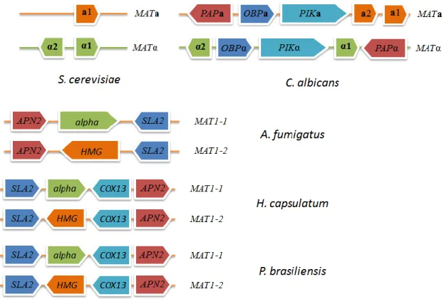
present in S. cerevisiae, but have no known function in sexual reproduction (Figure 5) [28, 47]. Both idiomorphs, MTLa and MTLα, also encodes three additional pairs of genes, poly(A) polymerase (PAPa and PAPα), phosphatidylinositol 4-kinase (PIKa and PIKα), and oxysterol binding protein (OBPa and OBPα), that are absent in S. cerevisiae and have no known function in mating [28, 34, 44]. In C. albicans, the transcription of α-specific genes is regulated by α1, and transcription of a-specific genes, contrary to what happens in S. cerevisiae, is activated by a2. The a1-α2 heterodimer inhibits the transcription of a- and α-specific genes for mating, and consequently, a/α diploids cells are unable to mate [28, 44].
Hull et al. [48] by the disruption of the genes of one MTL loci from a diploid a/α laboratory strain, SC5314, constructed both a- and α-type strains, demonstrating that C.
albicans is able to mate in vivo. Magee and Magee [49], constructed a/a and α/α
13
one copy of chromosome 5 (which contains MTL loci), and demonstrated that C. albicans can also mate in vitro, although at low efficiency rates.
Figure 5 – Organization of MAT locus from different Ascomycetes species.
Subsequent analyses by Miller and Johnson [50] showed that C. albicans switch from white to the opaque form to become mating competent. Opaque-form a and α cells mate about 106 times more efficiently than white-form cells [50]. White-form cells exhibit a round shape and dome-shaped white colonies, while opaque-form cells are elongated and form flatter and darker colonies [47, 51]. Besides the morphological differences between these two types of cells, they also show differences in virulence. White-form cells are more virulent in systemic infections while opaque-form cells are more efficient in colonization of the skin [52]. As described above, C. albicans is usually present in the gastrointestinal tract, oral and vaginal mucosa (at 37°C). However, opaque-form cells were also able to mate on the skin of baby mice, at a temperature of 31.5°C similar to the human skin. The fact that C. albicans opaque-form cells are transiently present on the human skin suggests that they can undergo mating and then either return to the same individual or be transmitted to a new host [28].
This phenotypic transition is negatively regulated by the a1/α2 heterodimer, which blocks diploid cells from mating, unless they undergo homozygosis to yield a/a or α/α cell types.
14
After the mating between two diploid strains, a tetraploid strain is produced. However, contrary to what is known in S. cerevisiae, instead of meiosis, the process responsible for generating 2N cells from 4N cells is a parasexual cycle, involving the loss of a chromosome [29, 45].
Microscopy analysis showed that, when opaque a and α cells are mixed, they communicate with each other through pheromone signaling. It was already reported the identification of a pheromone present only in α cells [44]. By homology to S. cerevisiae it was possible to identify in C. albicans Ste2 (α-pheromone receptor) and Ste3 (a-pheromone receptor) [44, 53]. Dignard et al. [54], were able to identify and characterize
MFa1, the gene responsible to encode a-pheromone.
It was shown that opaque a cells, when in contact with α peptide, form mating projections, and a quantitative polymerase chain reaction showed that, MFa and MFα were highly induced, contrary to what happens in S. cerevisiae [44, 55].
Alby et al. [55], reported that a cells produce both a and α pheromones, and when Bar1 (a protease present at a cells and responsible for the degradation of α pheromone) is absent, a cells produce and respond to α pheromone by an autocrine signaling. The a-a same-sex mating can also be stimulated when a minority of α cells is present as a source of α pheromone.
Taken together, these results indicated that C. albicans is also capable of homothallic mating between cells of the same mating-type, but that C. albicans strains do not undergo mating-type switching as is the case for S. cerevisiae.
1.2.1.3. Mating in Aspergillus fumigatus
The genus Aspergillus represents some of the most common fungi found in the environment, with only a small number of species related to human disease [34]. A.
fumigatus is an opportunistic human pathogen, that belongs to the phylum Ascomycota,
family Eurotiales [34]. It can cause potentially lethal invasive infections in immunocompromised individuals [56]. Like all the other Aspergillus species, it produces conidia that correspond to the infectious propagules, that when released into the atmosphere can be inhaled by the host [28, 29, 57].
In filamentous fungi, like A. fumigatus, the classification of the single mating-type locus is MAT1. Turgeon and Yoder [58] proposed a standard MAT nomenclature in which the two idiomorphs are designated by MAT1-1 and MAT1-2, highly divergent in sequence.
15
MAT1-1 and MAT1-2 are distinguished from each other by the presence a characteristic
alpha box motif, and a single ORF encoding a HMG gene, respectively (Figure 5).
Analysis of the completed A. fumigatus genome sequence showed the presence of a homologue of the MAT1-2 gene, which encodes a HMG-type protein showing high similarity with mating-type proteins of other filamentous ascomycetes that undergo sexual reproduction [57, 59]. However, MAT1-1 idiomorphs were not identified. Other mating-related genes were also found including a α pheromone precursor gene ppgA (highly homologues to Neurospora crassa and Sordaria macrospora pheromone precursor gene ppg1) as well as α and a-pheromone receptor genes homologous to those from S. cerevisiae. [56, 58] The genome organization of the MAT locus showed a conserved synteny with the MAT locus of A. nidulans, an Aspergillus species with a defined sexual cycle [60].
More recently, Paoletti et al. [56] demonstrated the existence of a MAT1-1 idiomorph. By performing a multiplex-PCR assay, they revealed the presence of MAT1-1 and MAT1-2 in similar proportions, on 290 clinical and environmental isolates. In this study, they demonstrate that the expression of pheromone receptors genes (PREA – a-pheromone receptor; and PREB – α-pheromone receptor) had no clear difference between the two mating isolates. In contrast, the expression levels of α-pheromone precursor were higher in the MAT1-1 isolates.
O’Gorman et al. [61] demonstrated that A. fumigatus can undergo sexual reproduction under certain conditions. They obtained mature cleistothecia after 6 months of incubation at 30°C in the dark.
The findings concerning the sexual reproduction in A. fumigatus are very important to understand the biology and evolution of the species, and gives new insights into the elucidation of sexual cycles of other fungi thought to be asexual.
1.2.1.4. Mating in Histoplasma capsulatum
Histoplasma capsulatum is a dimorphic fungus that produces airborne conidia or hyphal
fragments, which are reverted to the pathogenic yeast form at host temperature [62]. Once inhaled and hosted in the lung it can cause acute pulmonary disease and, in some cases, disseminated disease and death [63]. H. capsulatum can be found worldwide and its saprobic phase is associated with soil enriched with guano of bat and bird species,
16
specially of starlings (Stumus vulgaris) [62]. H. capsulatum belongs to the phylum Ascomycota, order Onygenales, and the new family Ajellomycetaceae.
Kwong-Chung [64] defined phenotypically, two mating-types of H. capsulatum, designated by (+) and (–). Mating between two opposite mating-type strains was confirmed, under laboratory conditions, using the mycelial form of fresh isolates [64]. An increase on pathogenicity of H. capsulatum seems to be related to the strains of the (–) mating-type. Samples from patients with acute pulmonary disease showed a higher frequency of strains with (–) mating-type, while samples from patients with disseminated disease and environmental samples had a balanced ratio of both mating-types [65, 66]. Recently, Bubnick et al. [63] correlated the differences of mating-types defined by phenotype with genotypic mating-types designation. Through comparative analysis of the
H. capsulatum genome sequence available at two websites and MAT loci already
identified in other Ascomycetes, they identified predicted MAT1-1 and MAT1-2 idiomorphs (Figure 5). The strains G217B and WU24, contained a region with high sequence identity and high similarity to the Aspergillus nidulans α1 region (found previously), designated by MAT1-1-1 [63, 67].
Since H. capsulatum is a heterothallic fungus and the strain G186AR did not show sequence similarity with A. nidulans α1 region, they performed a search for MAT1-2 idiomorph in its genome. In ascomycetes, the MAT idiomorphic regions are flanked by regions of homology (Figure 5). In this sense, performing a BLASTN analysis they found a region, flanked by regions with more than 95% of sequence similarity when compared to flanked regions from MAT1-1 sequence. The MAT1-2 idiomorph encode a predicted HMG DNA-binding domain. Based on this information they designated the mating-type of the strain G186AR as MAT1-2. T-3-1 is a known phenotypic (–) mating-type strain that contains the MAT1-2 idiomorph of the mating locus. By performing mating assays they could not obtain asci or ascospores, nevertheless they observed structures associated with the formation of the ascocarp [63, 64].
Laskowski et al. found homologous sequences of STE2 (α pheromone receptor) and STE3 (a pheromone receptor). They showed that strain G217B (MAT1-1) express STE2, but not
STE3, which could indicate that strains of MAT1-1 mating-type respond to α pheromone
[68]. They were also able to identify a putative pheromone gene, designated ppg1, but an a pheromone gene was not yet identified in H. capsulatum.
17
1.2.1.5. Mating in Cryptococcus neoformans
In basidiomycetes the sexual reproduction is also orchestrated by the MAT locus, however, while ascomycetes are bipolar (two mating-types), more than 50% of basidiomycetes are tetrapolar. Fungi with a tetrapolar system need two unlinked chromosomal loci, with differences in both alleles, to complete a sexual cycle. The MAT locus can be multiallelic, leading to thousands of different mating-types in some mushroom fungi [28, 69].
In basidiomycetes, after the recognition of two compatible mating-types and consequent cell fusion, the nuclear fusion is delayed. These fungi are able to have stable dikaryotic hyphae, which correspond to their predominant vegetative phase [28, 31].
Basidiomycota phyla include the specie Cryptococcus neoformans, which is one of the most common human fungal pathogen [34].
Cryptococcus neoformans is an opportunistic fungus, responsible for most cases of
meningoencephalitis in immunocompromised patients [69]. It can be found in pigeon guano and trees and the infection begins with the inhalation of airborne propagules that can colonize the host respiratory tract and after can spread to other body sites, specially the brain. However, the infection is usually asymptomatic and it can be either cleared or latent until the immune system of the host is compromised [70].
C. neoformans belongs to the phylum Basidiomycota, being more related to mushroom
fungi and Ustilago maydis (plant pathogen) than to ascomycetes, like S. cerevisiae and many other common fungal pathogens, including C. albicans, A. fumigatus, and H.
capsulatum [71]. It is a dimorphic haploid fungus that during the vegetative growth and
infection proliferates as budding yeast, and switches to hyphae during mating [72]. Sexual reproduction in C. neoformans is related to its virulence, since it is necessary for the production of spores, its infectious form [29]. The sexual cycle in C. neoformans is known for more than 30 years, and starts in response to nutritional limitations [64, 73]. It has a bipolar mating system, controlled by two opposite mating-types, MATa and MATα. Contrarily to ascomycetes, C. neoformans MAT locus encodes several mating-type specific genes, namely pheromone/pheromone receptor and homeodomain genes [69, 73, 74].
In C. neoformans three copies of the alpha pheromone gene were identified (MFα1,
MFα2, and MFα3) [74, 75]. The same study reported a MATα locus with a size of
18
reported that the MATα locus from C. neoformans encodes a putative pheromone receptor gene for MFa pheromone, designated by CPRα, which is located adjacent to STE12α. This gene showed a high degree of homology to pheromone receptors identified on other basidiomycetes fungi, such as Coprinus cinereus, Ustilago maydis, and Schizophyllum
commune. Subsequently, Chung et al. [76] isolated and characterized the CPRα gene,
showing that this gene plays an important role in mating. More recently, Chang et al. [77] identified and characterized a putative pheromone receptor from a MATa strain of C.
neoformans, designated by CPRa, which is located adjacent to STE12a. In order to
understand the role of CPRa, they performed mating assays, and the results suggested that
CPRa is involved in the mating pathway. They also demonstrated that the expression of at
least one of the pheromone receptors in either mating-type is required for C. neoformans undergo sexual reproduction [77].
MATa specific pheromones were identified and characterized by McClelland et al. [78].
The putative C. neoformans pheromone genes identified were designated by MFa1,
MFa2, and MFa3. The characterization of MFa1 showed that seems to be structurally
similar to the a-factor from S. cerevisiae and present conserved amino acid regions to
MFα1.
The MATα and MATa loci from C. neoformans, in addition to pheromone and pheromone receptors, also encode homeodomain (HD) factors that have been shown to be important in the control of cell identity and sexual development [79, 80]. Hull et al. [80] identified, in MATα cells, an α specific HD factor, named Sex inducer 1α (Sxi1α), and observed that the induction of the sexual development requires at least one a-specific component, however, they were not able to find it. More recently [79], using a combination of molecular genetics and bioinformatics, they identified an HD factor present at MAT locus from a cells, designated by Sxi2a. Moreover, the same study showed that the direct interaction between these two HD factors is essential to regulate the transcription of mating-related genes and to the induction of sexual reproduction.
Several studies correlated the mating-type with the virulence of C. neoformans. The
MATα idiomorph has a predominance of >95% in the C. neoformans population and it has
19
1.2.2.
Biogenesis of the mating pheromonesIn S. cerevisiae, the cells signal each other through the production of mating pheromones (a and α-factor) in order to stimulate the signal transduction pathway that leads to mating (detailed in the previous chapters). Before being secreted from the cell, both pheromones are generated from larger precursors, which are subsequently modified by posttranslational modifications and proteolysis to their mature form [82]. Despite their functional equivalence role in the cell response to mating, pheromones exhibit quite dissimilar biosynthesis.
S. cerevisiae mature α pheromone is a peptide of 13 amino acids
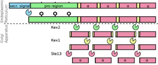
(WHWLGLKPGQPMY), generated from larger precursors encoded by two genes, MFα1 and MFα2, which contain tandem repeats of the mature α-pheromone [83-85]. In this sense, while MFα1 contains four α-factor repeats, encoding a precursor protein with 165 amino acids, MFα2 contains only two copies of the pheromone, encoding a protein with 120 amino acids [83, 84]. The precursor proteins are composed at the N-terminal, by a signal sequence followed by a proregion containing three recognition sites for N-glycosylation, and at the C-terminal, by the region containing the α-pheromone repeats separated from each other by connecting regions (Figure 6) [86-88]. The precursor proteins are translocated to the endoplasmic reticulum where the secretion signal is cleaved, producing the α pro-pheromone. The pro-region is extensively glycosylated and then, the α pro-pheromone is transported to the Golgi apparatus, where three proteolytic steps occur [86]. First, the Kex2 protease removes the proregion, by cleaving after the conserved lysine-arginine (KR) residues. Kex1 and Ste13 complete the maturation process by removing the connecting regions (Figure 6) [89, 90]. The mature α pheromone is then exported via the classical secretory pathway.
S. cerevisiae mature a pheromone is a peptide of 12 amino acids, which results from a
posttranslational maturation of a precursor protein encoded by two genes, MFa1 and
MFa2. The precursor proteins encoded by these two genes have 36 and 38 amino acids
length, respectively, containing a N-terminal extension, a conserved C-terminal CAAX motif (C =cysteine, A = aliphatic amino acid, X = any amino acid) and a single mature a-pheromone sequence. Following synthesis, the processing of the a proa-pheromone starts in the cytosol, by the farnesylation of a cysteine residue, in the CAAX motif, by Rma1 and Ram2 proteins. Subsequently, the action of Rce1 or Ste24 mediates the proteolytic
20
cleavage of AAX residues. Afterwards occurs the final step in modification of CAAX motif, which corresponds to the methylation of the cysteine, mediated by Ste14. The processing of the N-terminal starts with a proteolytic event, performed by Ste24, that removes the first seven residues, followed by the action of Axl1 that cleaves the others fourteen residues. The export of the mature a pheromone is mediated by the Ste6 transporter [87, 91, 92].
The presence of two types of pheromones, one prenylated and one unmodified, appears to be conserved amongst the ascomycetes. The presence of unmodified pheromones in several fungi is discussed below.
The mature α pheromone sequence from C. albicans has no conservation at the amino acid level compared with α pheromone from S. cerevisiae, however, the processing sites of the C. albicans pheromone are highly conserved. The conceptually translated protein of C. albicans contains a hydrophobic leader sequence, processing sites for the serine proteinase Kex2, and three copies of mature α pheromone. The processing of the α pheromone precursor requires the KEX2 gene to the C. albicans cells become mating competent [53].
The identification of a gene (ppgA) encoding a α pheromone precursor from A. fumigatus have shown similar flanking regions with S. cerevisiae precursor protein. Each one of the two copies of the mature α pheromone has at C-terminal a processing site for the Kex2 protease, the KR motif [57]. These repeats are preceded by the motif XP, which is a substrate for Ste13 in S. cerevisiae. At the N-terminal is possible to observe a hydrophobic signal sequence [57]. In addition, the mature nonapeptide pheromone (WCHLPGQGC) is highly similar to other filamentous ascomycetes pheromones [57]. In fact, the presence of several conserved amino acids found in fungal α pheromones overlaps the difficulties related to their small size and dissimilar sequence.
Analyzing the flanking regions of α pheromone locus in A. fumigatus it was possible to identify, by genome synteny, a putative α pheromone in H. capsulatum. The α pheromone sequence from H. capsulatum also show common features to known α pheromones from other fungi [93].
21
Figure 6 – Processing of S. cerevisiae α mating pheromone. The α propheromone is produced after the cleavage of the secretion signal. After the glycosylation, the α propheromone is transported from the endoplasmic reticulum to the golgi apparatus where three proteolytic steps leads to the production of the
mature α factor [87].
1.2.3.
Intracellular mating signaling pathwayThe pheromones produced by each mating-type are recognized by one of two specific G protein-coupled transmembrane receptors (GPCRs) – named Ste2 and Ste3 in S.
cerevisiae - that lead to the activation of a signal transduction cascade that includes a
MAPK pathway (Figure 7) [31, 37, 87]. In S. cerevisiae, this induction promotes a dissociation of the three subunits of G-protein, Gα(Gpa1), Gβ(Ste4) and Gγ(Ste18) [87].
While Gα subunit is responsible for the exchange of GTP for GDP, the Gβγ dimer recruits
Ste5 to the plasma membrane, promoting the activation of the MAP kinase cascade and mediating all the physiological responses induced by the pheromone-receptor interaction [40]. Ste5 is a scaffolding protein that has no catalytic domains and that supports Ste11, Ste7, and Fus3 [34, 40]. Ste20, a p21-activated protein kinase, is responsible for the direct phosphorylation and activation of scaffold-bound Ste11. This activation is also assisted by Ste50 that acts as an adaptor of Ste20 to the effector Ste11 [94]. The activated Ste11, a MEK kinase, phosphorylates and activates Ste7, a MAP kinase kinase, which is responsible for the phosphorylation and activation of Fus3, a MAP kinase (MAPK) (Figure 7).
Fus3 has two main roles: activate the expression of mating-related genes and promote cell cycle arrest. Accumulation in the nucleus leads to the inactivation of Dig1 and Dig2,
22
negative regulators of Ste12, a transcription factor that regulates the expression of mating-related genes, by binding the MCM1 to a and α gene-specific promoters [31, 34, 40, 87]. Fus3 also phosphorylate Far1, which has been reported to mediate cell cycle arrest in response to pheromone [95].
MAP kinase modules play a very important role in intracellular signal transduction pathways and are very conserved among diverse eukaryotes, including fungi [96]. Among these conserved MAP kinase pathways is the one involved in the mating response.
Even before the observation of a sexual cycle in C. albicans [46], some studies concerning the conserved mating signaling pathway cascade were performed in order to identify orthologs genes to S. cerevisiae mating pathway. A putative heterotrimeric G-protein gene, CAG1, with high homology to GPA1 from S. cerevisiae was identified and characterized [97]. They performed a mating assay, and concluded that CAG1 gene complemented the mating defects of gpa1 S. cerevisiae mutant.
Interestingly C. albicans encodes two homologous genes with sequence identity to S.
cerevisiae FUS3 and KSS1 (Cek2 and Cek1, respectively). CEK2 gene was able to
complement S. cerevisiae fus3/kss1 mutant, while the CEK1 gene was no able to do so. Therefore, it was concluded that CEK2 is a homolog of FUS3 from S. cerevisiae [98, 99]. The identification of a STE5 homolog (CST5) and its relation with pheromone response were recently published [100, 101].
Clark et al. [102] found a gene, HST7, which encodes a structurally similar protein to the
S. cerevisiae MAP kinase kinase Ste7. Performing mating assays, they observed that Hst7
can efficiently complement the ∆STE7 mutation. A subsequent study showed that C.
albicans CST20 gene is highly identical to the S. cerevisiae STE20 [103]. As it was
observed for CAG1 and HST7, CST20 had also the ability to fully complement the mating defect of S. cerevisiae ∆STE20. The same results were obtained to CPH1 gene from C.
albicans, homologous to STE12 from S. cerevisiae [104]. The identification of FAR1 in C. albicans showed that C. albicans FAR1 plays a central role in the pheromone response
[105, 106].
More recently, a study performed by Chen et al. [107] showed that the levels in which Cst20, Hst7, Cek1, Cek2 and Cph1 are required for mating in C. albicans are parallel to that of their homologues in the intracellular signaling pathway of S. cerevisiae (Figure 7). These findings means that C. albicans requires the same MAPK pathway as S. cerevisiae to respond to mating pheromones.
23
Figure 7 – Intracellular signaling pathway. (A, B) Pheromone response pathway in S. cerevisiae (adapted from [34]). (C) Homologous genes of the pheromone response pathway in C. albicans. Each of these genes has been functionally characterized for their role in mating. (D) Homologous genes of the pheromone response pathway in A. fumigatus. Contrarily to C. albicans, the functions in sexual reproduction of most of the genes involved in the pheromone response pathway identified in A. fumigatus were not confirmed. (E) Homologous genes of the pheromone response pathway in P. brasiliensis obtained by bioinformatics analysis. N.A. – not annotated.
Although there are some studies concerning the identification of genes involved in the intracellular signaling pathways from A. fumigatus, their functions in sexual reproduction are not clear. However, genomic analysis performed in A. fumigatus have shown genes homologous to S. cerevisiae and Aspergillus nidulans genes involved in the pheromone responsive pathway.
Liebmann et al. identified a gene encoding a G-protein α subunit, designated by GpaA [108]. The GPAA gene sequence is 98% identical to A. nidulans FADA [108]. The other two heterotrimeric G-protein components (SfaD [Gβ subunit] and GpgA [Gγ subunit]), a Ste20 homolog, SteC (Ste11 homolog), a Ste7 homolog, MpkB (Fus3 homolog), SteA (Ste12 homolog) were identified in A. fumigatus (Figure 7) and show high levels of homology with A. nidulans and S. cerevisiae orthologs [60].
During the sexual development of A. nidulans the expression levels of the subunits of the heterotrimeric G-protein FadA, SfaD, GpgA, SteC, Ste7 equivalent, MpkB and a protein regulator (Ste50 equivalent) were increased [109].
24
In this sense it can be predicted that some of these genes, important for sexual reproduction in A. nidulans, also plays a role in sexual reproduction and pheromone responses of A. fumigatus.
The discovery of C. neoformans sexual cycle led to an increase effort in the identification of regulatory molecules and mating inducing conditions. The identification of C.
neoformans MAT locus showed that it harbors more than twenty genes and that many of
them are involved in mating [69].
The GPCRs and their cognate heterotrimeric G proteins (Gpa1, Ste4 and Ste18 in S.
cerevisiae) are responsible to respond to a panoply of extracelular stimuli, including
pheromones. A study reported that as a consequence to the cell fate choice between budding yeast growth and asexual or sexual filamentous growth, C. neoformans have two Gα protein subunits involved in sexual development [110]. Both Gα subunits, designated by Gpa2 and Gpa3, were identified in the genome sequence and share homology with S.
cerevisiae Gpa1. They have shown that the principal role of activated form of Gpa3 is to
inhibit mating in environments in which there is no mating partner present, and the active form of Gpa2 contributes to the pheromone response that leads to mating [110]. The identified putative STE18 homolog gene, GPG2, in C. neoformans [111], is a γ subunit essential for pheromone signaling [110]. A Gβ subunit, Gpb1, was identified and is identical to S. cerevisiae Ste4. Wang et al. demonstrated that Gpb1 plays an important role in mating by activating the MAP kinase cascade that leads to conjugation tube formation in both MATa and MATα cells [112].
Nichols et al. found that Ste20a and Ste20α genes, which are located at the MATa and
MATα locus, respectively, play a role in mating since ste20 mutants are unable to undergo
sexual reproduction [113, 114]. It has been already shown that MATα cells are more virulent than MATa cells and a study showed that a clinical isolate deleted for STE20α is less virulent in animal models when compared to wild type [114].
A S. cerevisiae STE11 homolog gene was identified in the MATα and MATa locus, and C.
neoformans ste11α mutants were sterile, as is the case in S. cerevisiae [115].
By genomic sequence analysis it was possible to identify CPK1 and STE7 genes. Cpk1 revealed a significant identity with Fus3 (52%) and Kss1 (51%) of S. cerevisiae and Cek1 (56%) of C. albicans. Ste7 revealed a significant identity with Ste7 (39%) of S. cerevisiae and Hst7 (37%) of C. albicans. Contrary to other genes involved in the intracellular pheromone response (STE20a/α, STE11a/α, and STE12a/α), CPK1 and STE7 genes are
25
not mating-type specific, which means that both are present in MATa and MATα strains. In addition, CPK1 and STE7 gene are not contained within the MAT locus [116].
Molecular analysis to the hyphae production in MATα C. neoformans allowed the identification of a gene, designated STE12α, homolog to STE12 of S. cerevisiae and present only in MATα strains [117]. A STE12 specific of MATa strains, named STE12a, was also identified [118]. Both genes are related to the virulence of C. neoformans [117, 118].
Concerning P. brasiliensis mating intracellular pathway, recent genome annotations of three Paracoccidioides isolates showed the presence of conserved mating and meiosis specific genes [25]. However, no tests were performed to confirm the functional homology (Figure 7).
1.2.4.
Mating in Paracoccidioides brasiliensisThe sexual cycle in P. brasiliensis until now has not been described. However, there are some studies that show the presence of MAT loci in P. brasiliensis. Li et al. [119], identified two MAT idiomorphs, 1, which contains an α domain gene, and
MAT1-2, which contains an HMG domain (Figure 5). Torres et al. [120], identified the
mating-type idiomorphs in 71 P. brasiliensis isolates from various sources, and explored the basal expression of MAT gene in some strains in yeast/mycelial form, and found that their expression is low.
Taking advantage of GenBank database and BLAST tools, the sequence analyzes showed that MAT1-1 and MAT1-2 idiomorphs of P. brasiliensis show a high homology to homologous genes from Histoplasma capsulatum [120]. Synteny analysis also revealed common genome features of the MAT locus of H. capsulatum and P. brasiliensis, where
MAT1-1 and MAT1-2 are tightly linked with SLA2, COX13, and APN2 genes on both
species (Figure 5) [119].
Torres et al. performed mating assays with isolates from different mating-types, but they were not able to demonstrate in vitro mating [120]. However, equivalent distribution of the two mating-types in P. brasiliensis population [120], the presence of mating-related genes on P. brasiliensis genome [119] and the fact that some species phylogenetic related
26
(e.g. H. capsulatum) have a defined sexual cycle [28], lead us to consider that maybe P.
brasiliensis also has the ability to undergo sexual reproduction.
Having in mind all these subjects, the identification and knowledge concerning the MAT locus in pathogenic fungi and evolutionary studies are of main importance, not only to understand their pathogenicity but also to elucidate both the ancestral and evolving organization of mating systems [26].
1.3. Aims
The identification of sexual competence in the Paracoccidioides genus is an important issue for understanding the ecology and evolution of this fungus. Although the P.
brasiliensis genome encodes heterothallic mating loci and gene homologues of all
mating-signaling pathway components, as referred before, there has been no confirmation for the actual existence of a sexual cycle in this fungus. As it has been shown, mating in most fungi is regulated via the pheromone signaling MAP kinase pathway. We intend to characterize the functionality and activity of the mating pathway components in P.
brasiliensis through the:
i) Identification of a α-pheromone encoding gene;
ii) Evaluation of the basal expression levels of mating-related genes in P.
brasiliensis isolates of both mating-types in the yeast and mycelial forms;
iii) Functional complementation of Saccharomyces cerevisiae null mutants by the heterologous expression P. brasiliensis α-pheromone and its respective receptor (PreB).
Using these approaches we hope within this project to provide molecular evidence for the presence of a functional mating system in the Paracoccidioides genus. Such data will serve as a basis for further studies aimed at unraveling the basic biological and evolutionary aspects of sexual reproduction mechanisms in P. brasiliensis.
27
29
2.1. Strains and culture conditions
Strains of P. brasiliensis and S. cerevisiae used in this study are listed in Table 1.
For maintenance, S. cerevisiae strains were grown at 30°C on rich medium YEPD agar plates (0.5% [w/v] yeast extract, 1% [w/v] peptone, 2% [w/v] glucose, 2% [w/v] agar) or minimal medium dropout YNB agar plates (6,7% [w/v] Yeast Nitrogen Base without amino acids, 2% [w/v] glucose, 2% [w/v] agar), supplemented to meet auxotrophic requirements. For experimental procedures, S. cerevisiae strains were grown in YEPD or YNB broth at 26°C and 150rpm.
Table 1 – Strains used in this study.
a
– Plasmids listed in Table 2.
P. brasiliensis yeast strains were maintained at 37°C by periodic subculturing on brain
heart infusion (BHI) solid media (supplemented with 1% [w/v] glucose, 1.6% [w/v] agar). For subsequent analysis, P. brasiliensis strains were grown as yeast cells in BHI broth at
Organism Strain Genotypea Source
S. cerevisiae
BY4741 Wild type MATa EUROSCARF
BY4742 Wild type MATα EUROSCARF
BY4741 ∆STE2
MATa STE2::kanMX4 EUROSCARF
BY4742 ∆MF(α)1
MATα MF(α)1::kanMX4 EUROSCARF
BY4742 ∆MF(α)2
MATα MF(α)2::kanMX4 EUROSCARF
BY4741 ∆GPA1
MATa GPA1::kanMX4 EUROSCARF
BY4741 ∆STE4
MATa STE4::kanMX4 EUROSCARF
AGScα MATα MF(α)1::kanMX4;MF(α)2::hph This study
AGLPbα MATα MF(α)1::kanMX4;MF(α)2::hph pLPbα This study
AGMPbα MATα MF(α)1::kanMX4;MF(α)2::hph pMPbα This study
AGLPreB MATa STE2::kanMX4 pLPreB This study
AGLPbGpa1 MATa GPA1::kanMX4 pLPbGpa1 This study
AGLPbSte4 MATa STE4::kanMX4 pLPbSte4 This study
P.
brasiliensis
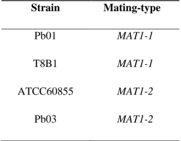
Pb01 MAT1-1 Chronic PCM [20]
T8B1 MAT1-1 Armadillo [121]
ATCC60855 MAT1-2 Chronic PCM [122]
![Figure 1 – Dimorphic fungi – Morphological forms at different temperatures of different fungus species [8]](https://thumb-eu.123doks.com/thumbv2/123dok_br/17910249.849532/17.892.123.757.101.440/figure-dimorphic-morphological-different-temperatures-different-fungus-species.webp)
![Figure 2 – Hypothetical biological cycle of P. brasiliensis (adapted from [14])](https://thumb-eu.123doks.com/thumbv2/123dok_br/17910249.849532/18.892.148.748.108.326/figure-hypothetical-biological-cycle-p-brasiliensis-adapted.webp)


![Figure 7 – Intracellular signaling pathway. (A, B) Pheromone response pathway in S. cerevisiae (adapted from [34])](https://thumb-eu.123doks.com/thumbv2/123dok_br/17910249.849532/36.892.153.771.112.431/figure-intracellular-signaling-pathway-pheromone-response-pathway-cerevisiae.webp)