Review
0103 - 5053 $6.00+0.00*e-mail: [email protected]
Biosensors Based on Gold Nanostructures
Marcio Vidotti,*,a Rafaela F. Carvalhal,b Renata K. Mendes,b Danielle C. M. Ferreirab
and Lauro T. Kubotab
aDepartamento de Química, Universidade Federal do Paraná,
CP 19081, 81531-980 Curitiba-PR, Brazil
bInstituto de Química and Instituto Nacional de Ciência e Tecnologia de Bioanalítica,
Universidade Estadual de Campinas, CP 6154, 13083-970, Campinas-SP, Brazil
O presente trabalho de revisão aborda os mais recentes avanços na tecnologia de biossensores alcançados através da montagem de biomoléculas associada com nanopartículas de ouro na construção de dispositivos analíticos. Esta revisão está dividida de acordo com a biomolécula
empregada no desenvolvimento de biossensores: (i) compostos imunológicos; (ii) DNA/RNA
funcionais; e (iii) enzimas e proteínas Heme. A im de facilitar a compreensão, cada seção foi
subdividida de acordo com o modo de transdução. Os imunossensores contendo nanopartículas de ouro têm uma ampla gama de aplicações nos campos alimentício, ambiental, farmacêutico, químico e de diagnósticos clínicos. As nanopartículas foram empregadas para melhorar o sinal analítico ou a imobilização dos imunocompostos. Em outra seção, os biossensores DNA/
RNA empregando nanoestruturas de ouro como labels e biossensores label-free associados
a nanoestruturas de ouro como transdutores foram sistematicamente relatados para a rápida identiicação de patógenos, espécies de interesse ambiental e diagnóstico clínico. A inclusão de nanopartículas de ouro em eletrodos modiicados aumenta a transferência de elétrons entre o transdutor e a biomolécula proporcionando um melhor desempenho quando enzimas e proteínas redox heme são usados. Biossensores para a detecção e quantiicação de glicose e peróxido de hidrogênio foram também discutidos.
The present review discusses the latest advances in biosensor technology achieved by the assembly of biomolecules associated with gold nanoparticles in analytical devices. This review is divided in sections according to the biomolecule employed in the biosensor development:
(i) immunocompounds; (ii) DNA/RNA and functional DNA/RNA; and (iii) enzymes and Heme
proteins. In order to facilitate the comprehension each section was subdivided according to the transduction mode. Gold nanoparticles based immunosensors have a wide range of applications in food, environmental, pharmaceutical, chemistry and clinical diagnostics. The nanoparticles were employed to improve whether the analytical signal or the immunocompounds immobilization. In another section, biosensors based on DNA/RNA biomolecules employing gold nanostructures as labels and label-free funtional DNA/RNA biosensors associated to gold nanostructures as tranducers were systematically reported for rapid identiication of pathogens, species of environmental interest and clinical diagnostics, respectively. The inclusion of gold nanoparticles in modiied electrodes itself enhances the electron transfer between the transducer and biomolecules leading to improved bioanalytical devices when redox enzymes and heme proteins are used. Biosensors for the detection and quantiication of glucose and hydrogen peroxide are discussed as well.
Keywords:gold nanoparticles, biosensors, bioanalytical devices, nanotechnology, biomolecules
1. Introduction
In the past few years an intensive effort has been done in the field of analytical science aiming the
Biosensors Based on Gold Nanostructures J. Braz. Chem. Soc. 4
properties, such as the blue-shift presented by some semiconductors nanoparticles.1-3 The second feature is
due to the huge relationship between surface area and volume,4-6 being more interesting for electroanalytical
purposes. Among the nanomaterials, gold has a special role. Many synthetic procedures can be found in the literature in order to control the size, monodispersion, morphology and surface chemistry of gold nanoparticles (AuNPs). The easy modiication of gold surface by thiol ended molecules makes then suitable for many different biological assemblies.7-9
Nanostructures present several advantages in analytical sciences when used as transducers or as a component of the recognition layer in a macrosized sensing device. In the irst case, the intrinsic properties of AuNPs, when used as transducers, several improvements are achieved such as mass transport, more availability of reactional sites, aggregation/dispersion optical effect and the increase of optical signal due to localized surface plasmon resonant properties of such added nanoparticles can be explored. In the later case, the improvement of the biosensor response can be achieved by the increase of the area/ volume relationship that increases the number of attached biocomponent in the sensing surface.
As mentioned above, one interesting point concerning AuNPs is related to the well known surface plasmon band (SPB), which consists in a broad absorption band in the visible region around 520 nm.9-12 This band is due to
collective oscillations of the electrons at the conduction band on the nanoparticle surface, which provides considerable information of the band structure in metals. This property has been a subject of many studies of optical spectroscopy properties.13
Nanoparticles have high surface areas and unique physical-chemical properties that can be easily tuned, making them ideal candidates for developing biosensing devices.14-16 AuNPs obtained from commercial sources or
conveniently produced in laboratories have attracted much attention in biological studies owing to their low toxicity, biocompatibility and unique optical properties. Biological tests measuring the presence or activity of selected analytes become quicker, more sensitive and lexible when nanoscale particles are put together, with numerous advantages over more traditional procedures.
Accordingly to earlier definitions, the biological recognition element of a biosensor ought to be of biological origin as an enzyme, an antibody, an antigen, a cell, a tissue, a DNA (deoxyribonucleic acid). However, not all recognition agents in novel devices are of biological origin, but synthetically produced elements such as crown ethers, cryptands, calixarenes and molecularly
imprinted polymers. The IUPAC (International Union of Pure and Applied Chemistry) had recommended that “the biological recognition element may be based on a chemical reaction catalysed by, or on equilibrium with, macromolecules that have been isolated, engineered or are present in their original biological environment”.17,18
The main objective on the utilization of biomolecules lies on the high selectivity for the analyte, where higher sensitivities can be reached by the development of new and effective transducers. In this sense, the utilization of nanostructured materials brought a new and exciting perspective to analytical sciences.
In present review, some of the latest advances in recognizing plataforms modification by using gold nanostructures and biomolecules assemblies are shown aiming the biosensor development. In Scheme 1 the organization of the present paper is shown: the biological detection element was subdivided in four classes such as redox proteins, immunocompounds, DNA/RNA-based (RNA: ribonucleic acid) molecules and other biomolecules. Each section is going to be organized according to the transduction element that was employed to prepare the biosensor. At Scheme 1 is summarized the effects and the role of AuNPs in the biosensor performance.
2. Immunosensors Based on AuNPs
The principle of the immunosensor takes into account the specific reaction between antibody and antigen performed on appropriate transducers. Immunosensors have a wide range of applications in food, environment, pharmaceutical, chemistry and clinical diagnostics, and one of the most important points relies on the appropriate choice of the immobilization method.19 In recent years, various
nanomaterials with physical and chemical properties have been applied to achieve improved immobilization of immunocompounds. The use of AuNPs on immunosensor development has recently received much attention20-24 being
the electrochemical, piezoelectric and optical the most studied transducers.
1.1. Optical
AuNPs have been used as a platform for the biomolecule immobilization, but their interactions with light makes possible to employ them to analytical purposes. The optical properties of nanoparticles changes according to their size and shape leading to different absorption bands. For example, gold nanorods have two principal plasmon absorption bands that are observed in the visible region of the electromagnetic spectra, these bands are related to the orientation of the rods. This characteristic was employed to the determination of human immunoglobulin G (IgG), which is used as a protein model by Wang et al.25 The authors have detected IgG by using silica-coated gold nanorods deposited on poly(4-vinylpyridine) (PVP) ilm on quartz substrates. The subsequent covalent bioconjugation of amino-functionalized gold nanorods-silica ilms with goat anti-human IgG is employed for the optical detection of antigen once there is a gradual absorbance change as the antigen concentration increases. The immunosensor demonstrated good sensitivity with color changes being observable by the naked eye.
Another work involving the human IgG optical detection employs the protein A, which is a cell wall component of Staphylococcal aureus that binds speciically to the Fc portion of IgG (anti-protein A) from many mammals.26 For
optical immunosensor construction, avidin was added in the biotin modiied cuvette, and then injected AuNPs, followed by the injection of protein A for oriented immobilization of human IgG on the cuvette surface of the resonant mirror. Avidin, in the pH used, is positively charged allowing the electrostatic binding with negatively charged AuNPs. The method shows good reproducibility with a detection limit of 8.7 µg mL-1.
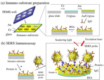
Using the speciicity binding between protein A and IgG, Lin et al.27 developed an immunosensor based on
AuNPs and employing surface-enhanced Raman scattering (SERS) technique. The preparation of immunoassay substrate was based on three steps, as illustrated in Figure 1. The elastomer polydimethylsiloxane (PDMS) ilm was formed on silicon wafer to create holes templates. After that, the PDMS cover and a glass slide with gold ilm were aligned and bonded together. In the last step, the IgG solution was pipetted onto each PDMS-surrounded gold well. For SERS assay, AuNPs were labeled with 5,5’-dithiobis(2-nitrobenzoic acid), which is a Raman-active molecule, followed by IgG immobilization. The Raman scattering increases as a function of the number of modiied-AuNPs link to protein A in the well. The SERS-based immunoassay was performed using a sandwich method and the detection limit was 1 pg mL-1.
Noble metal nano-structures presenting localized surface plasmon have recently shown to be suited for immunosensor purposes. This feature is advantageous when compared to conventional lat extended ilm formed in surface plasmon resonance (SPR) systems due to the simplicity of the optical coniguration and the enhanced oscillation of charge density between a metal substrate and the nanoparticle adlayer, since this oscillation is related to electromagnetic coupling of particle-particle and particle-substrate, strongly depends on particle size, particle distribution, distance between particle and substrate, shape of the nanoparticle and the type of substrate such as Pt, Ag and Au.28
An interesting application using this localized SPR technique is the quantitative determination of stanozolol, which is a doping substance, was enhanced by using the immunosensor based on AuNPs associated to SPR technique.29 In addition, the active area of AuNPs decreases
the minimum detectable number of molecules involved in the binding event, so in this case the detection limit reaches sensing down to pmol L-1. Another work that emphasizes
this improvement of SPR response is described by Kawaguchi et al.30 for TNT (trinitrotoluene) detection. The assay format of indirect competitive inhibition was used for the determination of TNT in the immunoreaction of the trinitrophenol-β-alanine immobilized via covalent bound by using poly(ethylene glycol) hydrazinehydrochloride (PEG-NH2) on AuNPs. Studies showed that the shift off resonance angle was approximately four times higher than those ones without AuNPs with a detection limit of 10 ppt for TNT. Another work employs AuNPs to construct a SPR immunosensor for detection of ibronectin by using the antibody immobilized on SAM (self-assembled monolayer)
Biosensors Based on Gold Nanostructures J. Braz. Chem. Soc. 6
modiied gold disk.31 The biomolecule was determined by
the methods of direct, sandwich and colloidal Au-enhanced immunoassays and the results were compared. The concentration range of ibronectin is higher when AuNPs are used on the immunosensor construction.
By employing another SPR immunoassay, a label-free biosensor was prepared by the spontaneous antibody immobilization on AuNPs trapped on poly-o-phenylenediamine nonconducting ilm was used to detect IgG.32 The ilm was electropolymerized on gold substrate
and the AuNPs were assembled on the modiied surface. Antibody molecules were adsorbed on surface to fabricate the immunoassay interface. The results demonstrated that AuNPs were uniformly dispersed on the porous surface of ilm, which formed a nano-structure biocompatible interface and a sensitive device for antigen determination with a detection limit of 1 µg mL-1.
2.2. Electrochemical
Electrochemical methods have been used in immunoassay due to their signiicant advantages over other approaches such as simple treatment procedure, inexpensive instrumentation, higher sensitivities, automated detection, easy handling for miniaturization and short detection times. For any electrochemical immunoassay protocol based on AuNPs the biocomponent immobilized has to maintain its bioactivity, a key factor for these measurements. The most of the works involving immunosensors is related to the diagnosis and/or monitoring of human diseases. The carcinoembryonic antigen is a protein used as tumor marker and has been frequently investigated in immunoreactions. A label-free amperometric immunosensor was fabricated by Ou et al.33 on a mercaptopropanesulfonic modiied gold
electrode surface based on LbL (layer-by-layer) assembly of AuNPs, multi-walled carbon nanotubes-thionine (Thi) and chitosan (Chit), {AuNPs/ MWCNT-Thi/Chit}n, and posterior anti-CEA immobilization via covalent bound. The detection is based on the variation of current responses before and after the immunoreaction. When the immobilized antibodies have bound with antigens, the antigen-antibody complex formed on the surface inhibited the electron-transfer. Since the formed ilm by LbL presents an electroative proile, the modiied electrode amperometric signal can be modulated by subsequent addition of target molecules. Then, it was verified a decrease of the amperometric signal as the concentration of antigen on surface increases. In another work, the CEA was determined by using a new amperometric immunosensor formed by nano-Fe3O4 -modiied carbon paste electrodes. The Fe3O4 nanoparticles
were employed due to their magnetic behavior avoidong the leaking of the functionalized nanoparticle from the composite electrode. Furthermore, the hydroxyl groups on the Fe3O4 surface were chemically modified with (3-mercaptopropyl)trimethoxysilane in order to attach the AuNPs / biomolecules. These modiications provided an improvement of the electrochemical signal.34 As
widely reported, the AuNPs provided a high supericial biocompatible plataform for CEA immobilization. The immunoassay was performed using a (horseradish peroxidase) HRP-labeled anti-CEA presents in solution.
Immunosensors based on AuNPs have been also employed in the agriculture field due to the high sensitivity when compared to other biosensors. Besides, immunoanalysis performed with biosensor are easier and faster than the conventional immunoassay methods, as radioimmunoassay (RAI) and enzyme-linked immunosorbent assay (ELISA), in which sample pretreatments are usually required. An interesting work was carried out by Tang et al.35 who developed an
electrochemical immunosensor for picloran (which is a widely used herbicide), using a substrate based on chitosan-AuNPs disposable membrane. The chitosan-AuNPs offered a large biocompatible speciic surface which have retained the bioactivity of immunocompound and promoted the electron transfer with the immobilized biomolecule. This strategy was employed in order to solve the regeneration ineficiency of the electrode surface after continuous use.
Another work developed for agricultural application was described by Valera et al.36 which described an
immunosensor for indirect conductimetric analysis of atrazine using an interdigitated microelectrode protected with N-acetylcysteamine and the pyrex substrate between the electrodes was modified with 3-glycidoxylpropyl trimethoxysilane (GPTS). The last one was used as an anchor to antigen (pesticide) followed by antibody immobilization. In order to generate the conductance signal, a secondary antibody-labelled with AuNPs was added upon the sensor surface. These kinds of electrodes array provide the possibility to obtain several replicates per assay. The use of AuNPs-labeled antibodies makes possible the antigen detection by means of simple DC (direct current) measurements.
AuNPs-based immunosensors for food analysis can be also found in the literature. An interesting work describes the detection of Escherichia coli in milk samples.37 In
was immobilized via cross-linking by using glutaraldehyde and after adsorption of antigen, a second HRP-labeled antibody was immobilized.
A novel experimental method for label-free detection of human IgG was proposed by Zhang et al.38 by immobilizing antibody on AuNPs/L-cysteine coated electrode using differential pulse voltammetry (DPV) for analysis. Due to the presence of AuNPs the modiied electrode exhibited an excellent electrochemical behavior toward the oxidation of dopamine (DA). Therefore, when the immobilized antibody is reacted with antigen in serum, they form a dense immunocomplex ilm. As a result the voltammetric response to DA decreases as the antigen concentration increases and the immunological reaction can be detected. The immunosensor showed a high stability and sensitivity with a detection limit of 0.25 ng mL-1.
2.3. Piezoelectric
The combination of QCM-based (quartz crystal microbalance) biosensors and AuNPs is a promising methodology of enhancement of the sensor response. The nanoparticle layer provides a three-dimentional architecture and increases the surface area, which can accommodate more ligand-molecules so by this way more target analytes can be attached. An interesting work that illustrates this behavior is related to the detection of antithrombin III by using a biosensor based on “in situ” growth of AuNPs on the electrode employing QCM technique. Heparin was immobilized on AuNPs by cysteamine self-assembled-monolayer (SAM). The frequency changes of antithrombin-heparin interaction were higher in AuNP “in situ” grown sensor than nanogold adsorption immunosensor.39
A reusable piezoelectric immunosensor using magnetic hydroxyapatite (HAP) nanocrystals embedded with γ-Fe2O3
and colloidal AuNPs for antibody immobilization is another example of human IgG detection.40 The capture of IgG was
carried out in a homogeneous bulk solution. Subsequently, the immunocomplexes formed were introduced into a laboratory-made detection vessel for magnetic-separation QCM measurement, where the immunosensor regeneration could be accomplished by canceling the magnetic ield. The nanocomposites provided a large loading capacity of biomolecules maintaining the well-preserved bioactivity, also the immunocompound structure exposed possess a good degree of lexibility and accessibility to the analyte. In general, the use of AuNPs in immunosensors as versatile and eficient substrates for antigen or antibodies immobilization showed not only the increased amount of adsorbed biomolecules on the metallic surface, but also the preservation of immunoactivity. Additionally, the AuNPs
can be used for enhancing the immunoreaction signals and improving the sensitivity.
3. Biosensors Based on AuNPs and DNA/RNA
Molecules
DNA can be used as well-characterized and controllable macromolecule in nanomaterials for programmable self-assembly, using the selective afinity of DNA pairs strands to form DNA nanostructures.41 Detection of specific
oligonucleotide sequences has important applications in medical research and diagnosis, besides the monitoring of environment and food and drug industries as well. Fluorescence-based DNA assays are the most widely used, but suffer from the presence of autoluorescence in some biological samples and substrates.42
The main useful feature of DNA as a linking agent is the variety of available sequences combined with the speciicity of base pairing. In fact, each different sequence acts as an independent linker. If a set of proteins is conjugated to a corresponding set of target ssDNA sequences, and a surface is patterned with the appropriate complementary oligonucleotide probes, sequence-speciic hybridization will direct the target conjugates to the appropriate spots on the surface.43,44 It is important to emphasize that double
and single-stranded oligonucleotides (dsDNA and ssDNA) have different electrostatic properties, because ssDNA can uncoil to expose its bases, whereas dsDNA has a stable double-helix geometry that always presents the negatively charged phosphate backbone.45
Numerous methods have been developed for identifying DNA-nanoparticle labels at array surfaces, including optical absorbance, light scattering, spectral and SPR shifts, electrical and electrochemical signals, and gravimetry. All of these procedures offer different advantages to the detection of speciic DNA sequence targets.
3.1. Optical
AuNPs-based diagnostics can happen by utilization of the color change of AuNPs upon aggregation, the best characterized example is the AuNPs functionalized with oligonucleotides which is able to speciically hybridize to a complementary target for the detection of speciic nucleic acid sequences in biological samples.46 The optical
properties of three-dimensional aggregations of AuNPs have been used to detect hybridization of speciic DNA sequences in solution and on surfaces as an alternative to luorescent labeling of DNA.47 The use of AuNPs and DNA
Biosensors Based on Gold Nanostructures J. Braz. Chem. Soc. 8
The use of oligonucleotide-modiied AuNPs for optical detection of DNA target represents an inexpensive and easy way to perform alternative assay to luorescence or radioactivity.46-48 Recognition methods based on these
materials have interesting features with respect to the enhancement of both selectivity and sensitivity as compared with many conventional tests that rely on molecular probes. This selectivity comes from the speciic regions of linkage exhibited by duplex deoxyribonucleic acid (DNA) structures formed between target strands of DNA and the nanoparticles probes or single AuNPs probes hybridized to capture DNA strands immobilized on matrix.49
One application of an optical AuNPs-based DNA biosensor can be found in the genetically modified organisms (GMO) analysis. Kalogianni and co-workers50
developed a biosensor in a dipstick coniguration for visual detection of GMO-related sequences (35S promoter and nopaline synthase terminator). In the present work, the migration of the buffer rehydrates the oligonucleotide-conjugated AuNP, which are then coupled with the DNA target giving a characteristic colour due to the accumulation of the nanoparticles at the end of the strip.
Another interesting DNA-AuNPs optical biosensor is related to the detection and quantiication of pathogens such as Helicobacter pylori, which is a bacterium that colonizes the human stomach and causes major diseases such as gastritis, peptic ulcers and stomach cancer. Gill detected Helicobacter pylori DNA by using the H. pylori ureC gene sequence for the ampliication of bacterial DNA resulting in two different DNA amplicons that were thiolated and linked to AuNPs. In the optical assay when both internal probes matches perfectly the speciic sequence, occurs the nanoparticle aggregation and the colour changes from red to purple indicating the presence of the bacterium DNA.51
This method showed high sensitivity and speciicity in comparison to histologic studies and also reduced the time and cost needed for the molecular diagnosis of H. pylori. It is possible to detect as little as 10 CFU (colony forming units) mL−1 of H. pylori in less than 1 h.
AuNPs were also used to recognize and detect speciic DNA sequences in a single step. Maxwell et al.52 developed nanobiosensors, where the AuNP was the core acting as both nano-scaffold and nano-quencher. Fluorophore labeled oligonucleotides were attached to AuNPs. This hybrid was found to spontaneously assemble into a constrained arch-like conformation on the AuNPs surface. Binding of target molecules results in a conformational modiication which restores the luorescence of the quenched luorophore. In this case, the gold nanocrystals have an important structural role once they interact with both thiol group and the luorophore that are attached to the two tops of an
oligonucleotide molecule. This interaction occurs on the nanometer scale and is essential for the organization of the oligos into an arch-like conformation on the particle surface.
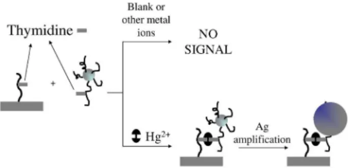
A different optical biosensor was developed by Lee and Mirkin53 to detect Hg2+ in lake water based on the
selective binding of a thymine-thymine mismatches through thymine-Hg2+-thymine complex formation upon
the surface of DNA/AuNPs probes. In order to improve the scanometric detection silver was deposited upon AuNP probes in a core-shell system. The chip-based scanometric method was able to discriminate mercuric ion from ifteen other environmental relevant metal ions. In addition, the nanoparticle-target complex enhances the sensitivity attributed to the scanometric method due to the existence of a cooperative effect between AuNPs and perfect complementary strands (Figure 2). The authors called this effect as catalytic once AuNPs promote an effective hybridization of complementary strands due to the multiple duplex interconnections between the network structures. The detection limit (1 nmol L-1 of Hg2+) of this assay in
lake water samples is according to the U. S. Environmental Protection Agency (EPA).54
Some short single stranded DNA or RNA sequences, known as aptamers, can speciically recognize non-nucleic acid targets such as small molecules, metal ions, proteins, whole cells and even viral particles. Functional DNA/RNA has great technological importance due to their potential use in diagnostics, especially in biosensing. DNAzymes and aptamers are collectively called functional DNAs.54-57
DNAzymes and RNAzymes are synthetic DNA-based biocatalysts or naturally occurring RNA molecules able to perform the same reactions like the protein enzymes, respectivelly. There are also a third functional DNA/RNA build up from the combination of a DNA/RNAzymes and an aptamer, the aptazyme that is an alosteric DNA/ RNAzyme.56
The recognition highly specific of any analyte is possible by functional DNA/RNA due to the development of a powerful combinatorial biology technique called “in vitro selection” or “systematic evolution of ligands by exponential enrichment” (SELEX).58-60 The combination of
the SELEX technology with the nanotechnology of DNA/ RNA functionalized with AuNPs is an opportunity to design high performance bioanalytical devices to discriminate a wide range of analytes.
There are great revision papers about the association of AuNPs with functional DNA/RNA in biosensing reported in the literature.54-57,60-62 The label-free functional
DNA/RNA-based biosensors belong to a class particularly interesting of bioanalytical devices that is emerging in the latest developments and it is going to be summarized in this section.
The intense colors exhibited by AuNPs solutions were related to the colloidal surface plasmon resonance phenomenon and are related to size, shape and local environment of the nanoparticles.9 Tamiya et al.63
explored the label-free detection of aptamer-protein interactions through the combination of localized SPR with interferometry in the relative relected intensity spectrum of the gold-capped oxide nanostructure; in this case, gold structures provided an enhancement of the localized SPR optical properties. The excitation of the optical characteristics and the detection were performed using only one optical iber coupled to a chip that was previously modiied with a thiolated TBA (thrombin-binding aptamer). The sandwich-type detection was employed and large dynamic ranges with a high sensitivity were veriied. The biosensor represents an alternative for the development of handheld diagnostic device.
By using SPR detection Wang and Zhou64 combined
the amplifying characteristic of AuNPs with the advantage of aptamer technique to design a “pseudo” sandwich reaction for detecting small molecules. The principle of this biosensor is based on surface inhibition detection: after the aptamer immobilization on a SPR gold ilm, a solution with different concentrations of adenosine was added to the SPR cell changing the structure of the aptamer. Interestingly, the aptamer possessing tertiary structure could not hybridize with its AuNPs-tagged complementary ss-DNA. Thus, the change of the SPR signal based on the hybridization reaction will decrease with the increase of the number of aptamers possessing tertiary structure, which is proportional to the adenosine concentration. The biosensor possesses good sensitivity and a high selectivity for adenosine. The main goal of this work is the possibility of detection of a high range of small molecules by using speciic aptamers once low weight molecules are dificult to detect by conventional SPR technique.
Several works were classiied as optical biosensors because they it the main request of a genuine biosensor: they present the recognition biomolecule intimately linked to a transducer element, the AuNPs.65-67 Liu and Lu65,66 prepared
optical biosensors based on the formation of aggregates of AuNPs functionalized with DNAzyme or aptazyme. In the irst work, a selective lead biosensor was developed based on the aggregation of “8-17” DNAzyme-AuNPs in the absence of Pb2+, resulting in a blue-colored nanoparticle assembly.
On the contrary, in the presence of Pb2+, the DNAzyme
catalyses the speciic hydrolytic cleavage of substrate strand, which disrupts the formation of the nanoparticle assembly, resulting in a red-colored individual nanoparticles. In the later manuscript, the employed aptazyme was composed by the “8-17” DNAzyme with an adenosine aptamer motif (the speciic elaborated sequence for the aptamer). In the absence of adenosine, the aptazyme is inactive and the substrate strands can serve as linkers to assemble functionalized AuNPs, resulting in a blue color. But in the presence of adenosine, the aptazyme is activated and cleaves the substrate strand, disrupting the formation of nanoparticle aggregates and changing the observed color. The performance of the biosensor was not evaluated in real samples and the effect of ions like Pb2+ could interfere in
the analysis. Authors claim that the employed strategy can be used in other biosensors for many analytes of interest regardless of whether the analytes are directly involved in the cleavage reaction or not.
The effect of the microenvironment around aptamers on the colloidal stability of AuNPs in which the aptamer was tethered was recently investigated. Zhao et al.67 have studied two different aptamers (adenosine and potassium aptamer) and discovered a unique colloidal stabilization effect associated to the conformation that aptamers adopt on the AuNPs surface. On the basis of this phenomenon, optical biosensors have been developed for the detection of adenosine, K+, adenosine deaminase and its inhibitors,
but just a few details about the analytical performance of each sensor was shown. The authors veriied that AuNPs with attached folded aptamer structures are more stable toward MgCl2-induced aggregation than those tethered to
Biosensors Based on Gold Nanostructures
J. Br
az. Chem. Soc.
10
Table 1. Label-free biosensors designed with functional DNA/RNA
Recognition biomolecule / transducer
Composition description Target Transduction Mode LOD Incubation time /
sample
Ref.
Adenosine Aptamer / Gold electrode
The aptasensor is obtained by annealing the aptamer probe with surface-immobilized capture probe, followed by an equilibrium with Methylene Blue. In the presence of adenosine, the double-stranded complex is dissociates, which results in the decrease in DPV signal
Adenosine Voltammetric / −245 mV vs.
Ag/AgClsat
1 nmol L-1 1 h / no 71
Thrombin-binding aptamer / Glassy carbon electrode
Thrombin aptamers was immobilized upon electrodeposited AuNP onto glassy carbon electrode. The change in electron transfer resistance of ferricianyde was monitore as the target interact with the immobilized aptamer
Thrombin Impedimetric / 200 mV biased
potential
0.06 nmol L-1 40 min / no 72
Thrombin-binding aptamer / Gold electrode
Thiolated aptamers are irstly immobilized on a gold substrate to capture thrombin molecules, and then the aptamer functionalised AuNP are used to amplify the impedimetric signal
Thrombin Impedimetric / 200 mV biased potential
0.02 nmol L-1 1 h in target solution
+ 120 min in AuNP/ TBA / no
73
Thrombin-binding aptamer / Au-coated porous anodic alumina
Thrombin-binding aptamer modiied Au-coated porous anodic alumina chip was developed and the localized surface Plasmon resonance and interferometric properties of the chip were monitored and optimized. Sandwich-type detection
Thrombin Optical (optical iber) / 400 to 800 nm
1 nmol L-1 1 h in target solution
+ 1 h in TBA II / no 63
Antiadenosine aptamer / AuNP
A speciic aptamer was immobilized upon SPR gold ilm. Then, different concentrations of the analyte was added to SPR cell. Last, the SPR cell was washed and AuNP tagged complementary DNA was added. The SPR angle-time curve is used to monitor the reaction progress between AuNP complementary ss-DNA and aptamer in the presence of adenosine.
Adenosine Optical (SPR) 0.748 nmol L-1 30 min / no 64
DNAzyme / AuNP An in vitro selected DNAzyme (named “8-17” DNAzyme) with high
selectivity for Pb2+ was immobilized upon AuNP. In the presence of Pb2+,
the substrate strand can be cleaved by the enzyme strand .
Lead Optical
(UV-Vis spectroscopy)
- 120 min / Leaded paint
65
Aptazyme / AuNp A “8-17” DNAzyme modiied with an adenosine aptamer was immobilized upon AuNP. In the presence of adenosine the aptazyme is inactivated, but in the absence of adenosine the substrate strand can be cleaved by the aptazyme, disrupting the formation of modiied nanoparticles aggregates.
Adenosine Optical (UV-Vis spectroscopy)
- 123 min / no 66
Adenosine aptamer / AuNP A biosensor for adenosine was developed based on the aggregation / redispersion of aptamer modiied AuNP in salt solution (MgCl2). The authors claim the development of biosensors for adenosine deaminase and also adenosine deaminase inhibitor, but they are not discussed here due to lack of analytical information in the manuscript.
Adenosine Optical (UV-Vis spectroscopy)
- - / no 67
Thrombin-binding aptamer / Gold electrode
The sandwich system of aptamer/thrombin/aptamer-functionalized Au nanoparticles was fabricated as the sensing platform. The change of the interfacial feature of the electrode was characterized by electrochemical impedance analysis with a redox probe and there was an enlargement of negatively charged aptamer-functionalized gold nanoparticles.
3.2. Electrochemical
Direct electrical detection is one of the simplest methods for bioaffinity sensing. The detection of a conductivity change can result in a detection limit of 500 femtomol L-1.68 The redox properties of AuNPs have led
to their extensive use in electrochemical biosensors for nucleic acid detection and other molecules, with numerous conigurations being explored.69
Other materials were also used in combination with AuNPs. For example, a novel DNA biosensor has been fabricated for the DNA hybridization detection based on layer-by-layer covalent assembly of AuNPs and multiwalled carbon nanotubes. The stepwise LbL assembly was characterized by electrochemical studies showing that the presence of AuNPs was linked to a change in the ilm conductivity. The hybridization events of the intercalated doxorubicin were monitored by differential pulse voltammetry (DPV). The electrochemical signal increased linearly with increasing target DNA concentration from 0.01 to 0.5 nmol L-1 and a detection limit of 7.5 pmol L-1
(signal/noise ratio of 3) was found.70
In general label-free techniques offer several advantages over conventional ones due to the simplification of operational procedures related to the compulsory step of labeling the analyte or the receptor in a biomolecular recognition event. Several techniques sensitive to changes in interfacial properties are suited to be combined with label-free functional DNA/RNA assays. The electrochemical approaches have been developed based on sandwich sensing and on target-induced displacement format.71-73
Yu et al.71 have developed an aptasensor with adenosine
as model system based on target-induced displacement by using an external electroactive indicator methylene blue employing DPV technique. The sensing substrate comprises a gold electrode modiied with a dithiol SAM that assemble an AuNPs ilm. A capture probe was loaded upon that nanoparticle ilm and an aptamer for adenosine was applied to hybridize in the presence of methylene blue, yielding a double-stranded complex. The interaction of adenosine with the aptamer displaces the aptamer sequence, which causes its dissociation from the interface releasing the electroactive indicator. This work demonstrates that the AuNPs ilm increases the surface loading of capture probe and, consequently, enhances the analytical signal.
An impedimetric biosensor based on a thrombin-binding aptamer as molecular recognition element was developed for the determination of thrombin. The signal enhancement was achieved by using AuNPs, which was a platform for thiolated aptamers to link.72 The biosensor monitors the
linear increase of the interfacial electron transfer resistance
using [Fe(CN)6]3−/4−as the concentration of thrombin raises
in the range from 0.12 up to 30 nmol L-1. Interestingly,
the sensitivity obtained with the proposed biosensor was compared with other ones prepared upon gold disk and gold thin ilm coated electrodes and the authors have veriied that the higher sensitivity was obtained with the former due to high surface density of thrombin-binding aptamers (TBA) immobilized on AuNPs. Another impedimetric aptasensor for thrombin was developed by Dong and et al.73 based on a sandwich sensing assay between thrombin and TBA. One thrombin molecule has two active sites, this way a sandwich protocol is easily fabricated: irstly, the thiolated TBA immobilized on gold electrode captures the analyte and then, the TBA functionalized AuNPs binds thrombin, amplifying the signal. Through such method, the detection sensitivity (0.02 nmol L-1) is higher when compared to
previously reported impedimetric biosensors.
3.3. Piezoelectric
Enterohemorrhagic Escherichia coli O157:H7 is a hazardous microorganism which causes human illness by several different mechanisms.74-76 Chen et al.77
presented a nanoparticle-ampliied DNA sensor using the hybridization of two specific probes using QCM. Although the mass of nanoparticles is small (a single, 10 nm diameter gold nanoparticle has a mass of a few attograms), nanomechanical resonator detectors are capable of sensing atto to femtogram masses of material.78,79 Nanoparticles
showed substantial improvement in the detection limits with sensitivity from 10-12 up to 10-14 mol L-1 of DNA77 by
a mass enhancement.
Since many projects are currently developing AuNPs-labelled DNA/RNA probes, it seems probably that future DNA/RNA sequence detection will involve metal nanoparticle-biomolecule conjugates. For example, Qiao et al.80 modiied AuNPs using IgG and ss-DNA in a combined dual bio-probe. The surface modiications of AuNPs related to antibody and DNA attachment was evaluated by techniques such as transmission electron microscopy (TEM), UV-Vis spectra and electrophoresis. This IgG/DNA based probe brings promising perspectives of diseases detection, early tumor and heart disease, and more important is that both antibodies and ss-DNA retained their bioactivities on the nanoparticle surface. A collection of other relevant biosensors based on DNA biomolecules and AuNPs are assembled in Table 2.
Biosensors Based on Gold Nanostructures
J. Br
az. Chem. Soc.
12
Table 2. Other biosensors designed with nucleic acids and AuNPs
Recognition biomolecule / transducer
Composition description Target Mode of Transduction LOD Incubation time /
sample
Ref.
20-base fragment of PAT gene sequence / a polyaniline nanoibers modiied carbon paste electrode
The surface of the polyaniline nanoibers/ carbon paste electrode was modiied with gold nanoparticles, carbon nanotubes and inally with a DNA probe by adsorption. The changes in the electron transfer resistance of the electrode surface after the hybridization of the probe DNA was used for the label-free detection of the target DNA.
Phosphinothricin acetyltransferase (PAT) gene
fragment
Impedimetric 5.6 ×10⁻¹³ mol L-1 600 s under the
application of +0.4 V vs. Ag/AgClsat
83
DNA oligonucleotides / Gold nanoparticles
The capture of gold nanoparticles on the test zone and control zone of the biosensor produced red bands enabling visual detection. It is a disposable nucleic acid biosensor (DNAB) for low –cost and sensitive detection of nucleic acid samples in 15 min.
DNA oligonucleotides
Optical 1.25 fmol L-1 - / Human genomic
DNA
84
Sythettic DNA sequences / Ruthenium complex
The surface of a gold electrode was modiied with a 1,6-hexanedithiol monolayer and AuNPs were immobilized upon it. Then, the ss-DNA was immobilized upon the AuNP. Hybridizaton was induced by exposure of the target ss-DNA to the solution of electrogenerated chemiluminescence probe consisting of complementary ss-DNA tagged with ruthenium complex. The performance of the developed biosensor was compared with a gold electrode modiied with ss-DNA.
DNA segments related to the cystic ibrosis
Optical (electrogenerated chemiluminescence)
6.7 ×10⁻¹² mol L-1 30 min / no 85
Peptide nucleic acid (PNA) / interdigitated electrodes
A chip-based biosensor was developed to detect a DNA sequence at low concentration. AuNPs were modiied with 3-mercaptopropionic acid that showed strong complex interaction with an inorganic linker, Zr4+, forming aggregates. The
formed aggregate was used as a conductive tag for the detection of DNA.
A DNA that was complementary
to PNA
Condutimetric 5 ×10-14 mol L-1 60-90 min / no 86
thiolated DNA / Rigid conducting gold nanocomposite (nano-AuGEC)
Firslly, the nano-AuGEC electrode was modiied with the thiolated DNA of interest. Then, it is performed the hybridization with a digoxigenin labeled complementary and non-complementary probe. At last, it is performed the enzymatic labeling using Anti-digoxigenin-HRP and the amperometric determination of the target is performed based on the enzyme activity.
Salmonella sp. Amperometric 9 fmol L-1 70 min / Puriied
Salmonella enterica
serovar typhimurium genomic DNA
87
20-base fragment of PAT gene sequence / a glassy carbon–modiied electrode
A poly-2,6-pyridinedicarboxylic acid ilm was electropolimerized upon a glassy carbon electrode . Then, gold nanoparticles were added to that ilm and then DNA probe (ss-DNA) was immobilized by the interaction of AuNPs with DNA. After the hybridization of the DNA probe with the complementary ss-DNA made the charge transfer resistance increase furher.
Phosphinothricin acetyltransferase (PAT) gene
fragment
Impedimetric 2.4 ×10⁻¹¹ mol L-1 60 min / no 88
ss-PNA / nano-gold modiied gold electrode
A label-free electrochemical biosensor based on the speciic signal of adsorbed ferrocene-containing cationic polythiophene after PNA-DNA hybridization. The selectivity and the sensitivity of the biosensor were increased by modifying a gold electrode with gold nanoparticles that were followed modiied with a HS-modiied peptide nucleic acid (PNA).
A DNA that was complementary
to PN
Voltammetric (differential pulse
voltammetry)
1.0 ×10 -11 mol L-1 60 min / no 89
Thiolated E. Coli O157:H7 DNA / Gold disc
A quartz crystal microbalance biosensor for real time detection of E. coli O157:H7 DNA based on nanogold particles ampliication was constructed. AuNPs were immobilized onto the thiolated surface of the gold electrode, then more speciic thiolated ssDNA probes could be immobilized. After the hybridization of the probe with the target DNA, the outer avidin-coated AuNPs could combine with the target DNA to increase the mass.
E. coli O157:H7 DNA
different ways have distinct features as oligonucleotide surface densities, availability for hybridization to targets and tendencies to nonspeciically bind to surfaces.81
4. Redox Protein Biosensors Designed with
AuNPs
The enzymatic biosensor performance depends on the immobilization of the biocatalyst. In this sense, several efforts have been done in order to ind an ideal arrangement to achieve high selectivity, stability, reliability and low detection limits. In this aspect, the incorporation of nanomaterials provides a huge advance, besides the great number of materials and architectures that can be tailored in an easy way. Among innumerous nanomaterials used for bioanalysis, AuNPs have a special role providing a stable immobilization of biomolecules retaining their bioactivity. Besides, AuNPs allow the enhancement of direct electron transfer between redox biomolecules and electrode surfaces, becoming the processes based on charge transfer faster and more effective. The most recent works concerning the modiication of electrodes for the construction of biosensors based on redox enzymes and heme proteins are described as follow.
Among electrochemical biosensors based on enzyme attachment, certainly, the glucose oxidase (GOx) assemblies are the most studied systems. This enzyme has attractive characteristics as its well known behavior, great stability and robustness.91 As hydrogen peroxide can
be formed by enzymatic reaction involving the glucose, its determination in several samples is very important for chemical, biological, clinical and other determinations, being the electrochemical methods the most employed. However, one barrier for direct electrochemical detection relies on the overpotential required for the peroxide reduction, a strategy to overcome this dificulty is the utilization of biosensors based on peroxidases.92-96
In order to construct enzymatic electrode employing the above system, probably the simplest assembly strategy consists in incorporating nanomaterials into a composite electrode, leading to an improvement of analytical response such as low background currents. One example is the simple mixing of gold colloidal nanoparticles and carbon paste, followed by the adsorption of GOx97 or HRP,98
providing a fast electron transfer between the electrode and biomolecules.
Taking into account the large surface area promoted by nanoparticles, another strategy for electrode modiication consists in a direct gold surface modiication, as described by Zhang.99 In this work, gold nanoparticles were linked
to gold electrode by dithiol via gold-sulphur bond, then a
cystamine monolayer was chemisorbed onto AuNPs and GOx could be covalently attached to the gold electrode, resulting in a biosensing interface stable for more than 30 days. In another strategy, AuNPs were attached to hollow porous thiol-functionalized polymeric nanospheres providing an active matrix for further immobilization of HRP.100
In order to improve the spatial arrangement of nanomaterials, the sol-gel science was employed to prepare a three-dimensional network able to encapsulate both enzymes and AuNPs. Thus, the tridimensional network can acts as a tiny conducting center improving the electron transfer.101 The construction of a glucose amperometric
biosensor by using this kind of modiication can be made by subsequent immersion of silica modiied Au electrode into different solutions: AuNPs, cysteamine and inally GOx.102 In a similar assembly, HRP was covalently attached
to AuNPs immobilized on a silica network.103
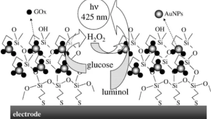
Recently it was reported an interesting work involving the assembly of AuNPs and GOx in a tridimensional silica network by means of chemioluminescent reaction between the H2O2 produced in enzymatic reaction and luminol catalyzed by AuNPs.104 The increase of luminescent signal
under different glucose concentration leads to a linear behavior from 1 µmol L-1 up to 5 m mol L-1. The electrode
modiication of this work is schematically shown in Figure 3.
Depending on its synthesis, AuNPs could present a negative charge on the surface, normally, due to citrate stabilization. This behavior can also be employed for electrode tailoring, by using the electrostatic deposition layer-by-layer (LbL) technique, reported by Detcher105
Biosensors Based on Gold Nanostructures J. Braz. Chem. Soc. 14
chloride) (PDDA),106 Prussian blue nanoparticles107 and
analogues108,109 and nickel hydroxide nanoparticles.110,111
Multilayer films of GOx/AuNPs on Au electrode surface using cysteamine as covalent attachment cross-linker, prepared by LbL technique, was performed by Yang et al.112 Bioelectrocatalytic response was directly
correlated to the number of deposited bilayers, due to the amount of active enzyme immobilized on the Au surface. Another interesting electrode modiication using LbL was recently reported by Crespilho et al.113 In this work,
irstly the AuNPs were chemically synthesized inside dendrimers [G4-PAMAN (poly(amidoamine) dendrimers of 4th generation)] from KAuCl
4 and formic acid solutions,
and then the composites were adsorbed onto ITO (indium tin oxide) electrode by alternating PAMAN-Au / PVS (poly(vinyl sulfonic) acid) bilayers. Electrocatalytic thin ilms of copper hexacyanoferrate were electrodeposited directly on AuNPS. Finally, enzymes were immobilized onto electrode by cross-linking method by adding a drop of a solution containing GOx, glutharaldehyde and bovine serum albumin (BSA). This electrode showed a sensitivity of 33.6 nA mmol L-1 cm-2 and a limit of detection
(S/N = 3) of 17 µmol L-1.
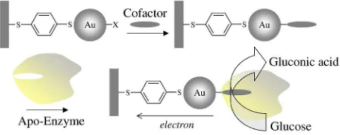
Another way to determine of glucose concentration can be made by the detection electrons generated in enzymatic reaction. But, the direct contact between the electrode and the enzymatic active site is not a simple task and the dificulty comes from the insulation of the biocatalytic redox centers by the protein organic shell. The cited assemblies of redox enzymes and AuNPs mentioned up to now already enhance the electronic transport through electrode and enzymes, but a further improvement can be achieved by the means of “molecular wiring”.114-117 One
of the most successful strategies for establish electrical communication between redox proteins and electrodes involve the extraction of the native redox cofactor from the protein and the reconstitution of the resultant apo-enzyme on a surface modiied with a monolayer consisting of a relay tethered to the respective cofactor unity,118 bellow, in
Figure 4 is shown a scheme of this method. The assembly this GOx enzymatic electrode is based on the modiication of AuNPs by aminlavin adenine dinucleotide cofactor and linked to the electrode surface by dithiol molecules. Apoglucose oxidase generated by exclusion of its native FAD cofactors was reconstituted on the cofactor-modiied AuNPs. The reported increase of anodic currents indicates that occur the electrical communication between its active site and the electrode.118,119 This system showed a huge
heterogeneous electron transfer constant (Ket) between
the cofactor site and the electrode being estimated as ca. 5000 s−1, which is about seven times higher than the
electron-transfer rate constant of native GOx with O2 as
electron acceptor. Accordingly to the authors this value can be attributed to the effective contact between the electrode and the redox cofactor that stimulates the effective electron transport to the electrode by means of the AuNPs, acting as a nano-electrode, increasing the maximum turnover rate.119
Heme proteins such hemoglobin (Hb), myoglobin (Mb) and cytochome c (Cyt c) do not undergo facile direct electron transfer reactions at bare electrode surfaces probably by unfavorable orientation of the redox site or due to its denaturation adsorption on the transducer. AuNPs as a biocompatible material provide an adequate microenvironment to support heme proteins for fabricating biosensors. Additionally, AuNPs have been used to achieve high uniformity and organization but with lack of restrictions in orientation of protein molecules in the biological recognition layer.
The direct heterogeneous electron transfer is possible from the protein to the transducer due to the high degree of order on a molecular level associated to the decrease of the insulating property of the protein shell promoted by the conducting tunnels made of AuNPs.
The effect of AuNPs on the electroactivity of heme proteins was mainly studied using hemoglobin and amperometric transducers as solid electrodes made of glassy carbon, gold and indium tin oxide (ITO) to a lesser degree. In general, due to their close similarities, heme proteins can be used as substitutes of peroxidases to catalyze the reduction of hydrogen peroxide, however higher potentials are required to carry out the analysis. Heme proteins associated with AuNPs have been used to build up highly sensitive biosensors for oxygen, nitrite and trichloroacetic acid.120-124 Dong et al.125 constructed
a voltammetric biosensor for hydrogen peroxide based on the immobilization of Hb upon AuNPs assembled on sulphydryl-terminated monolayer covalently bond to the glassy carbon surface. According to authors, the immobilization of Hb occurred due to the speciic interaction between amino-acids residues (like cysteine or
NH4+-lysine of Hb) and the AuNPs surface and this way,
it had maintained its biological activity during two weeks. Chen et al.126 studied the immobilization of Hb on AuNPs associated with a cysteamine monolayer on a gold electrode surface. The developed biosensor exhibits great electrocatalytic behaviour in the presence of H2O2, but retained its performance for only three weeks at 4 oC.
The maintenance of the bioactivity on immobilized redox proteins is an important issue that can be improved by creating a biocompatible protective environment, in this sense, the use of chitosan can be found in literature.127-130 In
another work, Chen et al.127 have studied the direct electron
transfer and H2O2 electrocatalysis of Hb, Mb and Cyt c
immobilized onto chitosan-stabilized gold nanoparticles self-sustained on a cysteine modiied gold electrode. The amperometric responses of the heme-proteins-modiied electrodes were monitored at −250 mV vs. SCE and the Mb-modiied electrode showed the best sensitivity towards H2O2 determination with a long-term stability attested
during three months.
Another approach for the construction of an amperometric H2O2 biosensor with Mb and clay-chitosan-stabilized gold nanoparticles was made by Zou et al.120 The electrocatalytic reduction of H2O2 was performed at
−260 mV vs. Ag/AgClsat with an adequate operational stability. Interestingly, Sun et al.128 developed a simple
biosensor based on the immobilization of Mb and AuNPs on a glassy carbon electrode using Naion® ilm. The immobilized
Mb exhibited an unmediated high electrocatalytic response to the reduction of H2O2 with high stability during 50 days, but on the other hand the amperometric applied potential was –405 mV vs. Ag/AgClsat.
Carbon nanotubes, as a new class of nanomaterials with unique electrical properties, have also been employed with AuNPs to the Hb immobilization in biosensors.121,129
Li et al.129 have fabricated a H
2O2 biosensor based on the
immobilization of Hb on multiwall carbon nanotubes and gold colloidal nanoparticles and Hu and Liu121 developed a
similar analytical device but entrapping Hb in a composite electrodeposited chitosan-multiwall carbon nanotubes ilm by assembling gold nanoparticles and hemoglobin step by step, in both works are observed a large background current of Hb FeIII/FeII redox couple in cyclic voltammograms,
low electrocatalytic towards peroxide reduction and high applied potential in amperometric monitoring. The immobilized enzyme exhibited more advantagenous analytical performance than the system without carbon nanotubes. This can be attributed to the synergistic action of both carbon nanotubes and AuNPs, since both of them have the ability to facilitate or promote the electron transfer between the proteins and the electrode surface.
AuNPs attached on ITO electrodes were applied for Hb and Mb immobilization by Oyama and Zhang.131,132 The
synthesis of gold nanoparticles was based on a surfactant-assisted seeding growth that was actually intended to prepare gold nanorods in solution. This synthesis provided both a short amount of rod-shaped gold nanoparticles and a large amount of spherical nanoparticles. The authors have adapted this method for ITO modiication. They established a two-step electrode modiication procedure. Firstly, 4 nm gold seeds consisted by citrate-stabilized gold nanoparticles were deposited on the ITO surface. Secondly, in a growth solution, surfactant capped gold ions were reduced when encountering the gold seeds on the electrode surface, leading to the growth of gold nanoparticles. The AuNPs attached to ITO surface was then composed by a mixture of both gold nanospheres and nanorods. It was veriied a catalytic behaviour towards the reduction of H2O2, and no characteristic peak for Hb appeared in the cyclic voltammogram recorded with Hb/AuNP/ITO. For Mb, the FeIII/FeII redox couple also appeared in the CV of
Mb/ITO electrode in buffer solution. In this later case, the presence of AuNPs onto ITO has just increased the surface area for Mb immobilization.
Layer-by-Layer ilms can be constructed by using the alternate adsorption of negatively charged AuNPs and positively charged Mb, {AuNPs/Mb}n, on pyrolytic graphite electrode surface as described by Hu et al.,122 where the size of AuNPs demonstrates distinct inluence on the properties of the modiied electrode. The smaller-sized AuNPs (6 nm) load more amount of Mb showing better electrochemical and biocatalytic activities toward the reduction of both O2 and H2O2 than those by employing larger AuNPs (40 and 90 nm). Recently, Hu and Zhang133
have assembled poly(propyleneimine)-Au nanoclusters and Mb, forming {PPI-AuNP/Mb}n (poly(propyleneimine))
multilayered ilms. This novel kind of inorganic-organic hybrid ilm with dendrimer stabilized gold colloids showed better electrochemical behaviour and catalytic performance than the previously reported biosensor.
Scheller et al.134 studied the adsorption and the
electrochemical behavior of Cyt c on AuNPs modiied carbon paste electrodes. The authors claim that the presence of AuNPs was essential to avoid denaturation as well as for the stable redox transformation of adsorbed Cyt c, however this system persists stable for just two days at 4 oC. The direct electron transfer of the protein
to the electrode was obtained with success, but showing a restricted electrocatalysis for the reduction of H2O2, promoted by Cyt c.
Biosensors Based on Gold Nanostructures
J. Br
az. Chem. Soc.
16
Table 3. Biosensors designed with Heme proteins and AuNPs
Heme protein/transducer Composition description Target Mode of Transduction/ potential vs. Ag/AgClsat
LOD / Sensitivity Stability Ref.
•Hb-Chit-AuNP-Cys / Gold electrode
♦Mb-Chit-AuNP-Cys / Gold electrode
♥Cytc-Chit-AuNP-Cys / Gold electrode
Heme proteins adsorbed onto chitosan (Chit) stabilized nanoparticles linked to a cysteine (Cys) SAM upon a gold electrode.
H2O2 Amperometric / − 250 mV •6.4 µmol L-1
♦1.8 µmol L-1
♥9.8 µmol L-1
3 months 127
Hb-AuNP-Cyste / Gold electrode Hb was immobilized on 24 nm AuNP associated with a cysteamine (Cyste) SAM upon a gold electrode.
H2O2 Amperometric / − 200 mV 0.12 µmol L-1 /
9.782 µA / mmol L-1
3 weeks 126
Clay-Mb-Clay / AuCS-GCE Chitosan-stabilized gold nanoparticles (AuCS) were synthesized and hybridized with oppositely charged clay nanoplates to produce a clay-chitosan-AuNPs nanohybrid material upon GCE.
•H2O2 ♦NO2–
Amperometric / − 260 mV •0.41 µmol L-1 /
0.62 µA / mmol L-1
♦0.05 µmol L-1 /
0.004 µA / mmol L-1
2 months 120
Hb-CS-CNT-AuNP / Gold electrode
Hb was entrapped in composite electrodeposited chitosan-multiwall carbon nanotubes (CS-CNT) ilm by assembling AuNPs and Hb step by step.
H2O2 Amperometric / − 360 mV 0.5 µmol L-1 /
0.032 µA / mmol L-1
1 weeek 121
Mb / AuNP-ITO An ITO electrode modiied with spherical and rod shaped AuNPs and Mb. H2O2 Amperometric / − 400 mV 0.48 µmol L-1 /
0.4070 µA / mmol L-1
40 days 131
Hb / AuNP-ITO An ITO electrode modiied with spherical and rod shaped AuNPs and Hb. H2O2 Amperometric / − 300 mV 4.5 µmol L-1 / 0.2977
mmol L-1 µA-1
40 days 132
{Hb-AuNP}n – Hb-CNT / GCE The {Hb-AuNP}n multilayer ilms were assembled onto Hb adsorbed onto multi-wall carbon nanotubes (Hb-CNT) through layer-by-layer technique.
H2O2 Amperometric / − 300 mV 0.08 µmol L-1 /
14.6 µA / mmol L-1
2 weeks 129
{Mb-AuNP}n / pyrolytic graphite {AuNP}n layer-by-layer ilms on pyrolytic graphite electrodes. H2O2 Amperometric / − 55 mV 0.4 µmol L-1 / 0.476
(mol/L)-1 A cm-2
- 122
{Mb-PPI-AuNP}n / pyrolytic graphite
{Mb/PPI-AuNP}n layer-by-layer ilms on pyrolytic graphite electrodes. PPI-AuNP =
poly(propyleneimine)-AuNP nanoclusters.
H2O2 Amperometric / − 55 mV - / 48 mmol L-1 µA-1 - 133
Hb-{AuNP-PDDA}n / PDA-GCE Hb was adsorbed on a {AuNP-PDDA}n layer-by-layer ilm on PDA modiied glassy carbon electrode. (GCE)
H2O2 Amperometric / − 250 mV 0.8 µmol L-1 2 weeks 136
Cyt c / AuNP-CP Cyt c was adsorbed on colloidal gold modiied carbon paste electrode H2O2 voltammetric / 0 mV - 2 days 134 Hb-AuNP-ATP-ABA / GCE AuNPs have been attached onto a GCE through sulfhydryl-terminated monolayer and
at the end Hb was adsorbed by AuNPs.
H2O2 Amperometric / − 250 mV 0.91 µmol L-1 /
0.125 µA / µmol L-1
2 weeks 125
Naion®-Mb-AuNP / GCE Mb and AuNP were immobilized upon GCE by a Naion ilm H
2O2 Amperometric / − 405 mV 0.5 µmol L-1 1 month 128
Hb-AuNP-Cys-AuNP-PtNP-CS / Platinum electrode
Hb/AuNP/L-cysteine (Cys)/AuNP/nanoparticles Pt (PtNP)–chitosan (CS) composite ilm-modiied platinum disk electrode has been prepared
H2O2 voltammetric / 0 mV 0.045 µmol L-1 /
17.6 µA / mmol L-1
2 weeks 130
Hb-AuNP-Cys-poly(pABSA) / Platinum electrode
Hb was adsorbed successfully on AuNP/L-Cysteine/poly(p-ABSA)-ilm modiied platinum disk electrode. pABSA = p-aminobenzene sulfonic acid
H2O2 Amperometric / 30 mV 0.07 µmol L-1 1 week 137
Hb-AuNP-TiO2 sol-gel ilm/ GCE Immobiliation of Hb and AuNP on glassy carbon electrode by a Titania sol-gel ilm NO2− Amperometric / − 750 mV 1.2 µmol L-1 50 days 123
Mb-AuNP-TiO2 sol-gel ilm / GCE
the analytical signal from the protein layer is restricted to a relatively low signal-to-background ratio, thus in some cases interfacial heterogeneity can result in superior protein adsorption at a surface. This way, assembled ilms of nonaqueous nanoparticles known as monolayer-protected clusters (MPCs) are interesting platforms for protein monolayer adsorption. Leopold et al.135 investigated the electrochemical properties such as electron transfer rate constants of Cyt c at MPC ilms of alkanethiol-protected AuNPs. The use of hydrophilic MPCs ilms as a terminal ilm layer results in adsorptive voltammetric behavior of Cyt c and the electron transfer rate constant was unaffected by the large electron transfer distance introduced by MPC assemblies when compared with traditional strategies to immobilize Cyt c. A compilation of approaches to study the direct electron transfer between Heme proteins and electrode surfaces with the aid of AuNPs are summarized in Table 3.
5. Conclusion and Future Trends
In this review some recent advances of AuNPs-based biosensors have been addressed. Although AuNPs have been known for more than two thousand years they are still a subject of an exponentially increasing number of interests such as optical, electronic, biomedical, magnetic and catalytic applications being a very important step of the development of nanoscience and nanotechnology. This fact could be related to both easy preparations with a large number of different synthetic methods where the shape and morphology of the particles can be easily tuned and their intrinsic physical-chemical properties, which lead to exciting new improvements of well known ields of chemistry as shown for biosensors.
Fantastic sensor devices for environmental or clinical applications can be constructed by the assembly of AuNPs and biocomponents such as enzymes, DNA / RNA, antibody, cells and others. The great afinity between gold and thiols allows the decoration of nanoparticles with a great number of macromolecules. The presence of the metallic gold itself enhances the signal of many transducers based on spectroscopy, luorescence, luminescence and electrochemical ways. Obviously these advances could be reached due to the development of characterization techniques such as SEM (scanning electron microscopy), AFM (atomic force microscopy), TEM (transmission electron microscopy), electrochemical and spectroscopy probes and others.
As stated previously, actually the development of composite inorganic-organic matrices based on nanostructures seems to be the next irreversible step
in the biosensor technology where new strategies of immobilization and interfaces are currently been investigated by hundreds of laboratories around the world, anyway aspects as durability, reliability, “in vivo” measurements, reproducibility, disposal of (nano)materials and biocompatibility are issues that still need to be best researched.
Marcio Vidotti obtained his MSc
in Chemistry in 2002 and the PhD in Physical Chemistry in 2007 both from the Chemistry Institute of the Universidade de São Paulo, Brazil. Following this he held a post-doc position at the Chemistry Institute of the Universidade Estadual de Campinas, Brazil under the supervision of Prof. Kubota (2008-2009). He also worked at the University of Warwick in the labs of Profs. Julie Macpherson and Patrick Unwin (2009-2010). Currently he has a position at the Chemistry Department in the Universidade Federal do Paraná. His interests are focused on nanomaterials, polymers and electrochemical devices.
Rafaela Fernanda Carvalhal
was born in 1980 in Londrina, Paraná. She graduated in Chemistry from the Universidade Estadual de Londrina in 2002 and was an internship at the Instituto Agronômico do Paraná in the laboratory of Dr. Mario Miyazawa (1999-2002). At the Universidade Estadual de Campinas she obtained her MSc in Analytical Chemistry in 2005 and she is currently a PhD student under supervision of Prof. Kubota at the same university. Her researches are focused on the development of disposable electrochemical devices for rapid analysis and diagnostics.
Renata Kelly Mendes graduated