FACULDADE DE MEDICINA DE LISBOA
E
VALUATION OF THE
I329L
AS A CANDIDATE TO
DELETE FOR PRODUCTION OF A VIRAL
ATTENUATED VACCINE AGAINST ASFV
A
NAC
ATARINAM
OTAC
ORREIAM
ESTRADO EMM
ICROBIOLOGIAC
LÍNICA4
ªE
DIÇÃOA impressão desta dissertação foi aprovada pela comissão
Coordenadora do Conselho Científico da Faculdade de
Medicina de Lisboa em reunião de 17 de Julho de 2008.
FACULDADE DE MEDICINA DE LISBOA
E
VALUATION OF THE
I329L
AS A CANDIDATE TO
DELETE FOR PRODUCTION OF A VIRAL
ATTENUATED VACCINE AGAINST ASFV
A
NAC
ATARINAM
OTAC
ORREIAM
ESTRADO EMM
ICROBIOLOGIAC
LÍNICA-
4
ªE
DIÇÃOD
ISSERTAÇÃO ORIENTADA PELOP
ROF.
D
OUTORM
ICHAELP
ARKHOUSETodas as afirmações efectuadas no presente documento são da exclusiva responsabilidade do seu autor, não cabendo qualquer responsabilidade à
I
NDEXAcknowledgements ... i
Abbreviations ... ii
Abstract... v
Sumário ... vi
1) General Introduction ... 1
1.1) Introduction ... 11.1.1) Virus inhibition of the Innate immune response ... 1
1.1.1.1) Virus inhibition of the complement system ... 1
1.1.1.2) Virus inhibition of interferon responses ... 2
1.1.1.3) Virus inhibition of apoptosis ... 2
1.1.1.4) Virus inhibition of Natural Killer cells ... 3
1.1.1.5) Virus inhibition of cytokines and chemokines ... 4
1.1.2) Virus inhibition of Acquired humoral and cellular immunity ... 5
1.1.2.1) Virus inhibition of antibody responses ... 5
1.1.2.2) Virus inhibition of MHC class I presentation ... 6
1.1.2.3) Viral inhibition of MHC class II presentation ... 7
1.1.3) The African Swine Fever Virus ... 7
1.1.3.1) Pathogenesis and epidemiology ... 7
1.1.3.2) Virus structure, entry, replication cycle and assembly ... 11
1.1.3.3) TLR signalling pathway ... 12
1.1.3.4) The I329L gene of the ASFV ... 16
1.2) Objective ... 19
2) I329L is a Non Essential Gene of the ASFV ... 20
2.1) Introduction ... 20
2.1.1) Inhibition of expression of I329L through siRNA technology ... 20
2.1.2) Construction of the I329L mutant deletion virus ... 22
2.2) Methods and Materials ... 23
2.2.1) Cell lines ... 23
2.2.2) siRNA Transfection and Ba71v infection ... 23
2.2.2.1) Treatment with siRNA oligos ... 23
2.2.2.2) Demonstration of siRNA inhibition of I329L expression by RT-PCR ... 25
2.2.2.3) Detection of intracellular and extracellular virus ... 25
2.2.3) Cloning of the mutant deletion virus construct ... 27
2.3) Results ... 29
2.3.1) Expression of I329L during Ba71v infection in Vero cells ... 29
2.3.3) siRNA titration in Ba71v infected Vero cells ... 31
2.3.4) Analysis of virus production from infected Vero cells transfected with I329L siRNA targets ... 32
2.3.4.1) Staining of p73, the major capsid protein of ASF virus ... 32
2.3.4.2) Analysis of viable virus production from siRNA recovered supernatants ... 34
2.4) Discussion ... 36
3) Failure to detect an impact of I329L expression in superoxide production
stimulated by LPS and dsRNA (poly i:c) in IPAM cells ... 38
3.1) Introduction ... 38
3.2) Methods and Materials ... 40
3.2.1) Cell lines ... 40
3.2.2) I329L expression in I329L lentivirus-transduced IPAM cells ... 40
3.2.2.1) Detection of I329L expression by RT-PCR ... 42
3.2.2.2) Detection of I329L through immunofluorescence ... 42
3.2.3) Assays of macrophage function ... 43
3.2.3.1) Measurement of nitric oxide ... 44
3.2.3.2) Measurement of nitric oxide in control and I329L -transduced IPAM cells . 44 3.2.3.3) Analysis of porcine cytokine gene expression through RT-PCR ... 45
3.3) Results ... 47
3.3.1) I329L expression in I329L lentivirus-transduced IPAM cells ... 47
3.3.1.1) Detection of I329L expression by RT-PCR ... 47
3.3.1.2) Detection of I329L expression by immunofluorescence ... 48
3.3.2) Superoxide production measurement on IPAM cells after LPS and PMA stimulation ... 48
3.3.3) Superoxide production measurement on empty lentivirus-transduced IPAM cells and I329L lentivirus-transduced IPAM cells after LPS stimulation ... 50
3.3.4) Superoxide production measurement on empty lentivirus-transduced IPAM cells and I329L lentivirus-transduced IPAM cells after poly(I:C) stimulation ... 51
3.3.5) Detection of cytokine expression by RT-PCR ... 53
3.4) Discussion ... 55
4) Construction of a transgenic mouse selectively expressing I329L in
macrophages ... 58
4.1) Introduction ... 58
4.1.1) Lenti-SP-EGFP, a synthetic promoter for Macrophage gene therapy ... 59
4.2) Methods and Materials ... 61
4.2.1) Cell lines ... 61
4.2.2) Luciferase reporter Assays ... 61
4.2.3) Mice ... 62
4.2.4) Gene amplication and Construction of the Lenti-SP-I329L-myc plasmid ... 62
4.2.5) Sequencing ... 64
4.2.6) Mouse genotyping by PCR ... 64
4.2.7) Culture of transgenic macrophage cells ... 64
4.2.8) Demonstration of transgene expression by RT-PCR ... 65
4.3.1) Confirmation of the impact of I329L on a mouse macrophage cell line ... 66
4.3.2) PCR analysis of transgenic mice ... 67
4.4) Discussion ... 70
5) Final conclusions... 73
A
CKNOWLEDGEMENTSFirst of all people I would like to thank Mike for the opportunity to work in his lab, under his supervision. Thank you for all the enthusiasm, support and also for your friendship.
Ao Professor Doutor Pedro Simas pela disponibilidade na co-orientação desta tese.
Ao grupo de Infecção e Imunidade, a todos os que lá estão e a todos os que já saíram: Ana Luísa, Carlos, Helena, Hugo, João Carreira, João Dias, Pedro, Rute, Sílvia Almeida, Sílvia Correia, e Vivian. Obrigada pelo apoio e por todas as sugestões em prol de melhorar este projecto.
À Sílvia Almeida, muito obrigada por toda a ajuda na bancada, nada disto teria sido possível sem ti. Mas quero especialmente agradecer-te por toda a Amizade, carinho e compreensão em todos os momentos.
Às minhas colegas de mestrado pelo companheirismo e pela partilha de ideias e conselhos. Um beijinho especial à Neuza e à Rita pela amizade, apoio e pelas muitas gargalhadas.
A todos os meus amigos, em particular à Filipa, Célia, Betty, Catarina, Tiago, João, Joana e Rubina por todo o apoio e por todas as gargalhadas e bons momentos partilhados.
Ao Manuel, obrigada por existires na minha vida e me dares todo o apoio, amor e carinho que alguma vez poderia desejar. Amo-te muito.
A
BBREVIATIONSASFV – African swine fever virus BSA – Bovine serum albumine CD – Cluster of differentiation cDNA – Complementary DNA cPPT - central polypurine tract DNA – Deoxirribonucleic acid dsRNA - Double-stranded RNA
EIAV - Equine infectious anaemia virus FCS – Fetal calf serum
FIV - Feline immunodeficiency virus HEK - Human embryonic kidney HIV - Human immunodeficiency virus IFN – Interferon
Ig - Immunoglobulin
IKKepsilon - Iκ B kinase epsilon IL - Interleukin
IRAK1 - Interleukin-1R-associated kinase 1 IRAK4 - Interleukin-1R-associated kinase 4 IRES - internal ribosome entry site
IRF3 - Interferon-regulatory factor 3 LPS - Lipopolysaccharide
LRRs - Leucine-rich repeats LTR - Long terminal repeat mAB – Monoclonal antibody miRNA – MicroRNA
MOI - Multiplicity of infection mRNA – Messenger ribonucleic acid NBT – Nitroblue tetrazolium
NF-κB - Nuclear factor-κB NGS – Normal goat serum NO – Nitric oxide
O2- - Superoxide
O.D. – Optical density O/N – Overnight
ORF – Open reading frame
PAMPs - Pathogen-associated molecular patterns PBS – Phosphate buffered saline
PCR – Polymerase chain reaction PFA – Paraformaldehyde
PMA - Phorbol myristate
Poly(I:C) – Polyinosinic-polycytidylic acid PRRs - Pattern recognition receptors RISC - RNA-induced silencing complex RNA – Ribonucleic acid
ROS - Reactive oxygen species RNS – reactive nitrogen species RRE - Rev-responsive element RT – Room Temperature
RT-PCR – Reverse-transcriptase polymerase chain reaction SCR – Scramble
SFFV – Spleen focus forming virus siRNA - Short interfering RNA
TBK - TRAF family member-associated NF-κB activator binding kinase TLR – Toll-like receptor
TNF – Tumor necrosis factor TRAF6 - TNFR-associated factor 6
TRIF - Toll/IL-1R domain-containing adaptor-inducing IFN-beta
A
BSTRACTViruses are obligate intracellular parasites and, as a consequence of many years of co-evolution with their hosts, have evolved genes/strategies to manipulate and/or evade host cell biology and immune responses. Many of these viral evasion genes code for proteins that are non-essential for virus replication in vitro. They may, however, be considered as valuable “ready made tools” to extend, explore and manipulate the regulation of the basic cellular processes that they manipulate.
African swine fever virus (ASFV) is a devastative acute pathogen of domestic pigs, principally in Africa where due to progressive urbanization and informal pig rearing, endemicity is growing.
In its wild life hosts, both vertebrate (warthog and bush pig) and invertebrate (soft tick), however, the virus has evolved many genes to escape the full ferocity of the host immune response and is non-pathogenic. This project has focused on one such strategy, specifically, the ASFV gene I329L, which has been demonstrated to inhibit TLR signaling. Two important future possibilities have been defined by this work:
1) The demonstration that I329L is a non-essential virus gene will permit construction of an I329L deletion-mutant virus and work towards this has commenced with the successful subcloning of the flanking regions of I329L into the necessary transfer vector. The I329L deletion virus will further our understanding of the role of I329L in the pathogenesis of the ASFV infection and may justify the testing of an I329L deletion mutant as a vaccine.
2) The demonstration that I329L functions in mouse macrophages and the subsequent construction of a macrophage restricted I329L transgenic mouse will provide an in vivo system to determine the role of I329L in healthy and infected macrophages.
S
UMÁRIOOs Vírus são parasitas intracelulares que como consequência de evoluírem em conjunto com os seus hospedeiros, desenvolveram estratégias/genes de manipulação e/ou evasão das células e resposta imunitária do hospedeiro. Muitos destes genes de evasão codificam proteínas que são não essenciais para a replicação do vírus in vitro. Podem, no entanto ser consideradas ferramentas úteis no estudo e manipulação da regulação dos processos celulares que manipulam. O vírus da Peste Suína Africana (PSA) é um patogénio que causa doença aguda em porcos domésticos, principalmente em África, onde o seu estado endémico é cada vez maior devido principalmente a uma urbanização progressiva e também à criação doméstica de porcos. No entanto, nos seus hospedeiros selvagens tanto vertebrados como invertebrados, o vírus desenvolveu estratégias e genes que lhe permitem evadir fortes respostas imunitárias, sendo não-patogénico. Este projecto foca-se no estudo de um gene do ASFV, o I329L, no qual já foram previamente feitos estudos no nosso laboratório que demonstraram que inibe a via de sinalização TLR. Foram definidas duas abordagens para o estudo deste gene neste projecto: 1) A demonstração de que o I329L é um gene viral não essencial que permite a construção de um vírus de delecção mutante, tendo o trabalho neste sentido começado pela clonagem bem sucedida das regiões que flanqueiam o gene no vector de transferência apropriado. O vírus mutante para o I329L permitirá a melhor compreensão das funções deste gene na patogenicidade da infecção e poderá justificar a construção de uma vacina mutante por delecção. 2) A demonstração de que o I329L mantém a sua função em macrófagos de ratinho e a subsequente construção de um ratinho transgénico com expressão de I329L restrita a macrófagos, proporcionam um sistema in vivo que permitirá avaliar o papel do I329L em macrófagos infectados e não infectados.
1)
G
ENERALI
NTRODUCTION1.1)
I
NTRODUCTIONViruses are among the most common and numerous microorganisms on Earth, having the ability to infect all types of cellular organisms. They are obligate intracellular parasites that can replicate independently of cell’s chromosomes but not of the cells themselves, which means they need a host. Their replication may be destructive to the host cell, an aspect that accounts for the fact that some viruses are agents of disease. Viruses co-evolve with their hosts which allows them to develop an intimate relationship with intracellular processes and, in turn, provide them with successful replication, assembly and transmission to new susceptible hosts. Viruses can also provide important new properties to host cells that are inherited when the host cells divide. These changes may be deleterious to the hosts, for example cervical cancer caused by the human papillomavirus, HPV. 60.
1.1.1) Virus inhibition of the Innate immune response
The immediate, or innate, immune response is multicomponent and serves to isolate and contain an initial virus infection.
1.1.1.1) Virus inhibition of the complement system
Viruses have evolved strategies for protection against complement activation and these can be classified in three general categories: 1) virus proteins which are homologous to mammalian complement regulatory proteins, 2) virus proteins which have no sequence homology, but
share functional characteristics with complement regulatory proteins and 3) viruses that incorporate host complement regulatory proteins into their envelope during virus maturation. Some examples come from inflammation modulatory protein (IMP), a complement inhibitor of cowpox virus (CPV) that inhibits the production of macrophage chemoattractant factors C3a and C5a 53. In doing so, no tissue damage occurs at the site of infection.
Other viruses, like herpes simplex virus (HSV) -1 and -2 and mouse cytomegalovirus (MCMV) encode Fc receptors; antibodies that bind either the virus particle or the infected cell might do it at the Fc region and will not activate phagocytes and complement 27.
1.1.1.2) Virus inhibition of interferon responses
The interferon response to virus infection is complex and multicomponent and begins almost as a virus enters a cell, where its induction activates the infected cell to an “anti-virus” state. Subsequent induction of secretion of interferons (IFNs) then alerts surrounding cells to the virus infection and these then, in turn, become activated to an anti-virus state. In addition, IFNs also play a part in the homeotasis of immune responses. The importance of the IFN response in resistance to viruses is emphasised by 1) The large number of virus genes that have been evolved for subversion of both the type I (IFNα and IFNβ) and the type II (IFNγ) IFNs and 2) The fact that mice with IFN responses deleted by knock-out of IFN receptors succumb to normally non-fatal virus infections 17, 80, 51.
1.1.1.3) Virus inhibition of apoptosis
Apoptosis is a process that is a prominent feature of all multicellular organisms and of some unicellular organisms such as yeasts, and as been highly conserved throughout evolution. Apoptosis can be triggered by a variety of inducers, including irradiation, cell-cycle
inhibitors, infectious agents like viruses and ligands of the TNF family. It is important in development and normal cell turnover processes, and also plays a key role in the innate response to and control of viral infections. Although one can consider apoptosis as an innate cellular response to limit viral propagation, later in the replicative cycle of the virus, apoptosis might facilitate virus dissemination. and that is why viral apoptosis regulators fall into two types: 1) Inhibitor proteins that function by blocking or delaying the onset of apoptosis to allow the viral replication cycle to be completed, and 2) Inducer proteins that stimulate apoptosis either by inappropriately interfering with cell cycle progression or by assisting virus release and dissemination.
The Bcl-2 proteins regulate ion permeability and membrane potential of mitochondria and inhibit the release of cytochrome c, which powerfully activates effector caspases, a family of cytosolic aspartate-specific cysteine proteases (CASPASES), which are crucial participants in the apoptotic cascade. Numerous viruses modulate the activity of bcl-2 proteins in the cell, either encoding viral homologues or changing protein levels. For example, Epstein-Barr virus (EBV), Human herpes virus-8 (HHV-8), African swine fever virus (ASFV), HSV, Murine herpes virus-68 (MHV-68), alcelaphine herpesvirus-1, adenovirus, and Bovine herpesvirus type 4 (BHV-4) code bcl-2 family homologues.
Some viruses have developed strategies to counteract hyperreactive oxides before they can exert their toxic effects on viral molecules or trigger a deleterious apoptotic response. These strategies protect viruses against attack by oxidative stress 18.
1.1.1.4) Virus inhibition of Natural Killer cells
NK cells are important effector components of the innate immune system and participate in the initial defence against viral infection, both by cellular cytotoxicity and by the production
of inflammatory cytokines. These cells are not MHC restricted and do not express T cell receptors. They are usually prevented from killing their targets by inhibitory signals provided through interaction of receptors on NK cells with self-MHC molecules on the surface of potential target cells 42. The missing-self hypothesis proposes that NK cells recognize the
absence of cell surface-expressed self-MHC class I product as a signal for attack and destruction of a target cell 59.
Some viruses might escape detection by NK cells through the production of surface-expressed MHC class I homologs that do not bind peptides, as indeed happens in both mouse and human CMV.
1.1.1.5) Virus inhibition of cytokines and chemokines
Cytokines are secreted polypeptides that coordinate inflammation, cellular activation, proliferation, differentiation and chemotaxis, and orchestrate the induction and maintenance of innate and adaptive antiviral responses. Therefore, targeting their function is of immediate value and contributes to virulence of viral pathogens. Several cytokines are of particular importance and are frequently targeted by viruses; these include IL-1, IL-12, TNF, IFN-α and -β, IFN-γ, and a number of chemokines.
Chemokines (CKs) are small disulfide-linked cytokines that are chemoattractants to leukocytes. They are divided in four different subfamilies according to conserved structural features, and their effects on leukocytes are mediated by changes in intracellular calcium and activation of second messenger systems, following binding of chemokines to G protein coupled receptors (GCRs) with seven-transmembrane regions 45.
One interesting mechanism some viruses have developed, namely large DNA viruses, is the mimicry of cytokines and their receptors. Herpesviruses secrete homologues of IL-16 and
Il-17, which could increase proliferation of the cells that are the targets of viral replication 4.
Poxviruses secrete cytokine receptors or cytokine binding proteins and these proteins were originally identified as homologues of TNFR, IL-1R and IFN-γR. Since then, secreted proteins that bind IFN-α and -β, CKs and IL-2 without any sequence homology have been described 4, 82.
1.1.2) Virus inhibition of Acquired humoral and cellular immunity
Antiviral immunity results from a combination of both cell mediated and antibody and complement immune responses.
The humoral immune, or antibody (Ab), responses will impede viruses from establishing an infection. Typically, protective immunoglobulins (Igs) will be secreted, bind to the virus surface and block interaction with cellular receptors. These Ab-virus complexes can then be recognized and eliminated by cells of the phagocytic lineage, which express Fc receptors. Also, Fc receptors will instruct NK cells to lyse Ab coated infected cells (Ab-dependent cellular cytotoxicity).
1.1.2.1) Virus inhibition of antibody responses
Receptors for the constant part of the immunoglobulin G (IgG), known as Fc receptors (FcγRs) are present on many mammalian hamatopoietic cells, such as monocytes, macrophages, neutrophils, eosinophils, platelets, B cells and certain classes of T cells 6. FcγRs
constitute an important bridge between the humoral and cellular immune systems because interaction of the IgG Fc domain with cell surface FcγRs results in the triggering of many effector functions, such as phagocytosis, cell proliferation, cell activation or inhibition, Ab
dependent cellular cytotoxicity (ADCC) and release of cytokines and inflammatory mediators. This is why some viruses express IgG Fc receptor homologues to bypass the effector consequences of Ab binding.
1.1.2.2) Virus inhibition of MHC class I presentation
Once inside a cell, viral mRNAs are translated using the cellular translation machinery. Like other proteins, cytosolic viral gene products are degraded by cellular proteases, as part of normal protein turnover among them, a multi-subunit catalytic protease complex known as the proteosome 26. Once degraded, the viral antigenic peptides are transferred to the lumen of
the endoplasmic reticulum (ER) by the transporter associated with antigen presentation (TAP) and “loaded” into the peptide binding groove of the MHC I complex, a molecule composed of a MHC I heavy chain and a β2m microglobulin light chain 40. Molecules of MHC I loaded
with viral peptide are transported from the ER, through the Golgi apparatus to the plasma
membrane, where they become available for surveillance by antigen-specific receptor CD8+ T cells, or CTLs, 90, 47. Upon recognition of the MHC I-peptide complex on the plasma
membrane of the virally infected cell or of a professional APC, the interaction between the T cell receptor complex and the class I molecule-peptide complex, activates the T cell to a host antiviral immunologic response.
Once the CTL precursors are activated, they proliferate and differentiate into effector cells. Viruses that attenuate or block viral peptide presentation on the surface of the infected cell could escape detection by CTLs. Several viruses use this strategy, both transcriptionally and post-transcriptionally, and viruses have evolved multiple mechanisms to interfere with antigen presentation, emphasising the importance of CTL responses in controlling virus infections.
1.1.2.3) Viral inhibition of MHC class II presentation
When pathogens enter the endocytic pathway by phagocytosis or receptor-mediated endocytosis, many of their proteins are degraded into antigenic peptides, mostly by endosomal proteases. These antigenic peptides are presented and integrated into MHC class II molecules, typically expressed by professional APCs, the macrophages, dendritic cells (DCs),
and also B cells. Once transported to the cell membrane, they are presented to CD4+ T cells, which then become activated 104, 74). Expression of MHC class II molecules can also be
induced in a variety of other cell types. The activated CD4+ T cells will then stimulate the development of CTLs and also help mount a serological antiviral response against the invader
46.
1.1.3) The African Swine Fever Virus
1.1.3.1) Pathogenesis and epidemiology
The African Swine Fever Virus (ASFV) was first observed in domestic pigs (Sus scrofa), in Kenya 67, 68 causing spleen enlargement and lethal haemorrhagic disease accompanied by
extensive lymphoid tissue destruction due to apoptosis 33, 50, 72, 73, 85. The mortality rates due to
the disease, were at this point of 100% 67, 68. Other immunological changes include
hypergammaglobulinaemia, as well as reduced responses to lectins by T cells from infected animals 81, 63.
The virus persists in sub-Saharan Africa in its natural hosts, the bush pig, Potamochoerus
porcus, and the warthog, Phacochoerus aethiopicus, but with total absence of clinical symptoms 29, 52. It also infects an invertebrate, the soft tick, Ornithodoros moubata and
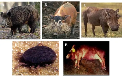
Ornithodoros erraticus, that inhabits warthog burrows (Figure 1). The soft tick parasites the warthog, thus creating a cycle of infection that maintains the virus in Africa. The virus is well adapted to these hosts producing infections that can persist for months or years. The soft tick can transmit the virus both to wildlife host and domestic pigs; porcine hosts can pass the virus between themselves through direct contact 52, 102, 105. The bush pig and warthog are thought to
be the natural reservoir of ASFV since they show no clinical signs of disease once infected with virulent and haemorrhagic isolates of the virus, that kill domestic pigs within 5-7 days post infection 52, 72, 73.
A B C
D E
A B C
D E
Figure 1 – Swine hosts of ASFV and vectors of infection.
(A) domestic pig, Sus scrofa, (B) bush pig, Potamochoerus porcus, (C) warthog,
Phacochoerus aethiopicus, (D) the soft ticks Ornithodoros moubata and Ornithodoros erraticus (E) domestic pig killed by ASFV with the characteristic haemorrhagic syndrome 108, 109.
After the original report of ASFV in Kenya in the 1920s, the virus was then detected in most sub-Saharan countries, where it is still endemic. It started spreading and it escaped from
Africa evolving towards less virulent strains that cause very attenuated and chronic forms of the disease 105. Once it reached Europe through infected pork products, it was detected in
Portugal and Spain in 1957, then again in 1960 where it remained endemic until the 1990s. Other European countries have been affected as well the Caribbean (Cuba, Dominican Republic and Haiti), and Brazil 102. The disease has been eradicated from most of these
countries outside Africa, with the exception of Sardinia where it still remains since it appeared in 1982. More recently, ASF has led to major economic losses after spreading through many West African countries, as is the case of Madagascar where it first appeared in 1998. Less virulent isolates have also been described in domestic pigs, as they cause a reduced mortality and, in some cases, chronic infections. Pigs that recover from this condition may remain persistently infected for long periods, thus providing a reservoir for infecting healthy pigs 55, 96, 101, 102.
The virus infects cells of the monocytic lineage, mainly the central cell of the innate immune response, the macrophage, both in vivo and in vitro. One report 94 described infection of
endothelial cells in vitro. Depending on the viral strain, the virus codes between 100-150 polypetides. About 40 of these have been described as being incorporated into the viral particle. A further 16 have been identified as enzymes and 30-40 as possible host modification genes 35, 52. In order to infect and replicate in macrophages, in vivo, the virus has
evolved strategies to modulate and/or interfere with the host immune response to infection. During disease, circulating lymphocytes and neutrophils show reduced numbers in acute infections 24, 98, 103. This is typically accompanied by hypergammaglobulinaemia. Although
as initially suggested by some studies 97, 98, there are no classical neutralizing antibodies
against ASFV 16, 107, 71, 37). For instance, serum from animals infected with a low virulence
viral isolate provided protection from infection with virulent ASFV isolates and against low passage tissue culture adapted viral variants 107. Also, the same protective or neutralizing
activity for several virulent isolates was observed when testing a monoclonal antibody (mAb) that reacts against a 72 kDa ASFV protein 16. However, neither the immune pig sera, nor the
anti-72 kDa ASFV protein mAb neutralized high passage tissue culture adapted ASFV isolates. It has been suggested that the adaptation of ASFV isolates to tissue culture conditions may be associated with the loss of determinants important for virus neutralization
107.
Another important observation in reinforcing the existence of protective serological immunity was that after passive transfer of anti-ASFV serum to pigs, these animals were partially protected against a highly pathogenic virus isolate. Not only there was a delay in the appearance of viraemia and a reduction in the virus titers, but some of the animals presented no clinical symptoms after secondary challenge with the pathogenic isolate. In contrast, in the control group, symptoms and death occurred 4 days after the challenge 71.
In spite of evident need, no vaccine is yet available, and disease control relies on rapid diagnosis, implementation of sanitary measures and movement restrictions. Diagnosis of disease poses a major complication due to the varying pathogenesis caused by different isolates, the similarity to other viral haemorrhagic fevers and the need to transport highly infectious samples to labs for testing. This policy is difficult to implement in many African countries since it requires good infrastructures that are lacking in many of these countries. ASF continues to pose a major threat to pig farming worldwide. An effective vaccine would help to control a major pathogen of pigs in large endemic areas in Africa, provide an
alternative to mass slaughter of animals and protect non-endemic from accidental entry of the virus.
1.1.3.2) Virus structure, entry, replication cycle and assembly
ASFV is a large icosahedral virus and the only known member of the Asfarviridae family, genus Asfivirus 28, 29. It has a linear double-stranded DNA genome with structural features
similar to iridoviruses and genomic organization similar to poxviruses 29, 52. The virus
replicates and assembles in the cytoplasm and encodes enzymes and factors that are required for replication and transcription of the virus genome. Depending on the virus isolate, the genome varies between 170 and 190 kb 15, and sequencing of the complete genome of the
Spanish tissue culture adapted isolate, Ba71v 105, of ASFV suggests that this virus may
encode 151 major open reading frames (ORFs) 52.
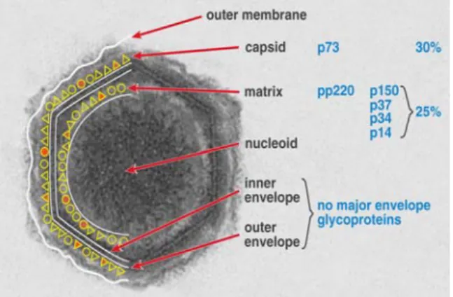
Extracellular ASFV particles have a diameter of approximately 200 nm and are formed by a DNA-containing nucleoid surrounded by three layers: an internal membrane, an icosahedric capsid, and an external membrane (Figure 2) 19. The virus enters cells through receptor
mediated endocytosis and is released from endocytic vesicles into the cytoplasm. This occurs through a mechanism of fusion of the virus envelope with the endocytic vesicles 2, 14, 93.
Studies involving the use of antibodies against virus proteins such as p12, p54, p72 and p30 have shown that these have important roles in the process of binding of the virus (p12, p72, p54) or internalisation (p30) 16, 35, 36, 37, 38.
Figure 2 – African Swine Fever virus structure. ASFV particles have a complex multi-layered structure. The nucleoprotein core structure is 70 to 100 nm in diameter. The nucleoprotein core is surrounded by a core shell and an internal envelope onto which the icosahedral capsid is assembled. The capsid is 170 to 190 nm in diameter. Although earlier reports suggested that this internal membrane consisted of a collapsed double membrane layer it has also been suggested that only one membrane layer is present. (Photograph from Linda Dixon, Institute for Animal Health, Pirbright Laboratory, UK)
Early virus mRNA transcription begins in the cytoplasm and is followed by virus replication through the use of host enzymes that are packaged in virions. These early transcripts encode proteins that include important enzymes such as DNA polymerase required for virus genome replication that occurs in the cytoplasm as early as 6 hours post infection. Late transcripts, on the other hand, largely encode structural proteins.
1.1.3.3) TLR signalling pathway
In vertebrates, the innate immune system is essential for host defence, as it constitutes the first line of defence against invading and potentially pathogenic microorganisms.
The host cells express various pattern recognition receptors (PRRs) that recognize pathogen-associated molecular patterns (PAMPs). For example, many pathogens contain carbohydrates and peptide derivatives, as well as methylated DNA, which the host can recognize as “non-self”. Once there is recognition of PAMPs by PRRs, an activation of intracellular pathways occurs, that leads to the induction of inflammatory cytokines, chemokynes, interferons (IFNs) and also upregulation of co-stimulatory molecules 49.
Innate immune receptors may be transmembrane signalling receptors such as Toll-like receptors (TLRs), or soluble components that opsonize microorganisms labelling them for recognition by complement receptors, Fc receptors or integrins. These receptors may trigger a variety of responses depending on the receptor and cell type: 1) through the mediation of microbe internalisation by phagocytic cells; 2) activation of antimicrobial mechanisms such as production of reactive nitrogen and oxygen molecules or species; and 3) stimulation of the production of inflammatory cytokines and chemokines that activate other immune cells 92.
In mammals, TLRs serve as key PRRs and play a crucial role in the induction of innate immune responses and consequent development of adaptative immune responses 49.
The Toll receptor was originally identified in Drosophila as an essential receptor for the establishment of the dorso-ventral pattern in developing embryos. In fact, Hoffmann and colleagues demonstrated, in 1996, that Toll-mutant flies were highly susceptible to fungal infection 56.
TLRs are abundantly expressed on antigen presenting cells such as dendritic cells and macrophages 49. The cytoplasmic portion of TLRs shows high similarity to that of the
Despite this similarity, the extracellular portions of both types of receptors are structurally unrelated. The IL-1 receptors possess an Ig-like domain, whereas TLRs bear leucine-rich repeats (LRRs) in the extracellular domain 87.
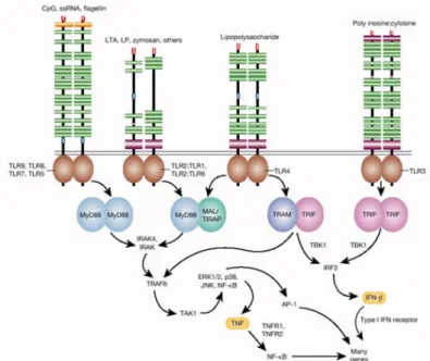
TLRs signal through two major pathways: the MyD88-dependent pathway and the TRIF (Toll/IL-1R domain-containing adaptor-inducing IFN-beta)-dependent pathway. The MyD88-dependent pathway triggers the release of pro-inflammatory cytokines, such as TNF-alpha, IL-6, and IL-12p40. MyD88 recruits various molecules that include IRAK4 (IL-1R-associated kinase 4), IRAK1 and TRAF6 (TNFR-associated factor 6) which in turn activate the nuclear factor-κB (NF-κB) that induces pro-inflammatory cytokine genes. The TRIF-dependent pathway is unique to TLR3 and TLR4 and contributes to pro-inflammatory cytokine responses as well as to the induction of type I IFN responses, particularly IFN-beta. To achieve this, TRIF recruits TRAF3, TBK (TRAF family member-associated NF-κB activator binding kinase) and IKKepsilon (Iκ B kinase epsilon), that activate IRF3 (IFN-regulatory factor 3) (Figure 3) 9, 25, 86.
The Toll family of proteins seems to be conserved, since homologous proteins have been described not only in Drosophila but also in other organisms, including plants. Thirteen TLRs (TLR1 to TLR13) have been identified in humans and mice together, and equivalent forms of many of these have been found in other mammalian species. However, equivalents of certain TLR found in humans are not present in other mammals 31, 21, 86.
Different TLRs exhibit specific responses. In fact, they have their own signalling molecules to manifest these specific responses and can be classified according to the types of PAMPs they are able to recognize (Figure 4) 87. For instance, TLR2, 4 and 6 recognize lipids. In addition,
TLR4 together with its extracellular components, MD-2 and CD14, can also recognize lipopolysaccharide (LPS) from Gram-negative bacteria. TLR2, in turn, forms heterodimers with TLR1, TLR6 and CD36 to discriminate molecules such as peptidoglycan, lipopeptides and lipoproteins of Gram-positive bacteria, mycoplasma lipopeptides and fungal zymosan. TLR5 recognizes protein ligands such as bacterial flagellin. Human TLR10 is thought to heterodimerize with TLR2 and TLR1, but the target ligand remains unknown 5, 49, 95.
One of the best ways to understand the central role for TLR in inflammatory responses is through mice lacking specific TLR or signalling molecules. For instance, mice lacking TLR4 are non responsive to LPS and, consequently, more susceptible to infection by Gram-negative bacteria such as Salmonella typhimurium, and mice deficient in TLR2 show a profoundly diminished response to Gram-positive organisms such as Staphylococcus aureus. In spite of these results, it is important to note that not all TLR deletions lead to severe infection phenotypes. This suggests that there may be a redundancy in recognition since probably all microorganisms are most likely to be recognized by multiple TLRs and not just one. In addition to being recognized by TLRs, microbes are also recognized by other innate recognition systems, as a way to prevent pathogens from easily subverting recognition and in consequence generating a controlled inflammatory response 92.
1.1.3.4) The I329L gene of the ASFV
ASFV is adapted to survive in vertebrate and invertebrate host cells. Both types of cells have defence mechanisms that are regulated by TLRs. In the porcine host, the virus mainly infects the macrophage, a key cell in innate immunity, as described earlier. For these reasons, the ASFV genome has been analysed using bioinformatic tools, and through this analysis the I329L gene was identified.
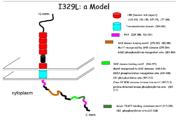
ORF I329L is a putative transmembrane protein of the ASFV of unknown function, with an extracellular domain of 222 amminoacids, a central transmembrane region formed by 21 residues, and 40% homology with the highly conserved 69 amminoacid intracellular C-terminal domain of mammalian Toll like receptors (TLRs). The protein coded by this gene shows a variety of potential glycosylation sites (Glc NAc) in the following amminoacids: 32, 39, 44, 76, 82, 101, 105, 219. These features suggest that I329L may be a candidate viral TLR
homologue. In fact, searches for similarities with other proteins in databases, in particular after alignment with TLR proteins, have shown a significant similarity between I329L and TLR3 from Drosophila melanogaster, specifically in a repetition site rich in leucine residues.
Figure 5 – Hypothetical model for I329L based of bioinformatic tools. TLRs are extremely rich in leucine residues as well as the conserved region of this protein. The extracellular portion of the molecule is formed by LRR repeats, an important motif for the interaction of these protein wit other molecules. Even though it is not clear whether I329L has a TIR domain, it resembles TRIFs in its cytoplasmatic domain. This suggests that this gene may modulate signalling through its interaction with TRIF. (Unpublished model developed by Vívian Oliveiraat the IGC)
The demonstration of interference of this gene with the activation of NF-κB, through a TLR-NF-κB reporter gene assay, has been performed in our lab by Vívian Oliveira (unpublished
data) and constitutes the stimulus for my work. The reporter gene assay consists on transfecting HEK-293T cells with three plasmids: one, containing a NF-κB-driven luciferase reporter, a second containing a CD4 extra-cellular domain co-ligated upstream of a set of TLR intracellular domains comprising TLR-1-6. As CD4 domains spontaneously dimerize when incorporated into the 293T cell membrane, the TLR intracellular domains are automatically brought into apposition. Therefore, there is constitutive signalling from the dimerized TLRs, and the resulting activation of NF-κB is detected by synthesis of luciferase. The effect of the co-transfection of the candidate gene cloned into the third plasmid provides an assay for interference of the viral protein with the TLR signalling pathway. Empty pcDNA3 serves as the negative control. It was observed that I329L inhibits NF-κB activation stimulated by multiple TLRs. This is of great importance since the NF-κB family of eukaryotic transcription factors is expressed in virtually all cell types and is involved in the regulation of expression of various genes important in immune response, inflammation and apoptosis, namely cytokines and its receptors, chemokines, growth factors and cell adhesion molecules 10, 11. Later
unpublished experiments by Vívian Oliveira showed that stimulating cells with the TLR3 ligand, polyinosinic-polycytidylic acid (poly(I:C)), a double stranded RNA analog, confirmed that I329L inhibited the TLR signaling transduction pathway.
These results strongly suggest that I329L is a host modification gene and thus a suitable candidate for the construction of a mutant deletion virus vaccine. Work towards such a vaccine and a deeper understanding of the role of I329L in host-pathogen interaction constitutes the objective of this project.
1.2)
O
BJECTIVEThis project is focused on exploring the function and utility of a host modification gene from the African Swine Fever Virus (I329L gene) manipulating toll receptor like (TLR) signalling. The goals of this project were to (1) determine whether I329L is an essential gene, (2) gain insights into function of the gene in the pig macrophage cell, and (3) assess of the impact of I329L in vivo by constructing a transgenic mouse selectively expressing I329L in macrophages. Such a transgenic mouse would be a model to understand how interference with TLR signalling might manipulate macrophage function in health and disease.
2)
I329L
IS AN
ONE
SSENTIALG
ENE OF THEASFV
2.1)
I
NTRODUCTIONOne of the possible approaches to produce an attenuated ASFV is through the deletion of genes known to be involved in evasion of the host immune system. As described earlier in this thesis, the gene I329L is a candidate for the construction of a mutant deletion virus vaccine. In order to test this possibility, it is first necessary to demonstrate that this gene is non-essential for the virus growth and replication in vitro. If the gene was essential to the virus, then its deletion would not yield a viable virus.
There are two possible approaches to test if this gene is essential to ASFV: 1) the knockdown of I329L through siRNA technology and 2) the construction of an I329L mutant deletion virus.
2.1.1) Inhibition of expression of I329L through siRNA technology
RNA interference (RNAi) is the process of mRNA degradation that is induced by double-stranded RNA (dsRNA) in a sequence specific manner. This process has been observed in all eukaryotes, from yeast to mammals. The RNAi pathway is thought to be an ancient mechanism for protecting the host and its genome against viruses that use dsRNA in their life cycles. This process plays a role not only in RNA and dsRNA stability/degradation but also in the regulation of translation, transcription, chromatin structure, and genome integrity. In the RNAi pathway, the dsRNA is processed to short interfering RNA (siRNA) which consists in 21-25 bp dsRNA with dinucleotide 3’ overhangs. Specifically, the guide strand of the siRNA
is assembled into an RNA-induced silencing complex (RISC) that cleaves the target mRNA
79, 84.
Due to its efficiency in specifically silencing the expression of any gene for which a sequence is available, it has been widely used and has become the cornerstone of many research projects.
The production of infectious virus by appropriately siRNA transfected cells will prove that I329L is a non-essential gene and thus a candidate for generation of an attenuated I329L-deleted ASFV vaccine.
Conventional transfection was used for transferring siRNA to Vero cells for gene silencing. The conditions for I329L specific siRNA delivery were optimized to avoid cytotoxicity. Two RNA regions were selected and the corresponding siRNA oligonucleotides were purchased. At first, these were tested separately, but later on both were combined, as this was more effective than a single siRNA, to achieve knock down. 24 hours post-transfection of siRNA, Vero cells were then infected with Ba71v. To confirm gene silencing, RT-PCR to detect I329L mRNA was performed on transfected Vero cells collected 18 hours post-infection. As a control, amplification of another ASFV gene (A238L) was also performed on the same samples in order to confirm the specificity of the silencing. The A238L gene of the ASFV is an inhibitor of transcription that encodes a bifunctional protein that inhibits both NF-κB and NFAT activation 65, 66, 75, 76. We chose this gene as a control, since it is expressed throughout
the cycle of the virus, including the time point that we were studying. A non-specific siRNA sequence from Dharmacon served as the negative control. Non-targeting, negative controls provide a baseline for measuring the effects of experimental siRNAs. Negative siRNA controls lack sequence complementarity to gene products and should have no effect in mRNA stability, mRNA expression, or protein expression. These controls are important for
distinguishing sequence-specific silencing from non-specific effects in the RNAi experiment. There are a variety of negative controls available, but we used a “scramble” control. This strategy consists in using a siRNA that has the same nucleotide composition, but not the same sequence, as the test siRNA. This is achieved in two ways: randomizing (also known as scrambling) the nucleotides in the siRNA or reversing the sequence of the siRNA. When their sequences fail to target a known gene or have a miRNA seed region match with a known gene, these siRNAs are appropriate as negative control siRNAs.
2.1.2) Construction of the I329L mutant deletion virus
The genome of ASFV can be manipulated through a process of homologous recombination between the virus genome and plasmid vectors transfected into the infected cell. Even though this kind of approach has been limited by the lack of suitable transfer vectors, García et al. described the construction of plasmid vectors with applications permitting the generation of recombinant viruses containing multiple deletions as well as viruses expressing foreign genes. In this work, the plasmid p72GUS10T (5.1 kb) was selected. This contains an ASFV chimeric gene formed by the fusion of the coding sequence of the gusA gene to the virus promoter p72.4 which was cloned into the polylinker region of plasmid pUC118 34.
2.2)
M
ETHODS ANDM
ATERIALS2.2.1) Cell lines
Vero cells (African green monkey epithelial cells) were cultured in Dulbecco's Modified Eagle's Medium (DMEM) supplemented with 10% Fetal Calf Serum, FCS, (heat inactivated), 2mM L-Glutamine, 100 Units/mL Penicillin, 100 μg/mL Streptomycin, all purchased from Gibco, Invitrogen Corporation.
Cells were routinely checked for mycoplasm contamination by performing a specific PCR.
2.2.2) siRNA Transfection and Ba71v infection
2.2.2.1) Treatment with siRNA oligos
siRNA targets were used to knock down expression of I329L in Vero cell lines. siRNA target sequences were purchased from Dharmacon. We chose two target sequences to be transfected into Vero cells: target 1, CAACCTACCTATATTACAA-3’ and target 2, 5’-TTATGCTCCTGTATATCAT-3’. These primers were kept at a 75 µM stock concentration. We first transfected each target separately but afterwards we had to combine the two in transfection in order to see an effect.
3x10⁵ cells/well were seeded on 6 well plates and left overnight. On the following day, cells were transfected using Lipofectamine 2000 (Invitrogen) according to the manufacturer’s instructions and left overnight. On day 3, 24 hours post-transfection, cells were infected with Ba71v using a multiplicity of infection (MOI) of 5. Ba71v is a tissue culture adapted Spanish isolate of the ASFV that encodes 151 major ORFs 105. All procedures regarding the use of
Ba71v were carried out at the P3 laboratory facility of the Instituto Gulbenkian de Ciência (Oeiras, Portugal) according to the in-house security rules.
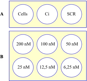
To perform the siRNA experiment we used the appropriate controls and also tested the minimum quantity of siRNA needed to observe gene knockdown. It is important to use as little quantity of siRNA possible as too much siRNA transfected into the cells may lead to off-target or cytotoxic effects. On the other hand, if too little siRNA is used, reduction of off- target-gene expression may be undetectable. The experimental scheme was according to figure 6.
Cells Ci SCR 200 nM 25 nM 100 nM 50 nM 12,5 nM 6,25 nM A B Cells Ci SCR 200 nM 25 nM 100 nM 50 nM 12,5 nM 6,25 nM Cells Ci SCR 200 nM 25 nM 100 nM 50 nM 12,5 nM 6,25 nM A B
Figure 6 – siRNA experimental design scheme. A) We used three controls: Vero cells only, Ba71v infected cells (Ci) and negative control scramble transfected and Ba71v infected Vero cells (SCR). B) Vero cells were transfected with different quantities of siRNA (200nM-6.25nM) as well as infected with Ba71v (MOI 5).
2.2.2.2) Demonstration of siRNA inhibition of I329L expression by RT-PCR
Total RNA was extracted from siRNA transfected and Ba71v infected Vero cells, using Trizol Reagent (Sigma). Samples of RNA were digested with DNase I (Invitrogen) and cDNA synthesis was performed using MMLV-Reverse Transcriptase (Invitrogen) according to the manufacturer’s instructions. I329L expression in the cDNA was detected by PCR, using Taq DNA Polymerase, on a PTC-100 Peltier-Effect Cycling apparatus. Primer sequences were I329Lup, 5’-GCTACTTCTTCTTGAACATGA-3’ and I329Llow, 5’-GCTTAGGAAGTG GCTTAACAGG-3’ and PCR conditions were dNTPs 200 µM, primers 1 µM, MgCl2 2 mM,
94ºC – 4’, 40X (94ºC – 1’, 50ºC – 1’, 72ºC – 2’), 72ºC – 10’. As a quantitative and qualitative control, tubulin was amplified using Taq DNA Polymerase with dNTPs 200 µM, primers 1 µM, MgCl2 2 mM, 94ºC – 4’, 40X (94ºC – 1’, 55ºC – 1’, 72ºC – 2’), 72ºC – 10’. Tubulin
primer sequences were the following: TubulinUp, 5’-GGTGGATCTAGAACCTGGG-3’ and TubulinLow, 5’-CCCAGTGAGTGGGTCAGC-3’. To control that only I329L expression was being targeted and not any other viral gene, the cDNA production for another ASFV gene, A238L, was also tested. A238L PCR was performed with Taq DNA Polymerase with dNTPs 200 µM, primers 1 µM, MgCl2 2 mM, 94ºC – 3’, 40X (94ºC – 1’, 54ºC – 1’, 72ºC – 1’), 72ºC
– 3’ and the primer sequences were A238Lup 5’-CTAGAATTCATGGAACACATGTTTCC A-3’ and A238Llow 5’-CTACTCGAGTTACTTTTCATACTTGTT-3’.
2.2.2.3) Detection of intracellular and extracellular virus
After knocking down the expression of I329L through siRNA we wanted to know if this gene was essential for the ASFV. With that purpose we 1) stained Ba71v infected Vero cells as well as siRNA transfected and Ba71v infected Vero cells, by immunofluorescence, using a
p73 specific monoclonal antibody; and 2) infected freshly plated Vero cells with the supernatants of each of the siRNA wells experiments, in order to count viral plaques.
For immunofluorescence, sterilized glass coverslips were put in the wells as the cells were being seeded for the siRNA assays. As some of the cells adhered to the coverslip surface, we used them for a staining with an antibody against p73, a major capsid protein of ASFV. Cells were fixed with 4% paraformaldehyde (PFA) in PBS for 20 minutes at room temperature (RT). Blocking was performed at RT for 30 minutes with PBS containing 0.05% Tween-20 and 5% Normal Goat Serum (NGS). The cells were permealized using PBS containing 0.1% Triton X-100. The staining with the primary monoclonal antibody 4H3 specific for p73 was performed at RT for 1 hour in blocking solution with a previously titrated concentration of the antibody. The secondary staining was also performed at RT for 1 hour with a goat anti-mouse IgG Alexa 568 antibody, purchased from Molecular Probes. To stain the nucleus of the cells, they were incubated in a DAPI solution (20 ng/µl) for 2 minutes. All washes between stainings were performed at RT in PBS containing 0.05% Tween-20.
Viruses were recovered from the supernatants of siRNA transfected and infected cells. These, were then titrated with the purpose of counting viral plaques, to determine if infectious progeny was being produced after knocking-down I329L.
In order to titrate the supernatants we used freshly plated Vero cells and infected them with a series of dilutions of the supernatants from pure supernatant to 10-1, 10-2, 10-3, 10-4 and 10-5 dilutions. DMEM medium with 0.5% agarose was added to the infected cells and left for five days. Afterwards, the cells were washed with PBS 1X and fixed with 4% PFA for 20 minutes
at RT. The cells were then stained with 0.1% toluidine blue for 30 minutes at RT and washed with tap water. After drying, the viral plaques were counted.
2.2.3) Cloning of the mutant deletion virus construct
In order to delete I329L from Ba71v the left and right flanking regions (~700 bp each) of the ASFV gene were cloned on either side of the p72.4 Gus 10T cassette. For instance, the amplification of the left flank of the gene was performed by inserting an EcoRI site at the 5’ end and a KpnI or SmaI site at the 3’end (in this case we used a KpnI site). These sites were cloned upstream of p72.4 Gus 10T. The right flank of the gene was also amplified by inserting a PstI site at the 5’end and a HindIII site at the 3’end, in order to clone it downstream of the p72.4 Gus 10T cassette.
KpnI/SmaI PstI
p72.4 Gus 10T
~ 2 kbp
I329L LF I329L RF
EcoRI KpnI/SmaI PstI HindII
p72.4 Gus 10T
~ 2 kbp
I329L LF I329L RF
EcoRI HindII
Figure 7 - Cloning strategy of I329L left (LF) and right flanks (RF) into p72.4 Gus 10T plasmid.
The right and left flanks (RF and LF, respectively) were amplified by PCR using Pfu DNA Polymerase, on a PTC-100 Peltier-Effect Cycling apparatus. Primer sequences were I329LRFup, 5’- GCCTGCAGCCTGTATATACTATTAAAAATT -3’, I329LRFlow, 5’- GCAAGCTTACCCTGATGGAAGACTATGTAT-3’, I329LLFup, 5’- GCGAATTCCATAA
AGTATG -3’. PCR conditions were dNTPs 200 µM, primers 1 µM, MgSO4 2 mM, 94ºC – 4’,
30X (94ºC – 1’, 55ºC – 1’, 72ºC – 2’), 72ºC – 10’. These fragments were restriction digested and cloned into p72.4 Gus 10T according to the scheme in figure 7.
After infection of Vero cells with Ba71v, followed by transfection of the I329LLF-Gus-I329LRF, progeny virus will be harvested, and the recombinants will be identified and purified by isolating viruses/plaques expressing the Gus gene (blue on X-Gluc substrate). Through the use of this strategy the Gus gene can be stably inserted into the Ba71v genome, replacing the I329L gene through recombinant homology.
The cells will then be infected with the mutant deletion virus to check that there is virus production and thus confirm that I329L is a non-essential gene. Even though the construct is already cloned and verified by sequencing, the remaining part in the construction of the deletion virus is ongoing work in our lab.
2.3)
R
ESULTS2.3.1) Expression of I329L during Ba71v infection in Vero cells
The ASFV ORF I329L has been described as a late expression gene 77. In order to know the
exact time point of expression of I329L during virus infection, we infected Vero cells with Ba71v and recovered the cells at different time points of infection: 8h, 10h, 12h, 14h, 16h, 18h and 20h and the expression of this I329L gene was determined by RT-PCR. As can be seen, I329L is indeed a late gene, its mRNA first being detected at 14h, 16h and 18h (Figure 8). 1 2 3 4 5 6 7 8 I329L Tubulin A B 1 2 3 4 5 6 7 8 I329L Tubulin A B
Figure 8 – Expression of I329L during Ba71v infection. A) RT-PCR for the I329L gene is negative for non infected cells (lane 1) as well as for time points 8h, 10h and 12h after infection (lanes 2, 3 and 4). I329L was expressed at 14h, 16h and 18h post infection (lanes 5, 6 and 7), but not at 20h post infection (lane 8). B) PCR for tubulin, as a control of the quantity and quality of the cDNAs.
After demonstrating transcription of I329L at 14h, 16h and 18h post infection, we decided to use the 18h time point for the next steps of the siRNA experiment.
All the following procedures regarding siRNA described in this thesis were performed at this time point of infection.
2.3.2) Transfection of siRNA targets in Ba71v infected Vero cells
We first decided to test the transfection of each siRNA target (1 and 2) separately in order to see if one target sequence was able to induce the knockdown of the gene. However, under these conditions, I329L expression was not suppressed (Figure 9A, 9B). On the other hand, when we combined both targets and transfected them into the cells, an inhibition of expression of I329L was observed with a concentration of siRNAs of 200 nM (Figure 9C). The inhibition was specific and the combined treatment with two siRNA probes did not inhibit transcription of A238L (Figure 9D).
I329L A238L Tubulin 1 2 3 4 I329L I329L A 1 2 3 4 B C D E (Target 1) (Target 2) I329L A238L Tubulin 1 2 3 4 I329L I329L A 1 2 3 4 B C D E (Target 1) (Target 2)
Figure 9 – Expression of I329L after transfection of siRNA targets and infection with Ba71v. A, B) Transfection of targets 1 and 2 respectively, non combined. RT-PCR for the I329L gene is negative for non transfected and non infected cells (lane 1), but positive for infected cells (lane 2) as well as for cells transfected with siRNA negative control Scramble (lane 3). Cells transfected with only one of the siRNA targets do not inhibit I329L expression (lane 4). C) RT-PCR for the I329L gene is negative for non transfected and non infected cells (lane 1), and positive for infected cells (lane 2) as well as for infected cells transfected with siRNA negative control Scramble (lane 3). Lane 4 demonstrates that I329L expression was knocked down by transfection of a combination of
both siRNA targets at a final concentration of 200 nM. D) RT-PCR for the A238L gene (lanes 2, 3 and 4) not only demonstrates that the cells are infected, but also that the siRNA target sequences are specific for I329L and have no effect in the expression of other viral genes. E) Tubulin amplification served as a control for the quantity and quality of the cDNAs.
2.3.3) siRNA titration in Ba71v infected Vero cells
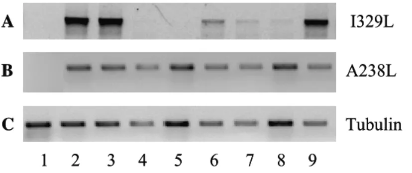
It is important to use the minimum quantity of siRNA as possible, as too high concentrations of siRNA transfected into the cells may lead to off-target or cytotoxic effects. On the other hand, if too little siRNA is used, the reduction of target-gene expression may be insufficient. Thus the amounts of siRNA tested in the transfection step were titrated (200 nM-6.25 nM). Effective knockdown was observed at oligonucleotide concentrations of 100 nM or 200 nM (lanes 5 and 4 respectively, Figure 10A). Lower quantities did not result in the inhibition of I329L expression (lanes 6-9, Figure 10A). Once again, there was no impact on transcription of the positive control, A238L.
I329L A238L Tubulin 1 2 3 4 5 6 7 8 9 A B C I329L A238L Tubulin 1 2 3 4 5 6 7 8 9 A B C
Figure 10 – Titration of the quantity of siRNA targets in infected Vero cells. A) RT-PCR for the I329L gene is negative for non transfected and non infected cells (lane 1), and positive for infected cells (lane 2), as well as for cells
transfected with siRNA negative control Scramble (lane 3). Lanes 4-9 correspond to 200 nM, 100 nM, 50 nM, 25 nM, 12.5 nM and 6.25 nM of siRNA target transfection titration respectively. I329L expression is knocked down at 200 nM and 100 nM concentrations of siRNA targets (lanes 4 and 5). Lower concentrations of siRNA target were unable to knockdown expression of I329L (Lanes 6-9). B) RT-PCR for the A238L gene not only demonstrates that the cells are infected, but also that the siRNA target sequences are specific for I329L and have no effect in the expression of other viral genes. C) PCR for tubulin, as a control of the quantity and quality of the cDNAs.
2.3.4) Analysis of virus production from infected Vero cells transfected with I329L siRNA targets
After confirming the specific knock-down of I329L through siRNA technology, we next investigated 1) the impact of knockdown of I329L on virus production and 2) the production of infectious virus by cells by cells with synthesis of I329L knocked down.
In order to achieve these objectives, we 1) stained the cells with an antibody against p73, a major capsid protein, to check that the cells were being infected and 2) recovered the supernatants from the siRNA transfected and for infection of Vero cells followed by counting the number of viral plaques.
2.3.4.1) Staining of p73, the major capsid protein of ASF virus
The major structural protein of ASFV is a capsid protein, p73. This protein is thought to associate with the membranes that form the envelope of the virus 23. Cobbold et al. generated
a mouse monoclonal antibody 4H3 to determine the onset of expression of p73 and the time course of packaging of capsid proteins into virions.
We used this antibody followed by a Goat anti-mouse IgG Alexa 568 secondary antibody in cells infected with the culture supernatants from the control and I329L specific knocked down Ba71v virus infected Vero cells.
As can be seen, there was no difference in the expression of p73 with or without siRNA knockdown of the I329L gene at all the concentrations of siRNA (200nM-6.25nM) tested. Only two of the images of the staining are presented in this thesis, one with just infected cells and one with both siRNA transfected (200nM) and infected cells (Figure 11).
A
B
A
B
Figure 11 – Staining of Vero cells with 4H3 monoclonal antibody. p73 can be observed in red and DAPI (blue) stains the nucleus. The staining was identical both in A) Ba71v infected cells and in B) siRNA transfected and Ba71v infected Vero cells (Magnification 40X).
2.3.4.2) Analysis of viable virus production from siRNA recovered supernatants
After confirming the specific knockdown of I329L through siRNA technology we used the supernatants of the Ba71v virus infected cells with and without siRNA mediated knockdown of I329L.
By comparing the number of viral plaques of Ba71v infected cells and Ba71v infected and transfected cells (with siRNA in the concentrations range defined) we could conclude that there was no impact in the production of infectious virus. Thus knocking-down I329L did not affect the production of infectious ASFV virus, and therefore I329L is truly a non-essential gene.
Cells only Cells
infected Scramble 200 nM 100 nM 50 nM 25 nM 12.5 nM 6.25 nM 0 0,5 1 1,5 2 2,5 3 3,5 4 4,5 pf u/mL / 1 0 3 1 2 3 4 5 6 7 8 9
Cells only Cells
infected Scramble 200 nM 100 nM 50 nM 25 nM 12.5 nM 6.25 nM 0 0,5 1 1,5 2 2,5 3 3,5 4 4,5 pf u/mL / 1 0 3 1 2 3 4 5 6 7 8 9
Figure 12 – Counting of viral plaques after siRNA supernatant titration. We observed no significant difference in the number of viral plaques by comparing the number of plaques counted both after infection with the supernatant of Ba71v infected cells (non siRNA transfected, sample 2) and the supernatant of Ba71v infected and scramble siRNA transfected cells (sample 3), with the number of
plaques counted after infection with the supernatants of both I329L siRNA transfected and Ba71v infected cells (samples 4-9).
2.4)
D
ISCUSSIONRNAi is an effective method of gene silencing, and is a very important tool in the study of genetics, molecular biology and physiology. It has been yielding considerable insights into the innate and adaptive immune systems, namely into the mechanisms that regulate the development, activation and function of cells that mediate immunity. Even though this technique may in the future be tested for clinical purposes, its limitations such as inefficient delivery in vivo, incomplete silencing of target genes, non-specific immune responses and off-target effects, must first be overcome. Nevertheless, it seems to be a powerful and promising tool applicable to vaccination and immunotherapy 61.
The I329L ORF is a host modification gene of the ASFV, as suggested by previous results in our lab, and therefore a suitable candidate for the construction of a mutant deletion virus vaccine. We decided to take two approaches to investigate whether this gene was essential to the virus or not.
In the first one, we used siRNA oligos in order to specifically knockdown the expression of I329L in Ba71v Vero infected cells. The I329L gene, already described as a late expression gene 77, was knocked down at the time of its expression at 18 hours post infection. Upon
siRNA oligo transfection we were able to see an inhibition of expression when using a combination of two oligos at 100 and 200 nM. Furthermore, supernatants recovered from cells infected with virus, but with expression of I329L knocked down, contained similar numbers of effective virus as control non-knocked down infected cultures. To further confirm that the cells had been infected, we performed immunofluorescence using a monoclonal antibody against p73, a major capsid protein of the virus. Once again, we observed no difference between the staining of control infected cells and cells that were both infected and
treated with siRNA to knockdown the expression of I329L. In addition, these results show that p73 expression is not affected by the knocking down of I329L and, in consequence, virus envelope formation is not altered.
The fact that the knockdown of expression of this gene does not affect assembly and production of infectious virus, not only shows RNA technology can be used to inhibit I329L synthesis in ASFV infected cells, but also constitutes the key finding of this study, namely that I329L is a non essential gene of the ASFV. Thus, the data presented in this chapter, together with the fact that I329L inhibits TLR3, makes this host manipulation gene a possible candidate to delete for the production of a viral attenuated vaccine.
Another approach to show that this gene was not essential to the virus was through the construction of an I329L mutant deletion virus. To this end, the necessary recombinant transfer plasmid for construction of the I329L deletion mutant has been constructed and the remaining work towards construction of the I329L deletion mutant is ongoing in our lab. Whether such a mutant will indeed prove to be a vaccine candidate will depend on its pathogenicity in the pig. If still pathogenic, one approach to achieve the vaccine would be to sequentially delete other genes known to be involved in evasion of the host immune system, such as I329L, or genes required for virus virulence or for efficient replication in the tick vector. The deletion of these immune evasion genes might change the balance in favour of the virus towards an effective host immune response and thus an increased chance of developing an effective protective immune response in vaccinated animals. This strategy has been shown to be efficient in Herpes simplex virus, where the blocking of virus immune evasion proteins improved the potency of the vaccine 48.