RESUMO.- [Identificação de novas flagelinas codifica
-das por fliC em Escherichia coli isoladas de animais
domésticos utilizando RFLP-PCR e sequenciamento.] A identificação da Escherichia coli requer conhecimento
sobre os sorotipos e fatores de virulência prevalentes per -mitindo a classificação em patogênico/não patogênico. No entanto, algumas destas bactérias não expressam o antíge -no flagelar in vitro. Neste caso, o PCR-restriction fragment
length polymorphism (RFLP-PCR) e o sequenciamento do gene fliC podem ser adequados para a identificação desses antígenos, substituindo a sorologia tradicional. Nesta pes -quisa foram estudadas 17 amostras de E. coli isoladas de
animais e que apresentavam antígeno H não tipável (HNT).
Identification of new flagellin-encoding
fliC
genes in
Escherichia
coli
isolated from domestic animals using RFLP-PCR and
sequencing methods
1Cláudia de Moura2*, Monique Ribeiro Tiba3, Marcio José da Silva4
and Domingos da Silva Leite3
ABSTRACT.- Moura C., Tiba M.R., Silva M.J. & Leite D.S. 2013. Identification of new fla
-gellin-encoding fliC genes in Escherichia coli isolated from domestic animals using RFLP-PCR and sequencing methods. Pesquisa Veterinária Brasileira 33(4):417-422. Uni-versidade Paulista, Av. Armando Giassetti 577, Vila Hortolândia, Trevo Itu/Itatiba, Jundiaí, SP 13214-525, Brazil. E-mail: [email protected]
Identification of Escherichia coli requires knowledge regarding the prevalent serotypes
and virulence factors profiles allows the classification in pathogenic/non-pathogenic. Ho -wever, some of these bacteria do not express flagellar antigen invitro. In this case the PCR --restriction fragment length polymorphism (RFLP-PCR) and sequencing of the fliC may be suitable for the identification of antigens by replacing the traditional serology. We studied 17 samples of E. coli isolated from animals and presenting antigen H nontypeable (HNT).
The H antigens were characterized by PCR-RFLP and sequencing of fliC gene. Three new flagellin genes were identified, for which specific antisera were obtained. The PCR-RFLP was shown to be faster than the serotyping H antigen in E. coli, provided information on
some characteristics of these antigens and indicated the presence of new genes fliC. INDEX TERMS: Escherichia coli, H antigen, PCR-RFLP, sequencing, serotyping.
1 Received on January 15, 2013.
Accepted for publication on February 14, 2013.
2 Instituto de Ciências da Saúde, Universidade Paulista (UNIP), Av. Ar
-mando Giassetti 577, Vila Hortolândia, Trevo Itu/Itatiba, Jundiaí, SP 13214-525, Brazil. *Corresponding author: [email protected]
3 Laboratório de Antígenos Bacterianos II, Departamento de Genética,
Evolução e Bioagentes, Instituto de Biologia, Universidade Estadual de Campinas (Unicamp), Cx. Postal 6109, Campinas, SP 13083-970, Brazil.
4 Centro de Biologia Molecular e Engenharia Genética, Instituto de Bio
-logia, Unicamp, Cx. Postal 6109, Campinas, SP.
Os antígenos H foram caracterizados por PCR-RFLP e se -quenciamento do gene fliC. Três novos genes da flagelina foram identificados, para os quais anti-soros específicos foram obtidos. A técnica PCR-RFLP mostrou-se mais rápida que a sorotipagem do antígeno H em E. coli, fornecendo in -formações sobre algumas características desses antígenos e indicou a presença de novos genes fliC.
TERMOS DE INDEXAÇÃO: Escherichia coli, antígeno H, PCR-RFLP,
sequenciamento, sorotipagem.
INTRODUCTION
Escherichia coli is the predominant member of normal hu -man and animal intestinal flora. This species also includes different virulence factors and serotypes associated with intestinal and extraintestinal diseases. The antigen O and H (O polysaccharide and flagellin, respectively) are the two major antigens of Gram-negative bacteria (Blanco et al. 2003, Hussein 2007, Mattsson & Wallgren 2008). Since the early 1940s, the gold-standard technique for O and H characterization has been the agglutination test for E. coli
-miological studies and has allowed the characterization of pathogenic E. coli serotypes (Mattsson & Wallgren 2008).
Several serotypes are associated with human illnesses and all of them are pathotypes associated with animals: O2:H5, 6, 7, 29; O8:H2, 19, 21; O20:H19; O22:H8; O25:H2; O26:H11, HNT; O45:H2; O91:H10, 21; O103:H2; O105:H18; O111:H8; O112ac:H19, 5; O113:H21; O118:H16; O119:H2, 6; O121:H19; O128:H2; O128ab:H2, 6; O145:H25, 28, O146:H21; O153:H25; O157:H7; O163:H19; O165:H25; O174:H2; 721; ONT:H2, 8, 11, 25, 28, 33, and 41 (Blanco et al. 2003, Hussein 2007, Mattsson & Wallgren 2008). How -ever, several difficulties have been observed in H antigen serotyping: (I) the expression of H-antigens can be depen -dent on various environmental signals; (II) the i-dentifica -tion of H antigen is a time-consuming process and requires the use of 53 specific antisera; and (III) there are a high number of cross-reactions among E. coli strains (Blanco et
al. 2003, Hussein 2007, Mattsson & Wallgren 2008). This procedure is important, because identification of a partic -ular H antigen saves time and reduces the number of an -tisera required to identify the O antigens in E. coli strains
(Blanco et al. 1992, Moreno et al. 2006).
The flagellum (the organelle responsible for motility) consists of repeated subunits of the protein flagellin that are expressed by fliC gene (Fields et al. 1997). Studies have demonstrated that PCR-restriction fragment length poly -morphism (RFLP-PCR) analysis could be used for identi -fying these antigens, replacing serology as a traditional technique (Machado et al. 2000). The polymorphism of the fliC gene reflects the structure of the flagellin molecule. Amino-acid sequences among the flagellin proteins from different H serotypes are well conserved in their N- and C-terminal regions, which bear the essential functions for protein export through the flagellum specific type III se -cretion machinery and for polymerization into the filament (MacNab 1992). On the other hand, the central regions are variable in length and amino-acid sequence, carrying H serotype-specific epitopes (Reid et al. 1999).
The RFLP-PCR for fliC gene has been developed by other authors who have shown that the restriction analy -sis of this gene could be used to type both O157:H7 and O157:H-Shiga toxin-producing E. coli strains (Fields et al.
1997). Subsequently, in a study involving strains isolated from human sources, a data base was constructed from all restriction profiles of H patterns, allowing to identify through this technique all the genes involved in expression of flagellins (Machado et al. 2000). These methods have shown to be important for serotyping, determining genetic relationships and for epidemiological studies (Fields et al. 1997, Machado et al. 2000, Moreno et al. 2006). However, some H antigen cannot be characterized by RFLP-PCR for
fliC, and for this reason, some authors use sequencing me -thods to identify new putative flagellins expressed by fliC
genes or other genes that can express flagellin (Machado et al. 2000, Prager et al. 2003, Tominaga 2004). In the present study, we characterized the flagellin genes from 17 E. coli
strains, using PCR-RFLP methods and sequencing. We pro -duced antisera for serology identification of new flagellin genes and also to confirm the presence of new antigens.
The fliC-RFLP technique proved to be faster than classic serotyping for determining the E. coli H antigen, characteri -zing the antigens in a few days and indicating new putative genes.
MATERIALS AND METHODS
Bacterial strains. A total of 53 Escherichia coli control strains
for H antigen were analyzed, as well as 17 E. coli strains belonging
to theE. coli collection of the Bacterial Antigen Laboratory, De
-partment of Genetic, Evolution and Bioagents, Institute of Biology, Unicamp, Brazil. The strains were isolated from sporadic diarrhea
cases in different time periods from bovine, swine and sheep (Ta
-ble 1). The strains were serotyped using O and H standard antise
-ra (Blanco et al. 1992) and all of them presented the non typeable H antigen (HNT).
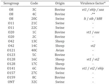
Table 1. Escherichia coli strains isolated from animals with the HNT pattern and their associated virulence factors
Serogroup Code Origin Virulencefactor* O8 3C Bovine vt1 / ehly / saa
O8 4C Bovine -**
O8 20C Swine lt / stb / k88
O11 21C Swine
-O11 22C Swine
-O20 1C Bovine vt1 / eae
O20 2C Bovine
-O42 13C Sheep
-O42 14C Sheep vt2
O121 40C Avian
-O123 5C Bovine
-O128 16C Sheep vt1 / vt2
O128 17C Sheep
-O141 6C Bovine vt1 / vt2 /ehly
O157 27C Swine
-O159 8C Bovine
-ONT 32C Swine
-* Virulence factor genes: lt = heat-labile toxin, stb = heat-stable toxin B, k88 = K88 fimbriae, vt1 = verotoxin 1, vt2 = verotoxin 2, ehly = Enterohe -molysin, saa = autoagglutinin protein, eae = gene for intimin. ** Negative
for all virulence factors studied.
DNA extraction, PCR and RFLP analysis. E. coli strains were
grown in 3ml of Luria-Bertani broth medium overnight at 37°C. The
genomic DNA was obtained using the Wizard® Genomic DNA kit (In
-vitrogen, USA). PCR for fliC gene and RFLP analysis was performed
according to the methods described previously (Fields et al. 1997,
Machado et al. 2000) using the FliCF1: 5’ATGGCACAAGTCATTAA
-TACCCAAC3’; FliCF2: 5’CTAACCCTGCAGCAGAGACA3’ and FliCM1: 5’CAAGTCATTAATAC(A/C)AACAGCC3’; FliCM2: 5’GACAT(A/G)TT
(A/G)GA(G/A/C)ACTTC(G/C)GT3’ primers (Fields et al. 1997, Ma
-chado et al. 2000). PCR was performed in the Thermal Cycler (Gene Amp PCR System 9700/Perkin Elmer Corporation, Norwalk CT/
USA), with 50 µL reaction volumes containing 2mM MgCl2, 1 µM
of each primer and 1.5 U of Taq DNA polymerase (Fermentas, Wal
-tham, USA). PCR was developed using cycles of denaturation for 1 min at 95°C, annealing for 1 min at 50-60°C and final extension step
for 7 min at 72°C. PCR-Fields products were digested with the RsaI
restriction enzyme (Invitrogen, USA) and PCR-Machado products
were digested with the HhaI restriction enzyme (Fermentas, USA)
according to the manufacturer’s instructions. The RFLP fragments were separated in 2% agarose gels by horizontal electrophoresis
for 3h at 10V/cm. The restriction fragments were stained with ethi
Pharmacia Biotech/ USA). Gel Compar II (Applied Maths/ Belgium) was used to identify RFLP patterns and to establish a database for
fliC fingerprinting. Fragments were considered identical if their si
-zes did not differ by more than 3.5% (allowable error).
Gene sequence analysis. Sequencing was carried out using the Big Dye kit (Amersham Biosciences, USA) and the 3700 DNA Analyzer (Applied Biosystems, Foster City, CA/USA) sequencer.
The sequence data were assembled using the ChromasPro packa
-ge (http://www. technelysium.com.au/chromas.html). Gene se
-quence searches were conducted using the BLAST and GenBank databases (NCBI website home page). Sequence alignments and comparisons were performed using ClustalW (http://www.ebi. ac.uk /Tools/clustalw2/index.html).
Nucleotide sequences and accession numbers. The DNA
sequences of the new fliC genes of the E. coli HNT were deposited
into the GenBank database under accession numbers HQ116826 to HQ116828.
Production of antisera of HNT antigen. The HNT antigen (non typeable antigens) suspensions used for the production of antisera were prepared according to the methodology described
previously (MacNab 1992). Bacterial strains were cultured in U
tu-bes in consecutive passages at 37°C for 18hs for E. coli motility. Af
-ter this, the strains were grown in Brain Hearth Infusion Broth (Di
-fco, Sparks, USA) at 37°C for 18hs and then were inactivated with an equal volume of formalin solution. These HNT antigens were inoculated in rabbits at serial doses of 0.5mL to 4.0mL and their blood was collected to obtain the antiserum and stored at -20°C.
Determination of HNT antisera titers and absorption of antisera. The titers of HNT antisera were determined by using
serial dilutions of antisera from 1:100 to 1:25,600. For the agglu
-tination tests, Kahn tubes were used containing 200µL of HNT of
each H antigen (homologous HNT antigen and all 53 H control an
-tigen) and equal volume of HNT antiserum. The tubes were incu
-bated at 45°C in a water bath for 3 hours. For unspecific reactions the antisera were absorbed against heterologous antigen using protocol described previously (Ewing´s 1986).
RESULTS
We began by testing the primers for PCR amplification for control strains and non-typeable strains. Then, the PCR products were submitted to RFLP analysis using specific restriction enzymes. To confirm the RFLP patterns, we se -quenced fliC genes and this allowed us to compare with an -tisera agglutination.
Detection of fliC genes in Escherichia coli strains by RFLP-PCR
With the exception of H17, H25, H53 and H54 (flagellin expressed by other genes), the fliC genes of 49 control E. coli H strains were amplified, digested, and submitted to
RFLP analysis. A common pattern was observed in RFLP --PCR for the fliC gene using primers FliCF1/2 and RsaI from
H1, H28 and H31 (P1); H2, H30 and H35 (P2), H3 and H8 strains (P3); H7, H19 and H27 (P7); H9 and H14 (P9); H11 and H47 (P11); H55 and H56 (P40). To further characteri -ze these alleles, we performed RFLP with the primers Fli -CM1/2 and HhaI endonuclease. The fliC genes encoding the
H3 and H8; H11 and H47; H19 and H27; H55 and H56 an -tigens were not differentiated by HhaI restriction analysis.
However, the H1, H28 and H31; H2, H30 and H35; H9 and H14; H7 strains were distinguishable when the PCR frag -ments were restricted with HhaI (Table 2).
Table 2. Results of RFLP-PCR assays of Escherichia coli H control strains
H an- PCR RFLP Pattern PCR RFLP Patern tigen (RsaI) bp (HhaI) bp
H1 1260 630, 330, 310 P1 1400 285, 195, 170, 70 P1 H2 1450 570, 410, 320, 120 P2 1650 1370, 180 P2 H3 1500 720, 320, 290, 150 P3 1540 360, 350, 150, 110 P3 H4 1105 440, 255, 230 P4 980 340, 285, 100, 60, 50 P4 H5 1290 1290 P5 1345 770, 260, 160, 120 P5 H6 1360 565, 335, 320 P6 1280 750, 150, 110, 70, 50 P6 H7 1260 570, 340, 330 P7 1290 790, 200, 150, 120, 105 P7 H8 1480 710, 330, 295, 150 P3 1520 360, 350, 150, 110 P3 H9 1970 1115, 315, 170 P8 2030 735, 470, 215, 120, 70 P8 H10 1260 540, 320, 310 P9 1250 740, 160, 115, 70, 50 P6 H11 1450 560, 300, 160 P10 1545 445, 435, 300, 220 P9 H12 1745 730, 410, 280, 160, 130 P11 1705 655, 410, 230, 175, 120 P10 H14 2035 1115, 315, 170 P8 1280 340, 245, 220, 110, 105, 60 P11 H15 1630 440, 325, 300, 230, 95 P12 1730 390, 360, 320, 215, 130 P12 H16 1610 390, 330, 300, 150 P13 1590 1220, 230, 140 P13 H17 -a 950 355, 305, 110, 70 P14
H18 1630 760, 420, 150, 120, 95 P14 1260 660, 250 P15 H19 1250 550, 335, 325 P7 1245 750, 150, 110, 70, 50 P6 H20 1800 385, 315, 300, 230, 200 P15 1870 710, 420, 200, 110, 60 P16 H21 1275 1275 P16 1325 720, 210, 110, 70, 55 P17 H23 1480 680, 390, 350, P17 1200 460, 320, 210, P18 300, 130 145, 105, 70
H24 1750 550, 440, 310, 275, 140 P18 1800 540, 340, 195, 145, 135 P19 H25 -a 1280 625, 195, 130, 125 P20
H26 1635 860, 570, 150 P19 1820 290, 260, 210, 180, P21
160,130, 100
H27 1230 560, 340, 330 P7 1330 740, 155, 110, 70, 50 P6 H28 1195 620, 335, 320 P1 1250 315, 235, 210, P22
110, 100, 80, 70
H29 1240 380, 340, 310, 175, 110 P20 1355 740, 280, 125, 80, 70 P23 H30 1730 590, 420, 310, P2 1800 410, 280, 240, 150, P24 120 115, 100, 85
H31 1250 610, 320, 310 P1 1255 380, 320, 285, P25 240, 215, 115, 65 H32 1690 760, 525, 305 P21 1760 430, 370, 300, 250, P26
210, 170, 130, 80
H33 1290 670, 420 P22 1150 235, 230, 210, 105 P27 H34 1665 640, 535, 415 P23 1650 670, 315, 160, 135 P28 H35 1425 570, 410, 310, 120 P2 1420 1210, 220, 195 P29 H36 2850 690, 560, 290, P24 2635 740, 595, 445, P30 210, 150, 105 305, 220
H37 1650 840, 330, 230, 130 P25 1770 680, 270, 240 P31 H38 1375 320, 180, 165, 150, 120 P26 1170 995, 130 P32 H39 1225 310, 280, 270, P27 1300 390, 250, 210, P33 210, 110, 90 170, 110, 105 H40 1530 315, 290, 250, 145, 85 P28 1595 380, 340, 195, 160 P34 145, 85
H41 1670 430, 320, 300, P29 1770 570, 440, 160, 130 P35 270, 215, 130
H42 1290 640, 320, 310, 95 P30 1245 320, 235, 210, 115, 70, 60 P22 H43 1510 390, 350, 300, P31 1575 465, 320, 215, 150, 115, 75 P18 290, 130 150, 115, 75
H44 1670/ 710, 610, 500, P32 925 335, 315, 275, P36 1035 300, 90 250, 190, 110, 70 H45 1730 430, 380, 315, 215, P33 1750 455, 410, 260, P37 140, 130, 110 250, 115
H46 1710 460, 315, 300, P34 1800 400, 310, 215, P38 250, 200, 105 180, 110, 80
H47 1510 575, 300, 155 P35 1540 445, 430, 300, 230 P9 H48 1470 630, 470, 290, 95 P36 1565 515, 290, 210, 125, 100 P39 H49 1680 410, 310, 290, 260, P37 1640 540, 350, 200, 150, 130 P40 210, 130, 70
H51 1790 360, 310, 270, 210, P38 1770 1000, 250, 205, 105 P41 150, 115
H52 1350 695, 375, 180, 90 P39 1200 335, 260, 220, 140 P42 H53 -a -a
H54 -a -a
H55 1300 900, 305, 105 P40 1250 440, 235, 165, 130, 80, 60 P43 H56 1300 900, 305, 105 P40 1290 440, 230, 160, 125, 80, 60 P43
Detection of fliC gene and RFLP analysis of non typea
-ble (HNT) E. coli strains
The fliC gene was amplified in all the analyzed strains (Ta -ble 3) and a common pattern was observed for the fliC gene with nine E. coli strains. In five bovine strains, two were se -rotyped as O20:H16 (P13), one as O123:H34 (P23), another as O141:H34 (P23) and one as O159:H34 (P23). Two strains isolated from pigs were classified as O128:H2 (P2). Two strains isolated from sheep had the gene fliC characterized as O157:H33 (P22) and O8:H44 (P36). Eight E. coli strains did not
present restriction patterns by both primers used (Table 3).
Table 3. Results of RFLP-PCR analysis of E. coli HNT strains and their restriction patterns
Strain Sero- PCR RFLP Pattern PCR RFLP Pattern group (RsaI) pb (HhaI) PB
1C O20 1580 380, 335, 305, 150 P13 -a - HNT
2C O20 1565 385, 330, 290, 145 P13 1670 1290, 245, 160 HNT 3C O8 1480 550, 410, 315 HNT -a - HNT
4C O8 1455 385, 320, 295, HNT 1560 985, 340, 260 HNT 235, 150
5C O123 1655 640, 530, 425 P23 -a - HNT
6C O141 1665 660, 530, 420 P23 1730 650, 330, 175, 145 HNT 8C O159 1625 650, 555, 430 P23 1625 675, 310, 160, P28
130, 95
13C O42 1290 960, 320 HNT 1380 900, 140, 130, 60 HNT 14C O42 1290 980, 320 HNT 1380 890, 130, 120 HNT 16C O128 1400 570, 410, 320, 120 P2 1400 1235, 240 HNT 17C O128 1435 570, 410, 320, 125 P2 1435 1240, 240 HNT 20C O8 1790 635, 605, 275, 245 HNT 1670 375, 325, 295, 225, P32
190, 160, 115
21C O11 1790 565, 530, 365, 255 HNT 1630/540 620, 350, 275, 200 HNT 22C O11 1700 550, 515, 355, 235 HNT 1705 635, 360, 210, 115 HNT 27C O157 1265 675, 435 P22 1130 255, 230, 118 HNT 32C ONT 1650 810, 735, 520, 320 HNT 1700 370, 240, 200, HNT
170, 120, 85
40C O121 1690 420, 305, 260, HNT 1560 700, 400, 240, 105 HNT 210, 125
a Not amplified by PCR.
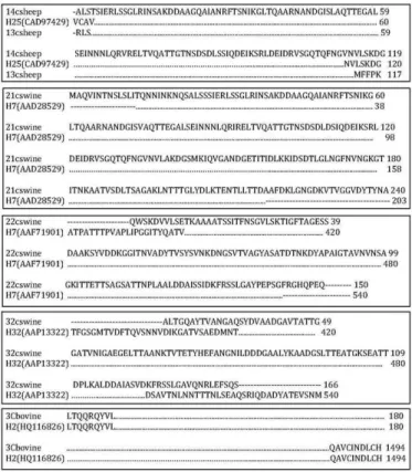
Fig.1. Inferred amino-acid partial and total sequence alignment
for the flagellin of E. coli HNT strains and protein control se
-quences. Black points represent the amino-acid identity.
Sequencing analysis of fliC gene in E. coli HNT
In five E. coli strains, it was possible to characterize the
antigen with partial sequences. Strains isolated from swi -nes were characterized, determining O11:H7 (two strains) and ONT:H32 (one strain). Antigens of two strains isolated from sheep were determined as O42:H25 (Figure 1). Three
E. coli strains, two isolated from bovine (serogroup O8) and
one isolated from avian (serogroup O121) were completely sequenced. The avian E. coli isolate showed close genetic
relations with the H45, up to 97% similarity, and with the
fliC gene of Shigella boydii, so, this strain was considered to
be associated with a new flagellin gene. Regarding the bovi -ne strains, o-ne of them showed total homology with the H2 antigen gene, but the antigen produced did not react with H2 antiserum. The second strain showed total homology with a sequence of the new fliC gene, recently described with the accession number CQ423574.
Antisera production and serology from the novel fliC antigens
Antisera against the flagellar antigens were produced from the strains HNT 3C, 4C and 40C. All antisera showed
positive results for the homologous H antigen when dilu -ted up to 1:12,800. However, none of them produced strong agglutination with the other 53 reference H antigens. The unspecific reactions disappeared when the antisera were absorbed with heterologous antigens.
DISCUSSION
The definition of Escherichia coli pathotypes according to
their virulence profile mainly implies a correlation betwe -en pathog-enic factors and specific serotypes. Therefore, serotype grouping (O- and H-antigens) of pathogenic E. coli strains remains the first line of characterization and is
considered the gold-standard approach in subtyping pa -thogenic bacteria. According to some authors, only when the serotype of clinical isolates is established can the other molecular methods for subtyping and fingerprinting be reasonably applied (Fields et al. 1997, Prager et al. 2003, Moreno et al. 2006).
With the purpose of streamlining the process of sero -typing of E. coli strains, alternative methods for subtyping
them have been widely applied, which focus on establishing relationships between the provoked human diseases and the specific serotypes of E. coli present in infections. Some
studies using RFLP-PCR have been successfully applied with strains isolated from humans, and all of them showed good results for characterizing H antigens (Botelho et al. 2003, Prager et al. 2003, Moreno et al. 2006).
Different E. colifliC PCR products could be detected in
analysis, some E. coli reference strains for H antigens (H17,
H25, H53 and H54) did not amplify the fliC gene. This result was expected because these flagellar antigens were not ex -pressed by fliC, but by other genes like flnA (H17) (Ratiner et al. 2010), flkA (H53) and flmA (H54) (Tominaga 2004). Other studies have already demonstrated that it is possible to classify the non typeable strains through conventional serology techniques using RFLP-PCR, demonstrating that this method can be more efficient (Botelho et al. 2003, Ba -dri et al. 2010). In our work, it was possible to characteri -ze nine H antigens through the RFLP method, confirming previous reports on the limitation of serological methods (Prager et al. 2003, Moreno et al. 2006, Badri et al. 2010). Moreover, beyond the RFLP-PCR, applying sequencing te -chniques, we could successfully serotype other strains, permitting the identification of three putative new genes of flagellin in HNT E. coli strains. These findings were con -firmed by the production of rabbit antiserum and by end -point agglutination tests with all known H-antigen referen -ce strains. Defining and establishing new H antigen types remains a main task for the International Escherichia and Klebsiella Centre (WHO).
Other authors characterized 43 of 53 H antigen expres -sed by fliC gene of E. coli by sequencing analysis (Wang et
al. 2003). Through the construction of these databases, the characterization of other H antigens became much more efficient with the use of partial or total sequences since N- and C- regions of the gene are conserved between all the different antigens (Wang et al. 2003). Using the fliC sequen-ce from the GenBank database website, it was possible to obtain information from the partial sequences, being the starting point for characterizing the remaining strains used in the present study. The total sequencing of the gene was not necessary since the partial sequence had demonstrated 100% similarity to the described H antigens already used in previous described reports (Wang et al. 2003, Feng et al. 2008).
The antisera produced against H antigens and not cha -racterized by the RFLP-PCR techniques of sequencing had shown to be highly specific to its homologous antigens; however, it demonstrated a low specificity to other control antigens (H1 to the H56), demonstrating that it is probably related to a new antigen that has not yet been described in the literature.
A number of concerns could be raised about the RFLP --PCR method. For instance, the fliC gene may not be am -plified by PCR due to inadequate primer homology. Howe -ver, we observed that the amplification could be obtained in most cases, warranting the use of this technique. Ano -ther potential limitation is that, since -there are unknown
fliC alleles, these alleles could not be matched with known RFLP-PCR profiles. However, if these genes were obtained by epidemiological studies, they may soon be determined as new patterns might be described for the diarrheagenic strains of E.coli, thus permitting a widespread use of this
technique for characterizing fliC genes, which can be used to determine the H antigen of the E. coli strains by sequen -cing techniques, as we did in this study. As previously men -tioned, classic serotyping methods presents stronger limi
-tations than the RFLP-CR sequencing methods. Moreover, the potential limitations of the classical serotyping techni -ques can be successfully complemented with the RFLP-PCR methods (Botelho et al. 2003, Prager et al. 2003, Amhaz et al. 2004, Moreno et al. 2006, Beutin et al. 2007, Badri et al. 2010).
The results observed in this study permitted us to con -clude that molecular methods (RFLP-PCR) showed to be more efficient in detecting H antigen of E. coli strains. The fliC-RFLP techniques proved to be faster than the classic serotyping methods for the detection of the E. coli H anti -gens. The RFLP-PCR/sequencing techniques were capable of rapidly determining H antigens, leading to the discovery of new flagellin genes produced by these bacteria.
Acknowledgements.- C. Moura, has a PhD fellowship granted by Coor -denação de Aperfeiçoamento de Pessoal de Nível Superior (CAPES). This study was supported by grants from FAPESP (Fundação de Amparo à Pes -quisa do Estado de São Paulo (Grant 2005/00713-0).
REFERENCES
Amhaz J.M., Andrade A., Bando S.Y., Tanaka T.L., Moreira-Filho C.A. & Marti -nez M. 2004. Molecular typing and phylogenetic analysis of enteroinva -sive Escherichia coli using the fliC gene sequence. FEMS Microbiol. Lett.
15:259-264.
Badri S., Fassouane A., Filliol I., Hassar M. & Cohen N. 2010. Sequence analysis of the gene encoding H antigen in Escherichia coli isolated from
food in Morocco. J. Microbiol. 48:184-187.
Beutin L. & Strauch E. 2007. Identification of sequence diversity in the
Escherichia coli fliC genes encoding flagellar types H8 and H40 and its use in typing of Shiga toxin-producing E. coli O8, O22, O111, O174, and
O179 strains. J. Clin. Microbiol. 45:333-339.
Blanco J., Blanco M., Alonso M.P., Blanco J.E., Garabal J.I. & González E.A. 1992. Serogroups of Escherichia coli strains producing cytotoxic necro -tizing factors CNF1 and CNF2. FEMS Microbiol. Lett. 15:155-159. Blanco M., Blanco J.E., Mora A., Rey J., Alonso J.M., Hermoso M., Hermoso
J., Alonso M.P., Dahbi G., González E.A., Bernárdez M.I. & Blanco J. 2003. Serotypes, virulence genes, and intimin types of Shiga toxin (verotoxin)-producing Escherichia coli isolates from healthy sheep in Spain. J. Clin.
Microbiol. 41:1351-1356.
Botelho B.A., Bando S.Y., Trabulsi L.R. & Moreira-Filho C.A. 2003. Identi -fication of EPEC and non-EPEC serotypes in the EPEC O serogroups by PCR-RFLP analysis of the fliC gene. J. Microbiol. Methods. 54:87-93. Ewing´s W.H. 1986. The genus Escherichia, p.93-134. In: Edwards P.R. &
Ewing’s W.H. (Eds), Identification of Enterobacteriaceae. 4th ed. Burgess,
Minneapolis.
Feng L., Liu B., Liu Y., Ratiner Y.A., Hu B., Li D., Zong X., Xiong W. & Wang L. 2008. A genomic islet mediates flagellar phase variation in Escheri-chia coli strains carrying the flagellin-specifying locus flk. J. Bacteriol.
190:4470-4477.
Fields P.I., Blom K., Hughes H.J., Helsel L.O., Feng P. & Swaminathan B. 1997. Molecular characterization of the gene encoding H antigen in Escheri-chia coli and development of a PCR-restriction fragment length poly -morphism test for identification of E. coli O157:H7 and O157:NM. J. Clin.
Microbiol. 35:1066-1070.
Hussein H.S. 2007. Prevalence and pathogenicity of Shiga toxin-producing
Escherichia coli in beef cattle and their products. J. Anim. Sci. 85:63-72.
Machado J., Grimont F. & Grimont P.A. 2000. Identification of Escherichia coli flagellar types by restriction of the amplified fliC gene. Res. Micro -biol. 151:535-546.
MacNab R.M. 1992. Genetics and biosynthesis of bacterial flagella. Ann. Rev. Genet. 26:129-156.
Mattsson S. & Wallgren P. 2008. Phenotyping of Escherichia coli serotypes
Moreno A.C.R., Guth B.E.C. & Martinez M.B. 2006. Can the fliC PCR-restric -tion fragment length polymorphism technique replace classic seroty -ping methods for characterizing the H antigen of enterotoxigenic Esche-richia coli strains? J. Clin. Microbiol. 44:1453-1458.
Prager R., Strutz U., Fruth A. & Tschäpe H. 2003. Subtyping of pathogenic
Escherichia coli strains using flagellar (H)-antigens: serotyping versus
fliC polymorphisms. Int. J. Med. Microbiol. 292:477-486.
Ratiner Y.A., Sihvonen L.M., Liu Y., Wang L. & Siitonen A. 2010. Alteration of flagellar phenotype of Escherichia coli strain P12b, the standard type
strain for flagellar antigen H17, possessing a new non-fliC flagellin gene
flnA, and possible loss of original flagellar phenotype and genotype in the course of subculturing through semisolid media. Arch. Microbiol. 192:267-278.
Reid S.D., Selander R.K. & Maniatis T. 1999. Sequence diversity of flagellin (fliC) alleles in pathogenic Escherichia coli. J. Bacteriol. 181:153-160.
Tominaga A. 2004. Characterization of six flagellin genes in the H3, H53 and H54 standard strains of Escherichia coli. Genes Genet. Syst. 79:1-8.
Wang L., Rothemund D., Curd H. & Reeves P.R. 2003. Species-wide varia -tions in the Escherichia coli flagellin (H-antigen) gene. J. Bacteriol.