RESUMO.- [Cinquenta anos num piscar de olhos: um es-tudo retrospectivo sobre lesões oculares e perioculares em animais domésticos.] Foi realizada uma investigação para obter-se uma visão geral das lesões oculares e perio-culares de mamíferos domésticos diagnosticadas ao longo de um período de 50 anos num laboratório de diagnóstico de patologia veterinária da Região Central do Rio Grande do Sul. Nesse laboratório, durante o período pesquisado foram
realizados 33.075 exames histopatológicos, 540 dos quais (1,6%) eram de lesões oculares e perioculares. Por várias razões, 90 espécimes foram excluídos do estudo. As 450 amostras restantes consistiam espécimes de cães (53,5%), bovinos (28,2%), gatos (11,1%), cavalos (5,1%), ovelhas (1,3%), coelhos (0,4%), e porco (0,2%). As pálpebras fo-ram o local mais prevalente (248/450) de ocorrência das lesões, seguidas da terceira pálpebra (73/450) e conjun-tiva (27/450). Em cães (241 diagnósticos) as lesões nas glândulas sebáceas (incluindo as glândulas meibomianas) consistiram nos achados mais comuns (75/241), seguidos dos tumores melanocíticos (52/241) e de conjuntivites inespecíficas (13/241). Neoplasmas de células escamosas, tanto benignos como malignos, foram achados relativa-mente comuns. Em bovinos, os locais anatômicos afetados por lesões perioculares e oculares, em ordem decrescente
Fifty years in the blink of an eye: a retrospective study of
ocular and periocular lesions in domestic animals
1Tessie Beck Martins2 and Claudio S.L. Barros2*
ABSTRACT.- Martins T.B. & Barros C.S.L. 2014. Fifty years in the blink of an eye: a retro-spective study of ocular and periocular lesions in domestic animals. Pesquisa Veteri-nária Brasileira 34(12):1215-1222. Programa de Pós-Graduação em Medicina Veterinária, Universidade Federal de Santa Maria, Camobi, Av. Roraima 1000, Santa Maria, RS 97105-900, Brazil. E-mail: claudioslbarros@uol.com.br
A survey was undertaken aiming to obtain an overview of ocular and periocular lesions diagnosed in domestic mammals over a period of 50 years in a veterinary pathology diag-nostic laboratory in the Central Region of the State of Rio Grande do Sul, Brazil. In this lab, 33,075 histophatological exams had been performed over the period surveyed, of which 540 (1.6%) concerned ocular and periocular lesions. For various reasons ninety speci-mens were excluded from the study and the remaining 450 consisted of samples from dogs (53.5%), cattle (28.2%), cats (11.1%), horses (5.1%) sheep (1.3%), rabbits (0.4%), and pig (0.2%). The eyelids were the most prevalent (248/450) site of lesions in each of the species studied, followed by third eyelid (73/450), and conjunctiva (27/450). In dogs (241 sam-ples) lesions in sebaceous glands (including Meibomian glands) were the most common findings (75/241), followed by melanocytic tumors (52/241) and nonspecific conjunctivitis (13/241). Squamous cell neoplasms, both benign and malignant, were relatively common. In cattle, anatomical sites affected by ocular and periocular lesions, in decreasing order of frequency, were eyelid, cornea and third eyelid. Squamous cell carcinoma (SCC) alone ac-counted for 80.3% of all diagnoses, while all neoplastic lesions made up for 85.0% of the lesions diagnosed in cattle. Neoplasia accounted for most of the lesions diagnosed in cats (39/50 cases); all of these were malignant, and SCC, hemangiosarcoma and fibrosarcoma were the most common types diagnosed. In horses, 19 out of 23 submissions were neoplas-ms and most were sarcoid (8/23) and SCC (8/23). There were six submissions from sheep with unpigmented skin, all of which represented SCC of the eyelids (5) and third eyelid (1). INDEX TERMS: Ophthalmology, palpebra, neoplasm.
1 Received on December 2, 2014.
Accepted for publication on December 15, 2014.
Part of Doctoral Degree of the first author.
de frequência, foram pálpebra, córnea e terceira pálpebra. Somente o carcinoma de células escamosas (CCE) perfez 80,3% de todos os diagnósticos, enquanto todas as lesões neoplásicas juntas perfizeram 85,0% das lesões diagnos -ticadas em bovinos. Em gatos, a maioria (39/50 casos) das lesões diagnosticadas era de neoplasia maligna e CCE, hemangiossarcoma e fibrosarcoma foram os diagnósticos mais frequentes. Em equinos 19 de 23 submissões eram neoplasmas e os mais comuns foram sarcoide (8/23) e CCE (8/23). Em ovinos foram encontradas seis submissões, to-das casos de CCE de pálpebra (5/6) ou terceira pálpebra (1/6) de ovinos de pele despigmentada.
TERMOS DE INDEXAÇÃO: Oftalmologia, pálpebra, neoplasma.
INTRODUCTION
The eye is a major special sense organ in vertebrate ani-mals (Fernald 1997), that depend on it to survive and inte-ract with the environment (Vorobyev et al. 2001, Williams 2010). Ocular pathology, a science that studies pathological processes that affect the eye bulb and its adnexal structures (Orellana & Pifano 2006), is a relatively new area in veteri-nary medicine, with its first publications dating from the beginning of twentieth century (Gelatt 2008).
In Brazil, veterinary ocular pathology is still modest when compared to ophthalmology, the specialty from whi-ch it derives, but the increasing number of veterinary es-tablishments and teaching institutions that offer ophthal-mologic service should soon depend, and luckily, impel, the development of ocular pathology.
It has been shown that the prevalence of diseases varies largely between countries and between regions within a country (Valentine 2006). Although important publications are available considering ocular diseases in specific animal species and ethiopathogenic entities, there is need for data about general prevalence of ocular lesions in our region. Lack of such data does not impair the diagnosis of lesions submitted to pathology labs, but their unavailability so-mehow forces local pathologists and students to confront their results with those in the foreign literature.
The purpose of this study is to determine the type and prevalence of ocular and periocular lesions in domestic mammals submitted to a veterinary pathology diagnostic laboratory in the Central Region of State of Rio Grande do Sul, Brazil.
MATERIALS AND METHODS
From January 1964 to December 2013, 33,075 histopathologic exams were performed at the Veterinary Pathology Laboratory, at the Federal University of Santa Maria (LPV-UFSM). All protocols were reviewed and cases pertinent to ocular and periocular le-sions were filtered. Protocols in which there was no diagnosis or description of the lesions, protocols regarding samples improper for diagnoses (due to autolysis or insufficient material), samples originating from research animals, and samples from normal tis-sue (those in which no lesions were observed) were excluded.
Lesions were grouped by animal species, site, type of primary process/etiology, and final diagnosis. Only mammals were taken into account for this study. Lesions were grouped firstly according to species - dogs; cattle; cats; horses; sheep; pigs; and other.
Sub-sequently, lesions were classified considering the main site affec -ted and/or site of origin, as follows: eyelid; third eyelid; lacrimal gland; eyeball; bulbar conjunctiva; cornea; uvea; lens; and “eye”. The latter category was created to accommodate diagnosis in which the site of lesion was not specified (eg. “tumor in the eye”). Such cases were not excluded because they accounted for a great portion of the total. The following criteria for classification were based on the primary pathologic process and etiology of the le-sions: alterations due to trauma; congenital anomalies; autoim-mune processes; infectious and parasitic diseases; degenerative diseases; metabolic and toxic diseases; neoplasia; and disturban-ces in cellular growth (DCG). The last category included lesions consisting of cellular hyperplasia and metaplasia, and cysts. In-flammatory lesions that could not be classified as infectious, pa -rasitic or autoimmune were grouped under the umbrella term
inflammation. Data related to animal breed, sex and age were not analyzed in this study.
RESULTS
During the fifty years encompassed by the current study, 33,075 histopathologic exams were performed at the LPV--UFSM. All protocols were reviewed and 540 cases (1.6%) concerning ocular and periocular lesions were filtered. Ninety of these (16.7%) were excluded because were not pertinent to the study, forty-eight of which related to birds and reptiles and forty-three, because their protocols lacked major data. In part of these protocols, information about description and/or diagnosis of the lesions was missing or not clear, and in other part, samples were not adequate for evaluation (due to small size or autolysis) or related to nor-mal tissues (no lesions observed). Four hundred and fifty (83.3%) cases were selected for this study. More than one half (53.5%) of the samples came from dogs, followed by cattle (28.2%), cats (11.1%), horses (5.1%) and other spe-cies [sheep (1.3%), rabbits (0.4%), and a pig (0.2%)].
Eyelid was the most prevalent site of lesion in each of the species studied, accounting for 55.1% of all samples submitted (248/450), followed by third eyelid (73/450; 16.2%), and conjunctiva (27/450; 6.0%). Regarding the type of disease, neoplasia was the most numerous process, with 79.1% of all samples (356/450) (Fig.1-4), followed by inflammatory lesions of uncertain cause (50/450; 11.1%) (Fig.5), and congenital anomalies (12/450; 2.7%) (Fig.6). Overall, squamous cell carcinoma (SCC) was the most com-mon entity, with a prevalence of 30.8% (139/450) (Fig.7).
and in another dog, it invaded the eyeball. In one situation, the dog had SCC affecting the conjunctiva and five other cutaneous sites along the body, as well as one cutaneous fibrosarcoma and one cutaneous mast cell tumor.
In cattle, sites affected by ocular and periocular lesions, in decreasing order, were eyelid (cutaneous side), cornea and third eyelid. SCC alone accounted for 80.3% of all diag-noses, while neoplastic lesions in total made 85.0%. Exten-sive invasion of the eyeball and orbit by SCC, with involve-ment of periocular tissues, was reported in seven cases. In five other cases, regional lymph nodes that were submitted with the ocular lesion had metastasis of SCC.
Neoplasia accounted for the major part of feline diag-nosis, with 39/50 (78%) cases. All neoplasms were malig-nant. SCC, hemangiosarcoma and fibrosarcoma were the most common diagnoses, with 16 (32.0%), 10 (20.0%), and three (6.0%) cases, respectively. Nine of sixteen (56.2%) cats diagnosed with SCC had similar lesions in other parts
of the head (ears; lips; nose; and/or contralateral eyelid), and in two occasions the neoplasm had already been remo-ved before. In another two cats, clinicians reported inva-sion of eyeballs by cutaneous SCC with origin in the eyelids. In horses, there were 24 submissions from 23 animals. Two out of 24 were samples from the same lesion, taken within a few weeks interval, and both were diagnosed as sarcoid, so they were condensed as a single diagnosis. When considering all samples, 19/23 (82.6%) accounted for neoplasms. Sarcoid and SCC were the most prevalent lesions, with 34.7% (8/23) each. There were six submis-sions from sheep, all of which represented SCC in the eyelid (5/6) or third eyelid (1/6). In all cases, animals had non--pigmented skin. In three sheep, there were SCC in another site (ear; third eyelid; and inferior eyelid).
Details about the distribution of lesions according to species, site (anatomical location), type of disease and diagnosis are presented in Tables 1-5.
Fig.1. Canine, product of excisional biopsy. Fragment of eyelid with large Meibomian gland adenoma (V1085-12). Alterations of the sebaceous glands corresponded to the most common di-agnoses in dogs, with 31.1% of all lesions.
Fig.3. Canine, product of exenteration. A trasmissible veneral tu-mor originating at the bulbar conjuntiva invaded eyeball and orbit. Sclera, optic nerve, third eyelid and eyelid (at the bot-tom) are barely recognizable (V1076-12).
Fig.2. Canine, product of exenteration. The orbit was partially filled by a mucinous mass, histologically diagnosed as a myx -oma, ventral to the eyeball. The dog was referred to the clini-cian because of exophtalmia (V0406-11).
Fig.4. Feline, product of enucleation. The architecture of eye struc-tures is markedly disrupted by a dense witish mass that out-grows the eyeball in all directions. Histologically, a diagnosis of fusiform ocular sarcoma was established. Lens (which is not observed here) was ruptured and surrounded by neutro-phils (V0003-08).
3 4
Fig.7. Presentation of ocular squamous cell carcinoma (SCC) on the eye and periocular tissues in domestic animals. Upper left: Bovine, prod-uct of exenteration. A corneal SCC extends and invades the anterior chamber of the eye with resultant phthisis bulbi (V0093-04). Upper right: Bovine, product of exenteration. Periorbital tissues of this bovine have been envolved with and partially replaced by an eyelid SCC (eyelid not shown in the picture). Sclera is disrupted in its caudoventral aspect, but there is no invasion of the posterior chamber by the mass at this stage (V1122-12). Lower left: Ovine before incisional biopsy. Upper and lower eyelids are distorted by a large exophytic and ulcerated mass. The lesion was complicated by a myiasis (V1276-11). Lower right: Canine, product of exenteration. There is an exophytic, focal mass in the bulbar conjunctiva, partially effacing the limbus. This dog had SCC in five other anatomical sites (V1565-13). Fig.5. Feline, right before excisional biopsy of a tumoral lesion in
the third eyelid. The lesion was bilateral. Histologically, the mass was diagnosed as eosinophilic conjunctivitis (V0113-08). This inflammatory lesion is an important differential di -agnosis for squamous cell carcinoma.
Fig.6. Feline, product of post-mortal enucleation. The cat died due to lower urinary tract disease. There are multiple centripetal projections that extend from the iris and attach to the lens ante-rior surfarce (posteante-rior synechia). Histologically, the projections consisted of fibrovascular tissue lined by pigmented epithelium. This lesion was present at birth, and final diagnosis was per -sistent pupillary membranes, a congenital anomaly (V0181-08).
DISCUSSION
Out of 33,075 histopathologic exams, four hundred and fif -ty cases of ocular and periocular diseases were identified. Dogs corresponded to more than half of the exams,
specially from cattle and sheep, with a large population of working horses and dogs, and local veterinarians used to specialize in large rather than small animal practice. Be-cause of that, this lab used to handle primarily samples from cattle along its first decade of actuation. As the de -cades went by, there was a gradual change in the profile of samples submitted for pathologic examination in the re-gion drained by the LPV-UFSM, when dogs rapidly became the most import species in terms of number of exams. This change probably relates to socio-cultural changes, such as the dramatic increase in the market of pets, but no study has been conducted to confirm that.
When considering anatomical location of lesions, eyelids accounted for the major part of submissions, with 55.1% of all samples, 86.3% of which were neoplasms. Third eyelid and conjunctiva accounted for 16.2% and 6.0% of the total, respectively, also with large proportions of neoplastic pro-cesses. One of the possible reasons why these sites are over-Table 1. Canine eyelid and third eyelid lesions diagnosed at
LPV-UFSMa (1964-2013)
Site Nb Type of Disease N Diagnosis N Eyelid (skin) 126 Neoplasia 107 Melanocytoma 43
Sebaceous gland 39
adenoma/
epithelioma
Squamous 8
papilloma
Melanoma 6
Cutaneous histiocytoma, 2
Mast cell tumor,
Sebaceous carcinoma
Fibrosarcoma, 1
Hemangioma, IKAc,
Sweat gland adenoma,
Undifferenciated
carcinoma
Autoimmune 9 DLEd, Uveodermatologic 3
syndrome
Alergic 2
dermatopathy
Pemphigus 1
erythematosus
Inflammation 5 Granulation tissue 2
Lipogranuloma 1
(chalazion), Cutaneous
lymphocytosis,
Chronic dermatitis
DCGe 4 Collagenous nevus, 1
Follicular cyst,
Sweat gland cyst,
Cartilagenous metaplasia
Metabolic 1 Zinc-responsive 1
dermatosis
Eyelid 38 Neoplasia 34 Meibomian gland 32
(margin) adenoma/ epithelioma
Meibomian gland 2
adenocarcinoma
Inflammation 3 Lipogranuloma 2
Sterile cutaneous 1
granuloma
Congenital 1 Mesenchymal 1
anomaly hamartoma
Eyelid 3 Infectious 1 Chronic bacterial 1 (conjunctiva) conjunctivitis
Inflammation 1 Granulation tissue 1
Neoplasia 1 TVTf 1
Third eyelid 37 Neoplasia 21 Third eyelid gland 6
adenocarcinoma
SCCg 5
Hemangioma 4
Third eyelid gland 3
adenoma
Basal cell carcinoma, 1
Hemangiosarcoma,
Lymphoma
Inflammation 15 Conjunctivitis 13
NGEh, Plasmacytic 1
conjunctivitis (plasmoma)
DCG 1 Glandular squamous 1
metaplasia
Total 204
a Laboratório de Patologia Veterinária da Universidade Federal de Santa Maria; b number of cases; c infundibular keratinizing epithelioma; d dis-coid lupus erythematous; e disturbances in cellular growth; f transmissi-ble veneral tumor; g squamous cell carcinoma; h nodular granulomatous episclerokeratitis.
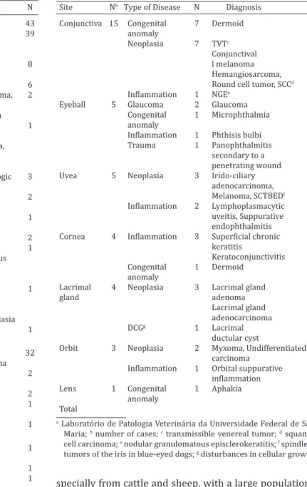
Table 2. Canine ocular and periocular (excluding eyelid and third eyelid) lesions diagnosed at LPV-UFSMa (1964-2013)
Site Nb Type of Disease N Diagnosis N Conjunctiva 15 Congenital 7 Dermoid 7
anomaly
Neoplasia 7 TVTc 2
Conjunctival 2
l melanoma
Hemangiosarcoma, 1
Round cell tumor, SCCd
Inflammation 1 NGEe 1
Eyeball 5 Glaucoma 2 Glaucoma 2
Congenital 1 Microphthalmia 1
anomaly
Inflammation 1 Phthisis bulbi 1
Trauma 1 Panophthalmitis 1
secondary to a
penetrating wound
Uvea 5 Neoplasia 3 Irido-ciliary 1
adenocarcinoma,
Melanoma, SCTBEDf
Inflammation 2 Lymphoplasmacytic 1
uveitis, Suppurative
endophthalmitis
Cornea 4 Inflammation 3 Superficial chronic 2
keratitis
Keratoconjunctivitis 1
Congenital 1 Dermoid 1
anomaly
Lacrimal 4 Neoplasia 3 Lacrimal gland 2
gland adenoma
Lacrimal gland 1
adenocarcinoma
DCGg 1 Lacrimal 1
ductular cyst
Orbit 3 Neoplasia 2 Myxoma, Undifferentiated 1
carcinoma
Inflammation 1 Orbital suppurative 1
inflammation
Lens 1 Congenital 1 Aphakia 1
anomaly
Total 37
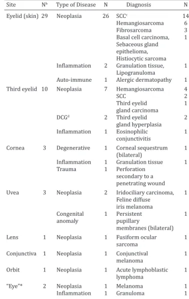
Table 4. Feline ocular and periocular lesions diagnosed at LPV-UFSMa (1964-2013)
Site Nb Type of Disease N Diagnosis N Eyelid (skin) 29 Neoplasia 26 SCCc 14
Hemangiosarcoma 6
Fibrosarcoma 3
Basal cell carcinoma, 1
Sebaceous gland
epithelioma,
Histiocytic sarcoma
Inflammation 2 Granulation tissue, 1
Lipogranuloma
Auto-immune 1 Alergic dermatopathy 1 Third eyelid 10 Neoplasia 7 Hemangiosarcoma 4
SCC 2
Third eyelid 1
gland carcinoma
DCGd 2 Third eyelid 2
gland hyperplasia
Inflammation 1 Eosinophilic 1
conjunctivitis
Cornea 3 Degenerative 1 Corneal sequestrum 1
(bilateral)
Inflammation 1 Granulation tissue 1
Trauma 1 Perforation
secondary to a
penetrating wound
Uvea 3 Neoplasia 2 Iridociliary carcinoma, 1
Feline diffuse
iris melanoma
Congenital 1 Persistent 1
anomaly pupillary
membranes (bilateral)
Lens 1 Neoplasia 1 Fusiform ocular 1
sarcoma
Conjunctiva 1 Neoplasia 1 Conjunctival 1
melanoma
Orbit 1 Neoplasia 1 Acute lymphoblastic 1
lymphoma
“Eye”* 2 Neoplasia 1 Melanoma 1
Inflammation 1 Granuloma 1
Total 50
a Laboratório de Patologia Veterinária da Universidade Federal de Santa Maria; b number of cases; c squamous cell carcinoma; d disturbances in cellular growth; *exact location not informed.
Table 5. Equine ocular and periocular lesions diagnosed at LPV-UFSMa (1964-2013)
Site Nb Type of Disease N Diagnosis N Eyelid (skin) 13 Neoplasia 10 Sarcoid 8
Fibroma, SCCc 1
Inflammation 3 Granuloma 2
Fibrinosuppurative 1
blepharitis
Third eyelid 5 Neoplasia 5 SCC 4
Undifferentiated 1
carcinoma
Eyeball 1 Trauma 1 Perforation secondary 1
to a penetrating wound
“Eye”* 4 Neoplasia 4 SCC 3
Fibroma 1
Total 23
a Laboratório de Patologia Veterinária da Universidade Federal de Santa Maria; b number of cases; c squamous cell carcinoma; *exact location not informed.
Table 3. Bovine ocular and periocular lesions lesions diagnosed at LPV-UFSMa (1964-2013)
Site Nb Type of Disease N Diagnosis N Eyelid (skin) 33 Neoplasia 30 SCCc 28
Squamous 2
papilloma
Inflammation 2 Chronic 2
blepharitis
Infectious 1 Dermatophytosis 1
Cornea 24 Neoplasia 16 SCC 14
Squamous 2
papilloma
Infectious 5 Keratoconjunctivitis 5
Inflammation 1 Bilateral exposure 1
keratitis secondary to
herpesvirus encephalitis
Toxic 1 Acute keratitis 1
secondary to
photosensitization
Trauma 1 Corneal perforation 1
secondary to a
penetrating injury
Third eyelid 20 Neoplasia 17 SCC 17
Inflammation 2 Conjunctivitis 2
Congenital 1 Dermoid 1
anomaly
Conjunctiva 11 Neoplasia 10 SCC 9
Sebaceous gland 1
carcinoma
Infectious 1 Lymphoplasmacytic 1
vasculitis (MCFd)
Orbit 2 Neoplasia 2 Lymphoma 2 Eyeball 1 Neoplasia 1 Melanoma 1 Uvea 1 Infectious 1 Bacterial 1
endophthalmitis
secondary to
encephalitis
“Eye”* 36 Neoplasia 36 SCC 35
Fibroma 1
Total 127
a Laboratório de Patologia Veterinária da Universidade Federal de Santa Maria; b number of cases; c squamous cell carcinoma; d malignant catar-rhal fever; *exact location not informed.
represented is that from a clinical perspective, the diseases of the eyelid and conjunctiva form a major part of what the primary care veterinarian is likely to diagnose and treat (Njaa & Wilcock 2010). When it comes to neoplasms, surgery is the typical therapy, and although each tumor has some characte-ristic or suggestive clinical features, only histological or cyto-logical examination of the specimen is definitively diagnostic (Maggs 2008). This might explain why 79.1% (356/450) of all samples in this study corresponded to neoplastic disease.
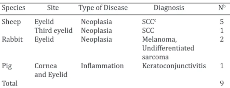
Table 6. Periocular lesions diagnosed in six sheep, two rabbits and one pig at LPV-UFSMa (1964-2013)
Species Site Type of Disease Diagnosis Nb
Sheep Eyelid Neoplasia SCCc 5
Third eyelid Neoplasia SCC 1
Rabbit Eyelid Neoplasia Melanoma, 2
Undifferentiated
sarcoma
Pig Cornea Inflammation Keratoconjunctivitis 1
and Eyelid
Total 9
a Laboratório de Patologia Veterinária da Universidade Federal de Santa Maria; b number of cases; c squamous cell carcinoma.
In this study, eyelid neoplasms accounted for 58.9% of all canine samples (142/241), half of which (52.1%; 74/142) were due to alterations of the sebaceous and mo-dified (Meibomian) sebaceous glands. That makes eyelid sebaceous neoplasms the most common canine lesion in our study, representing a third (30.7%) of all lesions, follo-wed by eyelid melanocytic tumors (49/241; 20.3%), and nonspecific conjunctivitis (13/241; 5.4%). There were originally 31 and 18 diagnoses of eyelid (malignant) mela-nomas and melacytomas, respectively, something in large disagreement with the literature (Njaa & Wilcock 2010). Cutaneous melanocytic tumors of dogs, including the eye-lid, are usually benign melanocytomas, unless they exhibit compelling anaplastic features and evidence of aggressive infiltration (Dubielzig et al. 2010). After reading the ma -croscopic and histologic descriptions, it was clear that in most cases, presence of junctional activity was used as the primary criterium of malignancy, usually regardless of ab-sence of mitotic figures and stromal invasion. Based on a study about a comparative approach to melanocytic neo-plasms (Smith et al. 2002), 25/31 melanomas were reclas-sified as melanocytomas.
In cattle, cats, and horses, differently than dogs, eyelid neoplasms are usually malignant (Stades et al. 2007). This corroborates the results obtained in this study, where eye-lid tumors were malignant in 93.3% of cattle, 100% of cats, sheep and rabbit, and 90% of horses.
SCC was the predominant type of eyelid neoplasm in cattle, cats and sheep, as has been described (Maggs 2008). The cause of ocular SCC is still poorly understood; howe-ver, there are several factors including genetic susceptibi-lity, nutrition levels, age, UV light, circumocular apigmen-tation and viruses that may contribute to its development (Tsujita & Plummer 2010). Besides the eyelids, SCC was observed in the third eyelid in cattle, horses, dogs, cats and a sheep; cornea, in cattle; and bulbar conjunctiva in cattle and one dog. Occasionally, the tumor invaded eyeball and orbit (especially in cattle and cats), was present in the skin in other parts of the head (ears; lips; nose; and/or contra-lateral eyelid) (cats and dogs) or body (dog), or draining lymph nodes (cattle); and/or had been removed before (especially cats). Indeed, SCC was the most common sin-gle entity in this study overall, with a prevalence of 30.8% (139/450).
Sarcoid and SCC were the the major lesions observed in horses, with 34.7% (8/23) of all diagnoses each. When
con-sidering eyelids only, sarcoid corresponded to 80% of sub-missions, whereas SCC was mainly observed in the third eyelid. It has been previously reported that approximately 10% of all equine neoplasms affect the eye or periocular structures, especially the eyelids, and that the most com-mon periocular masses include sarcoid, SCC, papilloma, lymphoma and melanoma (Giuliano 2010), the former two being the most important. There is difference in prevalence of equine sarcoid and cutaneous and mucocutaneous (in-cluding ocular mucosa) SCC according to geographic areas (Valentine 2006).
Sarcoid is believed to be a bovine papillomavirus-asso-ciated tumor with a genetic predisposition, where Quarter horses and Arabians appear to be at higher risk for develo-pment of this neoplams. The higher prevalence of sarcoid in some areas is apparently related to a higher exposure to bovine papillomavirus, where large number of beef cat-tle are in close proximity to horses and other equids, and to a higher density of predisposed animals or viral vectors (Giuliano 2010), whereas the increased incidence of equine ocular and cutaneous SCC may reflect increased exposure to solar radiation, especially of horses on pasture and of horses in high desert areas, non-pigmented ocular adnexa (particularly of the nictitating membrane), and pale perio-cular pigmentation (Valentine 2006).
There were six submissions from ovine, and all corres-ponded to SCC in the eyelid (5/6) or third eyelid (1/6). Ovine neoplasms are uncommon in general. SCC is the most common ocular and periocular neoplasm in sheep, but it tends to occur in this location only in animals that have non-pigmented skin or white colored head (Ahmed & Hassanein 2012). Other periocular tumors that have been reported in sheep are Meibomian gland adenoma (Rezaie et al. 2012), and basal cell tumor (Gorham et al. 1990).
CONCLUSIONS
From 1964 to 2013, eyelid neoplasms were the most com-mon pathologic diagnosis obtained from ocular and pe-riocular lesions of domestic mammals submitted to LPV--UFSM. Squamous cell carcinoma was the most numerous entity in this study.
REFERENCES
Ahmed A.F. & Hassanein K.M.A. 2012. Ovine and caprine cutaneous and ocular neoplasms. Small Rum. Res. 106:189-200.
Dubielzig R.R., Ketring K.L., McLellan G.J. & Albert D.M. 2010. Diseases of the eyelids and conjunctiva, p.143-200. In: Ibid (Eds), Veterinary Ocular Pathology: A Comparative Review. Elsevier, Edinburgh.
Fernald R.D. 1997. The evolution of eyes. Brain Behav. Evol. 50:253-259. Gelatt P.K.N. 2008. Veterinary ophthalmology: our past, present and
fu-ture. Bull. Acad. Vét. France 161:299-306.
Giuliano E.A. 2010. Equine ocular adnexal and nasolacrimal disease, p.133-180. In: Gilger B. (Ed.), Equine Ophthalmology. 2nd ed. Saunders Elsevier, Maryland Heights.
Gorham S.L., Penney B.E. & Bradley L.D. 1990. Basal cell tumor in a sheep. Vet. Pathol. 27:466-467.
Krehbiel J.D. & Langham R.F. 1975. Eyelid neoplasms of dogs. Am. J. Vet. Res. 36:115-119.
Slatter’s Fundamentals of Veterinary Ophthalmology. 4th ed. Saunders Elsevier, St Louis.
Njaa B.L. & Wilcock B.P. 2010. The ear and eye, p.1153-1244. In: Zachary J. & McGavin D. (Eds), Pathologic Basis of Veterinary Disease. 5th ed. El-sevier Mosby, St Louis.
Orellana M.E. & Pifano I.A. 2006. Patología ocular para el patólogo general. Revta Oftalmol. Venez. 62:16-31.
Rezaie A., Golshahi H. & Naddaf H. 2012. Coincidence of Meibomian ad-enoma and squamous cell carcinoma in the upper eyelid of a sheep: histopathological and immunohistochemical studies. Iranian J. Vet. Res. 13:343-346.
Roberts S.M., Severin G.A. & Lavach J.D. 1986. Prevalence and treatment of palpebral neoplasms in the dog: 200 cases (1975-1983). J. Am. Vet. Med. Assoc. 189:1355-1359.
Smith S.H., Goldschmidt M.H. & Mcmanus P.M. 2002. A comparative review of melanocytic neoplasms. Vet. Pathol. 39:651-678.
Stades F.C., Wyman M., Boevé M.H., Neumann W. & Spiess B. 2007. Eyelids, p.73-104. In: Ibid. (Eds), Ophthalmology for the Veterinary Practitioner. 2nd ed. Schluetersche, Hannover.
Tsujita H. & Plummer C.E. 2010. Bovine ocular squamous cell carcinoma. Vet. Clin. Food Anim. 26:511-529.
Valentine B.A. 2006. Survey of equine cutaneous neoplasia in the Pacific
Northwest. J. Vet. Diagn. Invest. 18:123-126.
Vorobyev M., Marshall J., Osorio D., Ibarra N.H. & Menzel R. 2001. Colourful objects through animal eyes. Col. Res. Appl. 26:214-217.