Full paper published online: November 30, 2007 ISSN 1678-9199.
NEURAL NETWORK-BASED SPECIES IDENTIFICATION IN
VENOM-INTERACTED CASES IN INDIA
MAHESHWARI R. (1), KUMAR V. (1), VERMA H. K. (1)
(1) Biomedical Research Group, Department of Electrical Engineering, Indian
Institute of Technology Roorkee, India.
ABSTRACT: India is home to a number of venomous species. Every year in
harvesting season, a large number of productive citizens are envenomed by such
species. For efficient medical management of the victims, identification of the
aggressor species as well as assessment of the envenomation degree is necessary.
Species identification is generally based on the visual description by the victim or a
witness and is therefore quite likely to be erroneous. Symptomatic identification
remains the only available method. In a previous published work, the authors
proposed a classification table for snake species based on manifested symptoms
applicable in Indian subcontinent. The classification table serves the purpose to a
great deal but as a manual method it demands human expertise. The current paper
presents a neural network-based symptomatic species identification system. A
symptom vector is fed as input to the neural network and the system yields the most
probable species as well as the envenomation severity as the output. The severity
status can be very helpful in calculating the antivenom dosage and in deciding the
species-specific prognostic measures for efficient medical management.
KEY WORDS: bites and stings, symptoms, species identification, neural network.
CONFLICTS OF INTEREST: There is no conflict.
CORRESPONDENCE TO:
INTRODUCTION
India, as a tropical country, has warm and humid climate suitable for a number of
arthropod and reptile species which can interact venom. In Indian terra firma,
scorpions and snakes are considered the major lethal venomous species of medical
importance.
The majority of Indian population resides in villages, where agriculture is the prime
occupation. Rainy season is the most productive time for farmers and is the peak
season of reptile and arthropod activities. The search for food and shelter brings
these species closer to human dwellings. The close proximity and survival instincts
often result in deadly encounters, resulting in the animal death or injury to the human
being. The situation is so gloomy that every year more than 15,000 people reportedly
die due to snakebites (1). However, unreported estimates range from 30,000 to
50,000 (2, 23) and scorpion sting deaths of infants and younger victims have not
been included in this figure.
Establishment of the species identity is very important in cases of venom interaction
for effective medical management. Many times, snake-bitten victims bring along the
dead snake for identification, but this method involves risk of a subsequent bite as
well as ecological issues, so this practice is discouraged. On the other hand,
description by a witness or the patient may not be authentic and thus the species
identification has to be done remotely based on a reliable technique. At the same
time, if the severity grade of the venom interaction is assessed, prognostic measures
can be taken together with a broad estimation of the dose and schedule. In the
absence of any other scientific method available at the grassroots level, symptomatic
identification remains the only alternative. However, with the symptoms
standardization, the system becomes precise and accurate.
Venom and Venomous Species
Venom is a mixture of proteins and peptides (6, 16). It is found in nature as one of
the earliest survival means. A number of species evolved venom and, in the
continuous quest for survival, improved their venom constituents and delivery
apparatus (5, 9). Venomous species use venom to control, kill and digest their prey.
Alternatively, it is used as a defense means against an aggressive enemy. Once the
venom enters the victim’s body at subcutaneous level, its different constituents start
tissues, proteolysis, synaptic blockades, cardiac complications, etc. (4, 20). Some
components in the venom target some specific organs or systems for faster
degeneration of the victim (18, 28).
With the course of time, the victim general condition deteriorates in all venom
interacted cases. The in vivo venom attacks specific target systems and a number of
complications may be triggered. The symptoms, the development rate of symptoms,
the clinical support and the required medicinal attention vary with the species. So,
identification of the aggressor venomous species and the envenomation degree play
an important role in medical management.
Snakes are the most familiar venomous reptiles all over the world. Being
poikilothermic, they prefer warm climates for inhabitation (11). Unlike most other
animals, they do not have any limbs. They have only teeth to attack and grab the
prey or defend themselves against an aggressor. A snake bites a human being in
order to defend itself when approached too close.
Snakes can be divided into three categories: aglyphs, opistoglyphs and
proteroglyphs (12). Aglyphs do not have venomous teeth and opistoglyphs have
posterior venom-delivery teeth. Proteroglyphs have their fangs located in the maxilla
and connected to the modified salivary gland, which produces highly toxic venom.
Venomous snakes belong to four families Viperidae, Elapidae, Colubridae and
Hydrophidae (10). The current discussion is related to the former two families only.
Viperidae venom is largely hemotoxic and primarily causes local symptoms (26),
whereas Elapidae venom is often neurotoxic and may cause neuropathy as well as
cardiomyopathy (13).
Scorpions, another medically important venomous species, belong to the phylum
Arthropoda, order Scorpionida, class Arachnida. Their venom delivery apparatus is a
pincer located in the caudal tip. Their venom is primarily neurotoxic, meant to
immobilize small insects.
Venom Interactions and Physiological Manifestations in India
Only four venomous snake species from the families Elapidae (cobra and krait) and
Viperidae (saw-scaled viper and Russell’s viper) have medical importance in India (8).
The topology of limited species has made identification simpler, but despite the
limited number, recognition of biting species is still a challenge. Research has
choice (22), but due to the non-availability of standard techniques for species
identification, administration of quadrivalent ASV serum catering to all venomous
snakes is the only method currently practiced.
India harbors many scorpion varieties as well and the most deadly scorpions are
those of Buthus or Mesobuthus genus. It is believed that the shorter the pedipalp, the
more lethal the scorpion. The Indian red scorpion (Mesobuthus tamulus) is
considered the most frightening scorpion in India.
Viperidae Envenomation
Saw-scaled viper (Echis carinatus) and Russell’s viper (Daboia russelli or Vipera
russelli) are common vipers in India. A victim of a viper bite manifests local symptoms
with minimum neurological signs. A few systemic symptoms like coagulopathy, renal
failure, etc, may be observed depending on the in-vivo amount of venom and the
delay in medical aid.
Saw-scaled viper is a smaller snake, compared with Russell’s viper, and so are its
anatomical features. The difference in anatomy may be observed in the fang marks
left at the bitten area.
The average gap between the fang marks of a saw-scaled viper is less than 1.75cm
(1.27±0.39cm), whereas the fang marks of a Russell’s viper have a wider spread
(2.07±0.43cm). The fang spread statistics allows the deduction that, in a Viperidae
bite, a fang gap of less than 1.75cm may be due to saw-scaled viper and a larger one
may be attributed to Russell’s viper. Also, Russell’s vipers have been observed to
have another pair of fangs protruding from the fangs’ base. In such a case, one or
two additional adjacent fang marks may also confirm a Russell’s viper bite. The sizes
of the puncture marks are obviously large in case of Russell’s viper bite.
Viper bites are often followed by edema development. In case of saw-scaled viper,
the edema is often centered at the fang marks and the back side of the limb has
lesser edema compared with the area around the fang marks. However, in Russell’s
viper bites, the edema engulfs the whole limb; it is more or less uniform in the
transverse plane with excessive fluid accumulation, termed as hyperedema. In
delayed cases, the pressure inside the limb is often so large that there may be small
fissures on the skin and there may be blood oozing from the bite marks and the
Viper bites cause minimal neurological complications, compared with developed local
symptoms. Systemic features have a slow onset targeting the platelets, the clotting
mechanism and the renal system. In delayed cases of viper bites, hematuria, oliguria
or renal failure is commonly seen. Viperidae envenomation can be confirmed with the
increase in bleeding time and clotting time.
Elapidae Envenomation
Elapidae envenomation in India is often more complex. Cobras have three variations,
binocled cobra (Naja naja naja), monocled cobra (Naja naja kaouthia) and black
cobra (Naja naja oxiana) whereas the kraits have two varieties, banded krait
(Bungarus fasciatus) and common krait (Bungarus caeruleus) (24). All these cobras
are biting species with similar venom constituents, and no separate ASV is
prescribed for each variety. Bites by banded krait have not been reported in literature
and this snake is believed to be a non-biting species for human beings. On the other
hand, common krait is a very aggressive snake in the night, but a very docile
creature during the day (14).
Cobra and krait-bitten patients manifest a few neurological features after bite. These
include dilatation of pupils, heaviness in the eyes, difficulty in respiration, irregular
speech, and a little later respiratory arrest. Cobra envenomation causes local
symptoms like edema, pain and necrosis near the fang marks. In krait bite, local
symptoms are often absent. As a result, the victim, often asleep, does not learn
about the bite immediately. A little later, when the neuromuscular symptoms start
developing, the victim awakes. This particular feature of krait bite is responsible for a
large number of snakebite mortalities in India.
Scorpion Envenomation
Scorpion sting victims feel a shooting pain locally and often only one sting mark is
seen. The neurological features start taking place soon and the pupils of the victim
get constricted. Other features include peripheral hypothermia, tachycardia,
respiration difficulty, renal failure, convulsions, hyperglycemia and coma. If the victim
is a child, these features develop rapidly. The only prescribed treatment is
Remote Species Identification
Enzyme-linked immunosorbent assay (ELISA) is a successful technique used in a
number of medical applications (22). ELISA performed on a sample from the bite site
is considered the most reliable and effective method for species identification
because there are vast variations in venom constituents among different species.
There have been attempts of making a field ELISA kit for snakebites in India too but
with limited success (21). In such a situation, the symptomatic identification remains
the only choice currently, which is often practiced by the medical staff, but due to a
large number of features, the method requires human expertise.
Thus, remote identification of the aggressor species by the manifested symptoms is
the only available method. The quest for species-specific symptoms has led to
analysis of the venom constituents of all venomous species. However, there might be
large variations in controlled environment laboratory experimentation and the
manifestations observed at the grassroots level. Hence, the authors have collected
clinical profiles of forty patients and enlisted the physiological manifestations as well
as supporting data of medical importance.
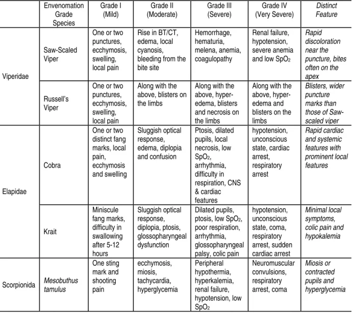
An attempt was made by the authors to categorize the snakebite symptoms in a
tabular form to be used at the healthcare centers, as given in Table 1 (15). But this
table did not include the scorpion sting cases, which also manifest neurological
complications. To avoid any possible misclassification and to make the classification
exhaustive, a modified table with scorpion sting symptoms is being proposed below.
It is pertinent to mention here that every envenomation grade includes symptoms of
Table 1: Symptomatic identification of venomous snakes in India. Envenomation Grade Species Grade I (Mild) Grade II (Moderate) Grade III (Severe) Grade IV (Very Severe) Distinct Feature Saw-Scaled Viper
One or two punctures, ecchymosis, swelling, local pain
Rise in BT/CT, edema, local cyanosis, bleeding from the bite site Hemorrhage, hematuria, melena, anemia, coagulopathy Renal failure, hypotension, severe anemia and low SpO2
Rapid discoloration near the puncture, bites often on the apex
Viperidae
Russell’s Viper
One or two punctures, ecchymosis, swelling, local pain
Along with the above, blisters on the limbs
Along with the above, hyper-edema, blisters and necrosis on the limbs
Along with the above, hyper-edema and blisters on the limbs
Blisters, wider puncture marks than those of Saw-scaled viper
Cobra
One or two distinct fang marks, local pain, ecchymosis and swelling Sluggish optical response, edema, diplopia and confusion Ptosis, dilated pupils, local necrosis, low SpO2,
arrhythmia, difficulty in respiration, CNS & cardiac features hypotension, unconscious state, cardiac arrest, respiratory arrest Rapid cardiac and systemic features with prominent local features Elapidae Krait Miniscule fang marks, difficulty in swallowing after 5-12 hours Sluggish optical response, diplopia, ptosis, glossopharyngeal dysfunction Dilated pupils, ptosis, low SpO2,
poor respiration, arrhythmia, glossopharyngeal palsy, colic pain
hypotension, unconscious state, coma, respiratory arrest, sudden cardiac arrest
BT=bleeding time, CT= coagulation time, SpO2= oxigen saturation level, CNS= central nervous system
Minimal local symptoms, colic pain and hypokalemia
Scorpionida Mesobuthus
tamulus One sting mark and shooting pain ecchymosis, miosis, tachycardia, hyperglycemia Peripheral hypothermia, hyperkalemia, renal failure, hypotension, low SpO2 Neuromuscular convulsions, respiratory arrest, coma Miosis or contracted pupils and hyperglycemia
MATERIALS AND METHODS
Parameter Selection for Remote Species Identification
Every year, more than a hundred patients report to healthcare centers in Kota
division of Rajasthan (India). They are victims of different species and have
envenomation of different degrees. These patients manifest various symptoms which
include local, neurological, systemic and circulatory abnormalities. In the current
medical practice in India, species-specific symptoms have not been given due
importance, but a systematic observation of manifested symptoms can reveal the
species.
Clinical profiles of forty patients have been studied which included all the possible
healthcare center without any medicine administered. The patients belong to all four
grades and five species (one patient bitten by a non-venomous snake has also been
included in the study and his psychosomatic symptoms were also observed). A
written and informed consent was obtained from the patients or the accompanying
relatives in all the cases. All the observations were taken without disturbing the
medical management. No experiment on animals was conducted.
The Manifested Symptoms
The manifested symptoms have been selected in such a way that either they are
manifested or are absent in cases of at least one species. It has also been kept in
mind that all the symptoms are obtainable with minimal instrumentation in minimum
time. There are twenty-nine symptoms which suit the constraints. All these symptoms
are considered in binary form in the feature vector with a numerical value of one or
zero.
i. Single fang marks: if there is only one fang/sting mark at the bite site.
ii. Paired fang marks: If there is a pair of fangs or more than two fang marks at the bite site, representing multiple bites.
iii. Gap in fang marks: If the gap between the fang marks from a bite is more than 1.75cm.
iv. Local edema: If there is edema developed, and if so, it is centering the fang marks alone.
v. Local pain: If there is pain at the bite/sting site.
vi. Hyper-edema: If there is excessive fluid pressure inside the affected limb and the whole limb is edematous.
vii. Ecchymosis: If the bite site and adjacent areas are getting discolored.
viii. BT/CT: If there is a marked rise in the bleeding time/coagulation time.
ix. Bleeding from the site: If the bite site is bleeding, incised and non-manipulated.
x. Blisters on limbs/trunk: If the body extremities or trunk present blisters/blebs.
xi. Hemorrhage: If there is bleeding from the gums or other openings of the body.
xii. Hematuria: If the urine is reddish.
xiii. Renal failure: If the subject is unable to urinate (even when the bladder is not empty).
xiv. Ptosis: If the eyelids are swollen and not opening fully.
xv. Pupil dilatation: If the pupils are dilated (mydriasis).
xvii. Colic pain/vomiting: If the subject reports pain in the abdomen and tendency to vomit.
xviii. Hemoptysis: If there is blood in the sputum.
xix. Glossopharyngeal paralysis: If the voice is obstructed or there is difficulty in speech.
xx. Neuromuscular paralysis: If the neuromuscular tone is abnormal.
xxi. Respiratory paralysis: If the ventilation is abnormal and oxygen level falls in the blood.
xxii. No local symptom: If there are no marked symptoms on the affected limb.
xxiii. Tachycardia: If the heart rate is higher than the normal limits.
xxiv. Bradycardia: If the heart rate is low than the normal limits.
xxv. Pupil constriction: If the pupils are constricted (miosis).
xxvi. Hyperglycemia: If the blood glucose level is above the normal limits.
xxvii. Peripheral hypothermia: If the extremities are getting cold (with excessive perspiration).
xxviii. Convulsions: If the subject is convulsive and hypertonic.
xxix. Coma: If the subject is in comatose condition.
Normalization of Age, Delay and Oxygen Saturation Level
To identify the severity of envenomation, the patient’s age, delay in transportation
and blood oxygen level were also accounted. These features were normalized
between zero and one.
The relationship between the age and the severity grade was exercised after a study
of different cases with a group of medical practitioners on different non-linear models.
The prescribed doses of ASV were considered as quantification of severity. The age
factor has been observed to be inversely proportional to the severity grade, and the
rate of development of symptoms is higher in juveniles, who are at higher risk when
all other conditions are the same. The age factor is less significant once the patient
crosses childhood. This method yielded a sigmoid relationship and the age can be
normalized as below:
5 age
age -1 factor age Normalized
+
= …(1)
The delay in transportation plays a vital role in the development of features prior to
the onset of treatment (3). Features develop very rapidly in the initial two hours in
Elapidae and scorpion envenomation (7). So, the delay was normalized as follows:
2 delay
delay delay
Normalized
+
= …(2)
where delay is expressed as hours.
Blood oxygen saturation (SpO2) is very significant for medical management.
Reduction in blood oxygen saturation causes tachycardia, seizure, confusion, coma
and eventually death in a few cases. So, it was considered inversely proportional to
the severity grade and normalized as follows:
100 100 SpO
Normalized 2 = −SpO2 …(3)
where SpO2 figure is expressed as percentage.
The Feature Vector
The combination between twenty-nine binary and three normalized parameters
resulted in a feature vector comprising of thirty-two distinct features. The values of
the features ranged between zero and one.
It would be pertinent to mention here that some features were interrelated as well as
opposite to each other, e.g. bradycardia and tachycardia. In early hours of Elapidae
as well as of Viperidae snakebites, the victim is in a state of shock and anxiety. Soon
after the bite, heart beats faster and later the heart may beat slower due to the
venom effect or depression.
The Neural Network
It was experienced that the species identification and the severity grading depend on
the presence and absence of certain symptoms. The perceptron-type neural network
model offers simplicity and proportional significance of the inputs with respect to the
output (19). So, a perceptron model was considered with no hidden layer and no
sigmoidal non-linearity at the output node. The perceptron type neural network would
demonstrate quantitative significance of manifested symptoms for particular species
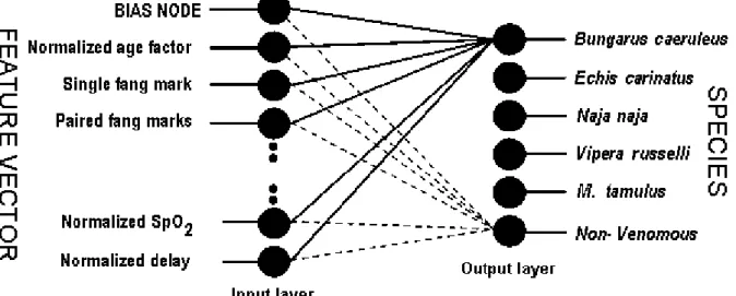
The proposed neural network consists of two layers only, one input layer and one
output layer. There are thirty-three input nodes, one input for each feature and one
bias node, which would have an inbuilt consistent input having value one. The output
layer is consisted of six output nodes; one output for each venomous species and the
sixth output node for non-venomous category. All the inputs are connected to the
outputs by means of weights. It is expected that the node corresponding to the most
probable species would yield the highest magnitude and this magnitude would be
proportional to the severity grade by the given relationship
i ij
j W I
O =Σ ∗ …(4)
where Oj is the jth output, corresponding to jth species
Ii is the ith input, corresponding to ith symptom
Wij is the weight connecting ith symptom and jth species.
Figure 1: Neural network topology, feature vector as input and species as output.
Forty cases of venom interaction were selected, in which the aggressor species were
well identified. Out of these cases, twenty-one were chosen for the training. This
group was comprised of all four severity grades of all five venomous species. One
case of bite by a non-venomous rat snake (Ptyas mucosus) was chosen; the patient
was under shock and exhibited some psychosomatic symptoms. The grades were
converted into numeral values with mild envenomation as one, moderate
envenomation as two, severe envenomation as three, and very severe envenomation
as four. Each case with symptoms, species and severity was taken as one training
The initial weight matrix was randomized and the symptoms were assigned at the
input nodes. The outputs were evaluated at the output layer. The resultant outputs
were compared with the desired output and error was fed back to readjust the
weights. This process was repeated exhaustively until the error was below a
predefined tolerance level, which is known as training of the neural network. Training
was repeated for all twenty-one training sets in random order exhaustively until the
error in each training cycle confined within a predefined tolerance band.
RESULTS
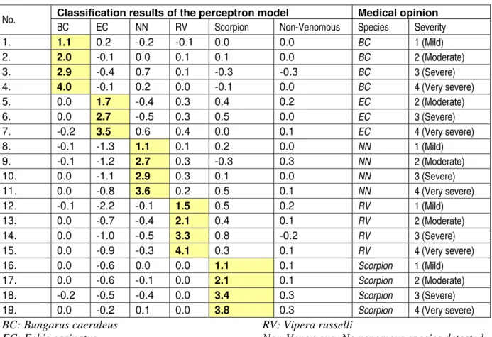
After a rigorous training and adjustment of weights, the model was tested on
nineteen cases in which the species identification and severity grading was available.
The neural network results are reproduced in Table 2.
No error was observed in species identification. However, the envenomation grades
had minor variations compared with the physician’s assessment. Training with more
number of cases is expected to improve the accuracy of envenomation grading.
Table 2: Classification results of the perceptron model and the medical opinion.
Classification results of the perceptron model Medical opinion No.
BC EC NN RV Scorpion Non-Venomous Species Severity
1. 1.1 0.2 -0.2 -0.1 0.0 0.0 BC 1 (Mild)
2. 2.0 -0.1 0.0 0.1 0.1 0.0 BC 2 (Moderate)
3. 2.9 -0.4 0.7 0.1 -0.3 -0.3 BC 3 (Severe)
4. 4.0 -0.1 0.2 0.0 -0.1 0.0 BC 4 (Very severe)
5. 0.0 1.7 -0.4 0.3 0.4 0.2 EC 2 (Moderate)
6. 0.0 2.7 -0.5 0.3 0.5 0.0 EC 3 (Severe)
7. -0.2 3.5 0.6 0.4 0.0 0.1 EC 4 (Very severe)
8. -0.1 -1.3 1.1 0.1 0.2 0.0 NN 1 (Mild)
9. -0.1 -1.2 2.7 0.3 -0.3 0.3 NN 2 (Moderate)
10. 0.0 -1.1 2.9 0.3 0.1 0.0 NN 3 (Severe)
11. 0.0 -0.8 3.6 0.2 0.5 0.1 NN 4 (Very severe)
12. -0.1 -2.2 -0.1 1.5 0.5 0.2 RV 1 (Mild)
13. 0.0 -0.7 -0.4 2.1 0.4 0.1 RV 2 (Moderate)
14. 0.0 -1.0 -0.5 3.3 0.8 -0.2 RV 3 (Severe)
15. 0.0 -0.9 -0.3 4.1 0.3 0.1 RV 4 (Very severe)
16. 0.0 -0.6 0.0 0.0 1.1 0.1 Scorpion 1 (Mild)
17. 0.0 -0.6 -0.1 0.0 2.1 0.1 Scorpion 2 (Moderate)
18. -0.2 -0.5 -0.4 0.0 3.4 0.3 Scorpion 3 (Severe)
19. 0.0 -0.2 0.1 0.0 3.8 0.3 Scorpion 4 (Very severe)
BC: Bungarus caeruleus EC: Echis carinatus NN: Naja naja
RV: Vipera russelli
DISCUSSION
The species symptomatic identification is usually performed by experts. A large
number of symptoms have been found and reported by different research groups.
The authors have concentrated on the most usual symptoms.
The proposed method identifies the aggressor species based on the manifested
symptoms, which can be very useful in the medical management. There were minor
variations in the severity grading provided by the physicians and calculated by the
neural network. Generally, the attending medical staff takes into account a number of
other physical parameters which have not been considered in the domain of this
network, like the general health state and the psychological state of the victim, the
first aid method used, etc, which may be responsible for the difference.
The results of the neural network are consistent in the classification. However, there
are grading differences regarding “mild”, “moderate”, “severe” and “very severe”.
Practically, it is difficult to draw crisp boundaries among different grades of
envenomation. Still, with more training on different cases, the error in severity
grading may be reduced.
CONCLUSION
The remote identification of venomous species is not a part of medical curricula in
India. Therefore, the medical community resorts to a quadrivalent ASV serum as the
standard medical practice in case of snakebites. However, this approach poses an
economical burden to the family of a poor victim and, at the same time, the attending
medical staff is not prepared to cope with rapid developments in symptoms in cases
of a few species. If this method is adopted by the medical community, cheaper and
specific monovalent ASV will be used as per the WHO guidelines, helping to reduce
the treatment costs (25).
The method proposed above is realizable in software, hardware or in a hybrid system.
Now, with the popularity of OS-based handheld gadgets, a handy solution using the
proposed method can be provided to the medical staff at the grassroots level for
species and severity grade identification.
ACKNOWLEDGEMENTS
The authors are highly indebted to Dr. Vinod Kumar Mahobia, Ex-Professor of
venomous animal species, their endemic behavior and scientific details. The authors
acknowledge the patients of bites/stings and their accompanying relatives who
consented to using their data for the research.
The authors are grateful to the generosity of Dr. B. L. Gochar and the administration
staff of Anurag Hospital and Research Center, Kota, for providing expert medical
opinion, access to the snakebitten patients and for the whole-hearted cooperation in
data collection. Words cannot express the efforts of Mr. Vishnu Shringi, Snake
Rescue Volunteer, who not only helped in taking notes, but also recorded vital
information as still photos and video footage. Finally, the authors wish to
acknowledge the opportunities extended by the Indian Institute of Technology,
Roorkee, India. Ranjan Maheshwari expresses his gratefulness to Engineering
College Kota, Kota (Rajasthan) for sponsoring him for obtaining of his PhD degree.
REFERENCES
1 AGRAWAL PN., AGGARWAL AN., GUPTA D., BEHERA D., PRABHAKAR S.,
JINDAL SK. Management of respiratory failure in severe neuroparalytic snake
envenomation. Neurol. India, 2001, 49, 25-8.
2 BAWASKAR HS. Snake venoms and antivenoms: critical supply issues. J. Assoc.
Physicians India, 2004, 52, 11-3.
3 CHAUHAN S., FARUQI S., BHALLA A., SHARMA N., VARMA S., BALI J.
Pre-hospital treatment of snake envenomation in patients presented at a tertiary care
hospital in Northwestern India. J. Venom. Anim. Toxins incl. Trop. Dis., 2005, 11,
275-82.
4 CHER CDN., ARMUGAM A., ZHU YZ., JEYASEELAN K. Molecular basis of
cardiotoxicity upon cobra envenomation. Cell. Mol. Life Sci., 2005, 62, 105-18.
5 FRY BG., WUSTER W., KINI RM., BRUSIC V., KHAN A., VENKATARAMAN D.,
ROONEY AP. Molecular evolution and phylogeny of elapid snake venom three-finger
toxins. J. Mol. Evol., 2003, 57, 110-29.
6 FUJINI TJ., NAKAJYO T., NISHIMURA E., OGURA E., TSUCHIYA T., TAMIYA T.
Molecular evolution and diversification of snake toxin genes: revealed by analysis of
intron sequences. Gene, 2003, 313, 111-8.
7 GAJRE G. Scorpion envenomation in children: should all stings be given
antivenom? Ann. Saudi Med., 1999, 19, 444-6.
9 HARRIS JB., GOONETILLEKE A. Animal poisons and the nervous system: what
the neurologist needs to know. J. Neurol. Neurosurg. Psychiatry, 2004, 75, 40-6.
10 HAZRA A. Poisonous snakebites in India. Community Dev. Med. Unit Ration.
Drug. Bull., 2003, 30, 1-12.
11 HICKS JW. The physiological and evolutionary significance of cardiovascular
shunting patterns in reptiles. N. Physiol. Sci., 2002, 17, 241-5.
12 JENA I., SARANGI A. Snakebite. In: JENA I., SARANGI A. Snakes of medical
importance and snakebite management. New Delhi: Ashish Publ. House, 1993:
99-105.
13 KIRAN S., SENTHILNATHAN TA. Management of snake envenomation. Word
Anaestesia, 2003, 16, 13-8.
14 KULARATNE SAM. Common krait (Bungarus caeruleus) bite in Anuradhapura, Sri
Lanka: a prospective clinical study, 1996-98. Postgrad. Med. J., 2002, 78, 276-80.
15 KUMAR V., MAHESHWARI R., VERMA HK. Toxicity and symptomatic
identification of species involved in snakebites in the Indian subcontinent. J. Venom.
Anim. Toxins incl. Trop. Dis., 2006, 12, 3-18.
16 MARKLAND FS. Snake venoms and the hemostatic system. Toxicon, 1998, 36,
1749-800.
17 MURTHY KR., HASE NK. Scorpion envenoming and the role of insulin. Toxicon,
1994, 32, 1041-4.
18 NIRTHANAN S., GOPALAKRISHNAKONE P., GWEE MC., KHOO HE., KINI RM.
Non-conventional toxins from elapid venoms. Toxicon, 2003, 41, 397-407.
19 RAO MA., SRINIVAS J. Hopfield, perceptron and related models. In: RAO MA.,
SRINIVAS J. Neural networks: algorithms and applications. New Delhi: Narosa Publ.
House, 2004: 35-62.
20 SAADEH AM. Case report: acute myocardial infarction complicating a viper bite.
Am. J. Trop. Med. Hyg., 2001, 64, 280-2.
21 SELVANAYAGAMA ZE., GNANAVENDHANA SG., GANESH KA., RAJAGOPAL
D., RAO PV. ELISA for the detection of venoms from four medically important snakes
of India. Toxicon, 1999, 37, 757-70.
22 VAN DONG LE., QUYEN LE K., ENG KH., GOPALAKRISHNAKONE P.
Immunogenicity of venoms from four common snakes in the South of Vietnam and
development of ELISA kit for venom detection. J. Immunol. Methods, 2003, 282,
23 WARRELL DA. Guidelines for the clinical management of snakebites in South
East Asia Region. New Delhi: WHO Southeast Asia Regional Office, 2005, 1-87.
24 WHITAKER R. Common Indian snakes: a field guide. New Delhi: National Book
Trust, 1995: 1-37.
25 WORLD HEALTH ORGANIZATION. Regional Office for South-East Asia.
Management of snakebites in South East Asia. The clinical management of
snakebites in the southeast region of Asia [serial on-line], 2003. Available from:
http://w3.whosea.org/bct/snake/5.htm.
26 YAMADA D., SEIKIYA F., MORITA T. Prothrombin and factor X activator activities
in the venoms of Viperidae snakes. Toxicon, 1997, 35, 1581-9.
27 YUGANDHAR B., MURTHY KRK., SATTAR SA. Insulin administration in severe
scorpion envenoming. J. Venom. Anim. Toxins, 1999, 5, 200-19.
28 ZHANG Y., TU AT. The effect of snake venom and their components on
adrenomedullary cells: catecholamine efflux and cell damage. Neurotoxicology, 2002,