Article
Printed in Brazil - ©2018 Sociedade Brasileira de Química*e-mail: [email protected]
A Water Soluble Naphthalimide-Based Chemosensor for Fluorescent Detection CN
−in Pure Water and Its Application in Practical Samples
Li Wang,a,b Wen-Ting Li,a Wen-Juan Qu,a Yan-Qing Fan,a Hong Yao,a Qi Lin,a
Tai-Bao Weia and You-Ming Zhang*,a
aCollege of Chemistry and Chemical Engineering, Northwest Normal University, 730070 Lanzhou, Gansu, P. R. China
bSchool of Chemical and Biological Engineering, Lanzhou Jiaotong University, 730070 Lanzhou, China
By rationally introducing aromatic carboxyl functionalized 1,8-naphthalimide, a water soluble fluorescent chemosensor (DA) was successfully synthesized. It could selectively and sensitively detect CN− in pure water via an intramolecular charge transfer to twisted intramolecular charge
transfer (ICT-TICT)state change mechanism. The detection limit of the chemosensor DA to CN−
was 1.38 × 10⁻⁸ mol L-1 which was lower than 1.9 µmol L-1 (set by the World Health Organization (WHO)). Notably, DA displayed rapid response (about 1 s) for recognizing CN− in pure water.
Furthermore, DA could be applied to monitoring CN− in tap water. Meanwhile, we prepared the
test strips based on DA, which could rapidly and efficiently detect CN− in water.
Keywords: water-soluble chemosensor, fluorescent, 1,8-naphthalimide derivative, CN−
recognition
Introduction
Cyanide (CN−) salts are extensively used in industry,
such as electric plating, gold mining, plastic production, and other areas.1 However, cyanide is greatly toxic to mammals with even a small amount of this species, leading to emesis, unconsciousness, and finally to death.2 Therefore, cyanide concentrations in drinking water must be lower than 1.9 µmol L-1.3 In this regard, the reasonable design and synthesis of effective sensors to selectively detect cyanide anion at the environmental and biological levels have received a lot of attention.4
Fluorescence sensors for specific anions play a fundamental role in medical, environmental and biological applications, what is more, it is one of the best tools with high sensitivity, rapid response and easy performance, much interest has been focused on the design and synthesis of fluorescent chemosensors for anions.5
To date, much effort has been drawn to develop the fluorescent chemosensors for CN−. However, most of them worked properly in a high percentage of organic solvent media for the detection of cyanide ions.6 As we know, the
use of organic solvents usually damages the normal function of biomolecules, thus, good water solubility are essential for practical use of the chemosensors in biochemical research.7 Although some of them could work in pure water,8 they did not display enough sensitivity for the maximum allowable level (1.9 µmol L-1) of CN− in drinking water. Thus, it is
highly challenging to develop new soluble fluorescent chemosensors forsensing CN− with high sensitivity and
excellent selectivity in 100% water solutions.
1,8-Naphthalimide derivatives are an excellent
environmentally sensitive fluorophore compounds, which is extensively applied to various fields of science and technology.9 As a consequence of their strong blue fluorescence and good photostability, 1,8-naphthalimide derivatives are often used for fluorescence sensors, switchers and ion probes.10 However, the application of naphthalimide sensors was limited due to poor solubility. We all know that carboxyl not only has a remarkable coordination ability,11 but also has an excellent hydrophilicity.
1,8-naphthalimide. As expected, chemosensor DA displays high sensitivity and rapid response (about 1 s) for recognizing CN− in pure water system. Compared with
most of the previously reported fluorescent chemosensors for CN− and naphthalimide sensors,13 the advantage of DA is that it could work in 100% water solutions. It is worth noting that compared with recently reported aromatic carboxyl functionalized 1,8-naphthalimide chemosensors, DA can be directly dissolved in pure water without changing COOH into COONa.
Experimental
Materials and apparatus
Fresh double-distilled water was used throughout the experiments. All other reagents and solvents (analytical grade) were commercially available and were used without further purification. 1H nuclear magnetic resonance (NMR) spectra was recorded on an Agilent DD2 at 600 MHz spectra. 1H chemical shifts are reported in ppm downfield from tetramethylsilane (TMS, δ scale with the solvent
resonances as internal standards). Fluorescence spectra were recorded on a Shimadzu RF-5310 (Japan). Melting points were determined on an X-4 digital melting-point apparatus (Beijing Tech Instrument Co., Ltd., China) and were uncorrected. Mass spectra (MS) were recorded on a Bruker Esquire 3000 plus mass spectrometer (Bruker-Franzen Analytik GmbH, Bremen, Germany) equipped with ESI (electrospray ionization) interface and ion trap analyzer.
Synthesis of the chemosensor DA
Compound DA was synthesized by condensation reaction of anthranilic acid and 1,8-naphthalic anhydride (Scheme 1).14 Anthranilic acid (0.55 g, 4.0 mmol) and 1,8-naphthalic anhydride (0.79 g, 4.0 mmol) were dissolved in absolute dimethylformamide (DMF, 20 mL) at 80 °C.
The reaction mixture was stirred and refluxed for 7 h. The mixture was still clear liquid after cooling down to room temperature. White precipitate appeared after adding distilled water, through filtration, washed with ethanol and dried under vacuum, then obtained the product DA in 85% yield (mp > 300 °C).
Characterization data for DA
1H NMR (600 MHz, DMSO-d
6) d 12.85 (s, 1H, COOH), 8.49 (m, 4H, Nap-H), 8.08 (m, 1H, Ph-H), 7.89 (t, J 6 Hz,
2H, Nap-H), 7.75 (t, J 6 Hz, 1H, Ph-H), 7.60 (t, J 6.6 Hz,
1H, Ph-H), 7.50 (d, J 6 Hz, 1H, Ph-H). ESI-MS m/z:
[M + Na]+ calcd. for C
19H11NO4Na: 340.06; found: 340.01.
General procedure for fluorescence spectra experiments
The stock solution of DA (0.2 mmol L-1) in pure water (pH = 7.2) was prepared and stock at room temperature, which diluted to a concentration of 20 µmol L-1 as the determined solution. The aqueous solutions of anions (10 mmol L-1) (F−, Cl−, Br−, I−, AcO−, SO
4−, H2PO4−, ClO4−, CN−, SCN−, S2− and OH−) were prepared via sodium salts.
The fluorescence spectra were obtained by excitation at 315 nm. The excitation and emission slit widths were both 5 nm.
General procedure for 1H NMR experiments
For 1H NMR titrations, sensor DA was prepared in DMSO-d6 and NaCN was prepared in double-distilled water. First of all, only sensor DA in DMSO-d6 was added
into the NMR tube, and then CN− anion was added at 0.1,
0.5, 1.0, 2.0 and 4.0 equiv. sequentially. All solutions were mixed directly in the NMR tube.
Practical samples analysis
For practical sample analysis, the tested water tap samples were prepared by adding known amounts of CN−.
The fluorescence spectrum of each sample was tested for three times, and the found concentration of CN− was
measured according to the developed calibration curve.
Preparation of the test strips
The test strips were carried out by submersing filter papers in the pure water solution of DA (2.0 × 10⁻⁴ mol L-1) and then dried in air. The CN− water solution (2.0
× 10−4 mol L-1) and
competitive anions water solution were dropped on the test strips. After drying, the images were recorded by a digital camera under UV lamp.
Results and Discussion
We researched the fluorescent response of chemosensor DA to various anions in 100% water solutions, including
F−, Cl−, Br−, I−, AcO−, SO
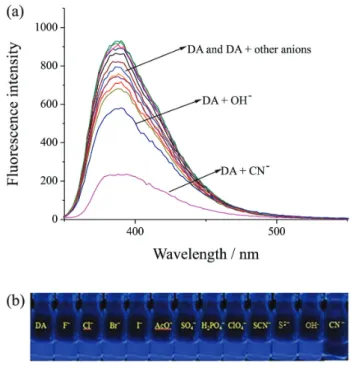
4−, H2PO4−, ClO4−, CN−, SCN−, S2− and OH−. As shown in Figure 1, the maximum fluorescent
emission band of free DA appeared at 390 nm. When 5 equivalents of CN− was added to the pure water solution of
chemosensor DA, the fluorescence emission band at 390 nm significantly reduced. The obvious color change from bright blue to colorless could be distinguished by naked-eyes through UV lamp (Figure 1). To further evaluate the selectivity of chemosensor DA, the same experiments were also conducted using F−, Cl−, Br−, I−, AcO−, SO
4−, H2PO4−, ClO4−, SCN−, S2− and OH−. Whether in the fluorescent spectra or by naked-eyes through UV lamp, nearly no obvious fluorescence responses were observed with these anions (Figure 2), demonstrating DA has excellent fluorescent selectivity toward CN− in pure water.
Furthermore, the quantum yields of DA and DA + CN−
were calculated using the following equation 1:
Yu = (Ys × Fu × As) / (Fs × Au) (1)
where Yu is the quantum yield of DA and DA + CN−;
Ys is the quantum yield of reference compound (quinine sulfate, Ys = 0.55); Fu is the fluorescent integral of DA and DA + CN−; Fs is the fluorescent integral of reference
compound; Au is the absorbance of DA and DA + CN−; As
is the absorbance of reference compound. The quantum
yield of DA and DA + CN− were 0.4183 and 0.0594,
respectively.
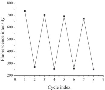
To get a quantitative idea of the sensing, the fluorescence titration experiment was further performed in pure water (Figure 3). Upon the addition of CN−, the fluorescence
emission intensity of chemosensor DA at 390 nm
decreased gradually and reached to minimum value by 3.8 equiv. of CN− (Figure 3a). The binding constant
value Ka, which was evaluated by the Benesi-Hildebrand method, was found to be 6.08 × 10⁵ mol-1 L (Figure 3b), and pKa = 4.17. Furthermore, the detection limit (LOD)
calculated according to the basis of 3σ/S (where σ is
the standard deviation of the blank solution and S is the slope of the calibration curve) was 1.38 × 10⁻⁸ mol L-1 (Figure S3, Supplementary Information (SI) section), which is superior to other cyanide chemosensors (Table 1) and far below the World Health Organization (WHO) guideline of 1.9 µmol L-1.3
The pH effects were measured to investigate whether the sensor is appropriate for the physiological detection (Figure 4). The detection of CN− can operate well in the
pH range of 4.0-8.0 in pure water, which confirmed that DA has also excellent sensing ability toward CN− under
physiological conditions.
We further examined the sensor’s reactivity toward CN−
(Figure 5). While DA (2.0 × 10⁻⁵ mol L-1) was treated with CN− (5 equiv.) in pure water, the fluorescence intensity at
390 nm decreased rapidly and reached a plateau in about 1 s. The kinetic test result proved that our sensor could rapidly detect CN− in pure water.
We further explore the selectivity of chemosensors DA for cyanide when DA was treated with other competitive anions, such as F−, Cl−, Br−, I−, AcO−, SO
4−, H2PO4−, ClO4−, SCN−, S2− and OH− (Figure 6). The fluorescence emission
Figure 1. Fluorescence spectra of DA (20 µmol L-1) in pure water media in the presence of CN− (5 equiv.) (λex = 315 nm). Inset: color change of
DA, DA + CN− (5 equiv.) under UV lamp in pure water media.
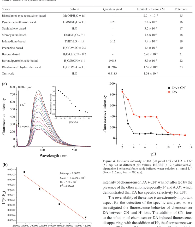
Table 1. Sensors for cyanide determination
Sensor Solvent Quantum yield Limit of detection / M Reference
Bis(salamo)-type tetraoxime-based MeOH/H2O = 1:1 – 8.91 × 10⁻⁷ 15
Pyrene-benzothiazol-based DMSO/H2O = 1:1 0.23 2.8 × 10⁻⁷ 16
Naphthalene-based H2O – 3.2 × 10⁻⁷ 17
Merocyanine-based EtOH/H2O = 9:1 – 1.6 × 10⁻⁶ 18
Indanedione-based THF/H2O = 1:9 0.12 9.4 × 10⁻⁷ 19
Phenazine-based H2O/DMSO = 7:3 – 1.4 × 10⁻⁶ 20
Boronic-based H2O/CH3CN = 8:2 – 6.45 × 10⁻⁸ 21
Borondipyrromethene-based H2O/EtOH = 1:1 0.015 5.9 × 10⁻⁸ 22
Rhodamine-B hydrazide-based H2O/DMSO = 1:1 0.8916 1.59 × 10⁻⁷ 23
Our work H2O 0.4183 1.38 × 10⁻⁸
Figure 3. (a) Fluorescence spectra (λex = 315 nm) of DA (20 µmol L-1) in the presence of different amounts of CN−. Inset: a plot of fluorescence
intensity at 390 nm vs. number of equivalents of CN−; (b)
Benesi-Hilderbrand plot of DA with CN−.
Figure 4. Emission intensity of DA (20 µmol L-1) and DA + CN−
(50 equiv.) at different pH values. HEPES (4-(2-hydroxyerhyl) piperazine-1-erhanesulfonic acid) buffered water solution (1 mmol L-1) (λex = 315 nm, λem = 390 nm).
intensity of chemosensor DA + CN− was not affected by the
presence of the other anions, especially F− and AcO−, which
demonstrated that DA has specific selectivity for CN−.
The reversibility of the sensor is an extremely important aspect for the detection of the specific analyses, so we investigated the fluorescence behavior of chemosensor DA between CN− and H+ ions. The addition of CN− ions
to the solution of chemosensor DA induced fluorescence disappearing, with the addition of H+, the fluorescence was turn on. This reversible process of “on-off-on” could be repeated at least three times with a little loss of fluorescent intensity (Figure 7).
On the basis of the spectral results, the recognition
mechanism of chemosensor DA with CN− is proposed
indicating that the recognition of CN− is a deprotonation
process. In addition, we prepared the carboxylate salt of DA using sodium carbonate, and investigated its photophysical properties. The results show the fluorescence spectrum of DA− and DA + CN− have significant overlaps (Figure S4,
SI section), and prove that the recognition of CN− is a
deprotonation process.
To study its practical applications, the chemosensor DA was used to measuring CN− in tap water. By adding a known
quantity of standard CN− to tap water and calculating its
recovery, the accuracy of the method was evaluated. The recoveries of different known quantities of CN− added were
gained from 96.6 to 103.6% with a favorable analytical precision (relative standard deviation (RSD) ≤ 5.0%), which indicated the chemosensor DA was able to determine CN− in tap water. The results were presented in Table S1
(SI section).
Motivated by the favorable characteristic of chemosensor DA in solution, test strips were carried out
Figure 5. Time-dependent fluorescence intensity of DA (20 µmol L-1) in the presence of 5 equiv. CN− in pure water (λex = 315 nm, λem = 390 nm)
was recorded after 0, 1, 2, 4, 6 and 8 s.
Figure 6. Emission intensity of DA (20 µmol L-1) in the presence of 5 equiv. of other anions and DA + CN− in the presence of 5 equiv. of other
anions in pure water (λex = 315 nm, λem = 390 nm).
Figure 7. Fluorescence intensity of DA (20 µmol L-1) upon the alternate addition of CN− and H+ in pure water.
intramolecular rotation was restricted by the hydrogen bond. In this case, the sensor is in the intramolecular charge transfer (ICT) state and displays a very strong fluorescence. With the increasing addition of CN−, the hydrogen bond
is destroyed, the benzene ring of chemosensor DA could take place of an intramolecular rotation, and change to twisted ICT (TICT) state and show a weak fluorescence emission at 390 nm. With further adding H+ to solution of DA + CN−, the fluorescence intensity was recovered to an
ICT state (Figure 8). Meanwhile, the 1H NMR titration experiments were conducted to demonstrate the proposed recognition mechanism. As shown in Figure 9, upon the gradual addition of CN−, the –COOH peak at 12.85 ppm
disappeared, and the signal peak of the hydrogen atoms of the benzene ring and the naphthalene ring shifted upfield,
by submersing filter papers in the pure water solution of DA (2.0 × 10⁻⁴ mol L-1) and then dried in air. The test strips
coated with DA were utilized to sense CN− and other anions,
similar to the pH test paper. As shown in Figure 10, when one drop of CN− (2.0
× 10−4 mol L-1) water solution was added on the test kits, the fluorescence turn off response can be observed by naked-eye under UV irradiation. The same procedures were done for competitive ions. However, potentially competitive ions had no influence on the detection of CN− by the test strips. Hence, DA also
possesses excellent selectivity for CN− in test strips.
Conclusions
In summary, by rationally introducing aromatic carboxyl and 1,8-naphthalimide moieties, we designed
a novel water soluble fluorescent chemosensor (DA). As expected, DA displays high sensitivity and rapid response (about 1 s) for recognizing CN− in 100% water
solutions via an ICT-TICT state change mechanism. The detection limits of the chemosensor DA toward CN− was
1.38 × 10⁻⁸ mol L-1, which was far below the WHO limit (1.9 µmol L-1). Furthermore, the sensor DA was able to determine CN− in tap water with good recovery. Meanwhile,
we prepared the test strips based on DA, which could rapidly and efficiently detect CN− in water.
Supplementary Information
Supplementary information is available free of charge at http://jbcs.sbq.org.br as PDF file.
Acknowledgments
This work was supported by the National Natural Science Foundation of China (NSFC, No. 21661028; 21662031; 21574104), the Program for Changjiang Scholars and Innovative Research Team in University of Ministry of Education of China (IRT 15R56).
References
1. Xu, Z.; Chen, X.; Kim, H. N.; Yoon.; J. Chem. Soc. Rev.2010,
39, 127; Mannel-Croise, C.; Probst, B.; Zelder, F.; Anal. Chem.
2009, 81, 9493; Vallejos, S.; Estevez, P.; Garcia, F. C.; Serna,
F.; de la Pena, J. L.; Garcia, J. M.; Chem. Commun.2010, 46, Figure 10. Photographs of DA on test strips (A) free DA; (B) after
submersing in pure water solutions of DA + CN−; (C) after submersing
in pure water solutions of DA + other anions; (D) after submersing in pure water solutions of DA + CN− + other anions using UV lamp at room
temperature.
Figure 9.1H NMR spectra of free DA (600 MHz, DMSO-d
7951; Kim, D.-S.; Chung, Y. M.; Jun, M.; Ahn, K. H.; J. Org. Chem.2009, 74, 4849; Rahman, S.; Assiri, Y; Alodhayb, A. N.;
Beaulieu, L. Y.; Oraby, A. K.; Georghiou, P. E.; New J. Chem.
2016, 40, 434.
2. Baskin, S. I.; Brewer, T. G. In Medical Aspects of Chemical and Biological Warfare; Sidell, F.; Takafuji, E. T.; Franz, D.
R., eds.; TMM: Washington, 1997, p. 271.
3. World Health Organization (WHO); Guidelines for Drinking-Water Quality; World Health Organization: Geneva, 1996. 4. Hu, J. H.; Sun, Y.; Qi, J.; Pei, P. X.; Lin, Q.; Zhang, Y. M.; RSC
Adv.2016, 6, 100401; Jung, H. S.; Han, J. H.; Kim, Z. H.; Kang, C.; Kim, J. S.; Org. Lett. 2011, 13, 5056; Kumar, S.; Singh, P.;
Hundal, G.; Hundal, M. S.; Subodh Kumar, S.; Chem. Commun.
2013, 49, 2667; Sun, Y.; Hu, J. H.; Qi, J.; Li, J. B.; Spectrochim. Acta, Part A 2016, 167, 101.
5. Cort, A. D.; Forte, G.; Schiaffino, L.; J. Org. Chem. 2011,
76, 7569; Li, Y. P.; Yang, H. R.; Zhao, Q.; Song, W. C.; Han, J.; Bu, X. H.; Inorg. Chem. 2012, 51, 9642; Hu, J. H.; Li, J.
B.; Qi, J.; Chen, J. J.; New J. Chem. 2015, 39, 843; Dong, W. K.; Akogun, S. F.; Zhang, Y.; Sun, Y. X.; Dong, X. Y.; Sens. Actuators, B 2017, 238, 723; Hu, J. H.; Li, J. B.; Qi, J.; Sun, Y.; Sens. Actuators, B 2015, 208, 581.
6. Guliyev, R.; Buyukcakir, O.; Sozmen, F.; Bozdemir, O. A.;
Tetrahedron Lett.2009, 50, 5139; Gee, H. C.; Lee, C. H.;
Jeong, Y. H.; Jang, W. D.; Chem. Commun.2011, 47, 11963; Sharma, N.; Reja, S. I.; Bhalla, V.; Kumar, M.; Dalton Trans.
2014, 43, 15929; Chawla, H. M.; Shukla, R.; Goel, P.; New J. Chem.2014, 38, 5264; Jo, H. Y.; Park, G. J.; Na, Y. J.; Choi, Y.
W.; You, G. R.; Kim, C.; Dyes Pigm.2014, 109, 127. 7. Li, X. H.; Gao, X. H.; Shi, W.; Ma, H. M.; Chem. Rev. 2014,
114, 590.
8. Chung, S. Y.; Nam, S. W.; Lim, J.; Park, S.; Yoon, J.; Chem. Commun.2009, 2866; Chen, X.; Nam, S. W.; Kim, G. H.; Song, N.; Jeong, Y.; Shin, I.; Kim, S. K.; Kim, J.; Park, S.; Yoon, J.;
Chem. Commun.2010, 46, 8953; You, G. R.; Park, G. J.; Lee, J. J.; Kim, C.; Dalton Trans.2015, 44, 9120; Xu, J. F.; Chen,
H. H.; Chen, Y. Z.; Li, Z. J.; Wu, L. Z.; Tung, C. H.; Yang, Q. Z.; Sens. Actuators, B2012, 168, 14.
9. Ma, T. H.; Dong, M.; Dong, Y. M.; Wang, Y. W.; Peng, Y.; Chem. Eur. J. 2010, 16, 10313.
10. Duke, R. M.; Veale, E. B.; Pfeffer, F. M.; Kruger, P. E.; Gunnlaugsson, T.; Chem. Soc. Rev. 2010, 39, 3936; Zheng, S.
P. L.; Lynch, P. L. M.; Rice, T. E.; Moody, T. S.; Gunaratne, H. Q. N.; de Silva, A. P.; Photochem. Photobiol. Sci. 2012, 11,
1675; Banerjee, S.; Veale, E. B.; Phelan, C. M.; Murphy, S. A.; Tocci, G. M.; Gillespie, L. J.; Frimannsson, D. O.; Kelly, J. M.; Gunnlaugsson, T.; Chem. Soc. Rev. 2013, 42, 1601; Reger, D. L.; Leitner, A. P.; Smith, M. D.; Cryst. Growth Des. 2016, 16,
527.
11. Sun, Y. X.; Dong, W. K.; Wang, L.; Zhao, L.; Yang, Y. H.; Chin. J. Inorg. Chem.2009, 25, 1478; Dong, W. K.; Sun, Y. X.; Zhao,
C. Y.; Dong, X. Y.; Xu, L.; Polyhedron2010, 29, 2087; Dong, X. Y.; Sun, Y. X.; Wang, L.; Li, L.; J. Chem. Res.2012, 387; Wang, P.; Zhao, L.; Spectrochim. Acta, Part A2015, 135, 342; Dong, W. K.; Ma, J. C.; Zhu, L. C.; Sun, Y. X.; Akogun, S. F.; Zhang, Y.; Cryst. Growth Des.2016, 16, 6903.
12. Cheng, X.; Li, H.; Zheng, F.; Lin, Q.; Zhang, Y. M.; Yao, H.; Wei, T. B.; Dyes Pigm. 2016, 127, 59; Wang, L.; Li, W. T.; Qu, W. J.; Su, J. X.; Lin, Q.; Wei, T. B.; Zhang, Y. M.; Supramol. Chem.2017, 29, 489, DOI: 10.1080/10610278.2016.1277586; Chang, J.; Cai, Y.; Wei, T. B.; Sens. Actuators, B2015, 213,
501; Lin, Q.; Zhu, X.; Fu, Y. R.; Yang, Q.; Sun, B.; Wei, T. B.; Zhang, Y. M.; Dyes Pigm.2015, 113, 748; Qu, W. J.; Gao, G.
Y.; Shi, B. B.; Wei, T. B.; Zhang, Y. M.; Lin, Q.; Yao, H.; Sens. Actuators, B2014, 204, 368.
13. Hu, J. H.; Sun, Y.; Qi, J.; Li, Q.; Wei, T. B.; Spectrochim. Acta, Part A2017, 175, 125; Wang, F.; Xu, Y. L.; Aderinto, S. O.;
Peng, H. P.; Zhang, H.; Wu, H. L.; J. Photochem. Photobiol., A
2017, 332, 273; Aderinto, S. O.; Zhang, H.; Wu, H. L.; Chen,
C. Y.; Zhang, J. W.; Peng, H. P.; Yang, Z. H.; Wang, F.; Color. Technol. 2017, 133, 40; Wu, H. L.; Aderinto, S. O.; Xu, Y. L.;
Zhang, H.; Fan, X. Y.; J. Appl. Spectrosc. 2017, 84, 25; Yu, B.; Li, C. Y.; Sun, Y. X.; Jia, H. R.; Guo, J. Q.; Li, J.; Spectrochim. Acta, Part A2017, 184, 249.
14. Reger, D. L.; Debreczeni, A.; Horger, J. J.; Smith, M. D.; Cryst. Growth Des.2011, 11, 4068.
15. Wang, F.; Gao, L.; Zhao, Q.; Zhang, Y.; Dong, W. K.; Ding, Y. J.; Spectrochim. Acta, Part A2018, 190, 111.
16. Li, J. J.; Wei, W.; Qi, X. L.; Zuo, G. C.; Fang, J. K.; Dong, W.;
Sens. Actuators, B 2016, 228, 330.
17. Shi, B.B.; Zhang, P.; Wei, T.B.; Yao, H.; Lin, Q.; Zhang, Y. M.; Chem. Commun. 2013, 49,7812.
18. Xiao, K.; Nie, H. M.; Gong, C. B.; Qu, X. X.; Tang, Q.; Chow, C. F.; DyesPigm.2015, 116, 82.
19. Hu, J. W.; Lin, W. C.; Hsiao, S. Y.; Wu, Y. H.; Chen, K. Y.;Chen, H. W.; Sens. Actuators, B2016, 233, 510.
20. Yang, L.; Li, X.; Qu, Y.; Qu, W.; Zhang, X.; Hang, Y.; Ågren, H.; Hua, J.; Sens. Actuators, B 2014, 203, 833.
21. Wang, S. T.; Sie, Y. W.; Wan, C. F.; Wu, A. T.; J. Lumin. 2016,
173, 25.
22. Yu, Y. H.; Shu, T. T.; Yu, B. J.; Deng, Y.; Fu, C.; Gao, Y. G.; Dong, C. Z.; Ruan, Y. B.; Sens. Actuators, B 2018, 255, 3170. 23. Pei, P. X.; Hu, J. H.; Ni, P. W.; Long, C.; Su, J. X.; Sun, Y.;
RSC Adv. 2017, 7, 46832.
Submitted: November 24, 2017 Published online: February 23, 2018