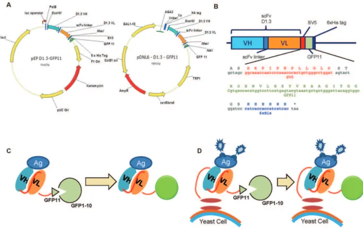
Fluorescent labeling of antibody fragments using split GFP.
Texto
Imagem

Documentos relacionados
The structure of the remelting zone of the steel C90 steel be- fore conventional tempering consitute cells, dendritic cells, sur- rounded with the cementite, inside of
(iii) By using quantitative fluorescent beads labeled with a known number of phycoerythrin (PE) molecules, the curve of fluorescence intensity can be extrapolated to evaluate the
In a second stage of this study, Antibody fragments (Fabs) specific for hDLL1-DE3 were generated by phage display, using the produced protein as target, in which one
If the fluorescent protein is specifically accumulated at the division septum (which contains two membranes) then the fluorescence ratio should be higher than two,
A recombinant yellow fever (YF) 17D virus expressing the reporter green fluorescent protein (GFP) with the stem-anchor (SA) region of E protein fused to its carboxy terminus was
Epifluorescence micrograph showing the colonization and reproduction of Colletotrichum gloeosporioides (40x magnification). a) Hypocotyls at 15 days after inoculation
The present study compared the bioavailability of crude protein and lipid from biofloc meals generated with an activated sludge system using two water sources: wastewater
