UNIVERSIDADE DE LISBOA
FACULDADE DE MEDICINA DE LISBOA
DOUTORAMENTO em MEDICINA
ESPECIALIDADE de NEUROLOGIA
2015
COGNITIVE FUNCTIONS DURING
MIGRAINE ATTACKS
UNIVERSIDADE DE LISBOA
FACULDADE DE MEDICINA DE LISBOA
COGNITIVE FUNCTIONS DURING MIGRAINE ATTACKS
RAQUEL SANTOS GIL GOUVEIA
Dissertação orientada pela Professora Doutora Isabel Pavão Martins
Tese especialmente elaborada para obtenção do grau de
Doutor em Medicina, Especialidade de Neurologia
JÚRI
- Presidente: Professor Doutor José Luís Ducla-Soares
- Vogais: Professor Doutor José Fernando da Rocha Barros
Professora Doutora Sara Marta Pereira Santos Cavaco
Professor Doutor Carlos Alberto Fontes Ribeiro
Professor Doutor José Manuel Morão Cabral Ferro
Professora Doutora Maria Isabel Segurado Pavão Martins
Professor Doutor Tiago Vaz Maia
i
A b s t r a c t
Background: Attack-related cognitive symptoms in migraine are frequent yet scarcely
characterized and undervalued as contributors of disability. Conflicting evidence arose about an increased risk of cognitive decline in older migraine patients.
Objectives: (1) to study the occurrence of cognitive symptoms in migraine attacks; (2)
to evaluate objective evidence of cognitive dysfunction in migraine attacks and its neuronal correlates and (3) to study the effect of persisting migraine in cognitive function or cognitive decline in older adults.
Methods: Occurrence of attack-related cognitive symptoms was detailed by systematic
literature review and a cross-sectional clinical-based systematic survey; their relevance to disability was studied prospectively using headache diaries. An instrument (Mig-SCog) was developed, validated and tested to identify and quantify attack-related subjective cognitive symptoms. Cognitive function during attacks was evaluated by a systematic literature review and a clinical-based prospective two-period randomized cross-over study using an extensive neuropsychological battery. A briefer battery was tested in repeated applications in interictal patients and controls. Brain perfusion during attacks was studied with arterial spin labeling magnetic resonance imaging (ASL-MRI) and cortical response to a working memory task with blood-oxygen level dependent functional magnetic resonance imaging (BOLD-fMRI). A prospective controlled cross-sectional population-based study of neuropsychological performance of older adults with persisting migraine and non-migraine headache was followed by a 5 years re-evaluation of the same sample, to screen for cognitive decline.
Results: Cognitive symptoms were the most frequent non-migraine defining symptoms
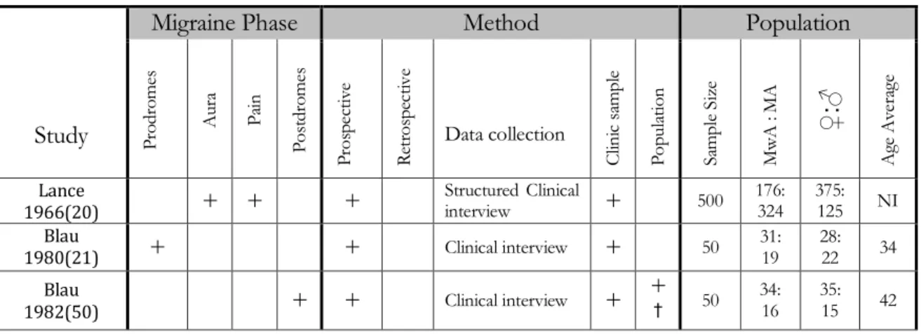
reported in the prodromic(37%) and headache(38%) phases of migraine attacks in a systematic review of 28 series, with a total sample of 8392 patients. Cognitive symptoms are also present in the postdromic or resolution phase, although fatigue (71%) is reported more often. Of 165 patients prospectively surveyed, 87% reported an average
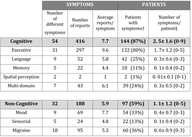
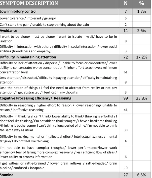
of 2.5 attack-related symptoms, over two-thirds executive (attention, processing efficiency and speed). Cognitive symptoms were ranked prospectively by 34 migraine patients recording 229 attacks, being second only to pain in terms of intensity and attack-related disability. An instrument to quantify migraine attack-related symptoms was constructed from a set of 43 candidate items, using factor analysis. The reduced 9 item Mig-SCog is fast to apply covering executive functions and language, having good internal consistency (Cronbachs’ alpha 0.82) and reliability (Cohen’s kappa 0.55) and high correlation with external validity measures such as the 43-candidate item list (rho=0.69) and the Cognitive Failures Questionnaires(rho=0.61). The Mig-SCog presents negligible recall bias (no difference in scores obtained during an attack or while headache free) and Migraine patients score it higher for migraine higher for migraine (7.9±4.6) than for non-headache pain (2.3±2.9, p<0.0006) or pain free (1.6±2.4, p<0.0006). Comparing Mig-SCog scores in migraine and tension-type headache patients, those were higher for migraine in all scale items (p<0.0001) except those related to naming (8 and 9). The AUC of Mig-SCog score for the diagnosis of Migraine was 0.835 (95% CI of 0.763-0.906, p< 0.0001) reinforcing specificity for migraine.
Ten studies of neuropsychological evaluation during migraine attacks are available in the literature, only half had data allowing comparison of cognitive performance within and outside attacks (encompassing 163 migraine patients). All these were able to demonstrate some type of impairment (most often executive) although some bias could not be excluded from their study design. In our sample of 24 patients which completed an extensive neuropsychological evaluation in these two conditions (attack and headache-free) controlling for the majority of relevant bias (in particular the practice effect), performance was worse during the attack in the majority of cognitive tests, in particular in word reading speed (p=0.013), verbal learning (p=0.01), short term verbal recall with (p=0.01) and without (p=0.013) semantic cueing and delayed recall with (p=0.003) and without (p=0.05) semantic cues. Another sample of 24 interictal migraine patients and 24 matched controls performed equally in a shorter battery focused on executive functions that was applied twice with a short interval (average 45 days) to test the practice effect of repeated evaluations that was demonstrated in all tests, being significant in Stroop Interference test (p=0.002, multiplicity corrected); a meaningful
iii
We were unable to find any relevant brain perfusion nor brain activation differences evoked by a working memory task during a spontaneous migraine without aura attack of an average intensity of 6.8 on a 0-10 VAS scale and an average duration of 16 hours in a sample of 13 women, compared to being headache-free.
Persistent migraine or headache after the age of 50 related to worse performance in some neuropsychological tests (attention and processing speed in migraine patients, n=61; sematic memory and memory retrieval in non-migraine headache, n=50) in a population sample of 478 individuals tested extensively. After 5 years, 275 (57.5%) of the same sample were screened for cognitive decline, that occurred in 14.9% of the sample. Neither migraine nor non-migraine headache influenced the odds of decline.
Discussion: Attack-related cognitive symptoms are very frequent, mostly executive and
contribute to disability, supporting that they should be addressed as endpoint in clinical trials of acute migraine treatments and included in disability assessments. An efficient way to assess attack-related subjective cognitive symptoms in clinical practice or research is now available – the Mig-SCog. Although migraine-related reversible cognitive dysfunction was demonstrated during attacks, no advances on potential brain mechanisms underlying these findings were made. Interest is focused to obtain more functional data, with studies of evoked activation paradigms, functional connectivity and combined imaging and neurophysiological studies. Although persisting headache in older adults seems to influence executive performance, these changes are most likely adaptive and do not seem to influence the process of brain degeneration and associated cognitive decline.
Key Words
R e s u m o
Introdução: Os sintomas cognitivos que ocorrem durante a fase álgica das crises de
enxaqueca são muito frequentes, no entanto foram pouco estudados e sobretudo pouco valorizados como potenciais contributos para a incapacidade funcional verificada durante as crises. A relação entre sintomas referidos pelos doentes e a efetiva disfunção cognitiva nas crises é inconsistente; a mais, não existe uma explicação fisiopatológica evidente nas já conhecidas alterações do funcionamento cerebral associadas à crise de enxaqueca que sejam potencialmente relacionáveis com este tipo de disfunção. A potencial persistência de queixas cognitivas na fase intercrítica em doentes com enxaqueca é controversa mas, a existir, poderá contribuir para um potencial aumento de risco de declínio cognitivo nestes doentes.
Objetivos: Este projeto de investigação apresenta 3 objetivos principais: (1) estudar a
ocorrência de sintomas cognitivos durante as crises de enxaqueca; (2) avaliar a ocorrência de disfunção cognitiva objetivável e do seu substrato neuronal e (3) estudar o efeito da persistência da enxaqueca até idades tardias (acima dos 50 anos) na função cognitiva e no risco de declínio cognitivo.
Métodos: A ocorrência de sintomas cognitivos durante a crise de enxaqueca foi estudada
inicialmente através da realização de uma revisão sistemática da literatura que permitiu recolher e classificar os principais sintomas já descritos. Em seguida procedeu-se à aplicação de um inquérito sistematizado numa amostra de base clinica de doentes com enxaqueca episódica. A perceção da relevância destes sintomas para a incapacidade atribuível à crise de enxaqueca foi estudada prospectivamente com diários de enxaqueca numa outra amostra de base clínica de doentes com enxaqueca episódica. Foi desenvolvido e validado um novo instrumento (Mig-SCog) que permite identificar e quantificar de forma sistemática os sintomas cognitivos subjetivos associados às crises de enxaqueca. Este instrumento foi testado de forma a avaliar a sua especificidade para a enxaqueca e a sua reprodutibilidade.
v
De forma a avaliar a efetiva ocorrência de disfunção cognitiva na fase álgica das crises de enxaqueca foi efetuada uma revisão sistemática da literatura avaliando todos os estudos que efetuaram avaliação neuropsicológica em doentes durante as suas crises de enxaqueca. Atendendo às limitações encontradas, foi realizado um estudo prospetivo cruzado e randomizado efetuando uma avaliação neuropsicológica extensa de um grupo de doentes durante uma crise de enxaqueca sem aura e no seu estado normal, livre de dor. Adicionalmente foi montada e testada num estudo transversal controlado uma bateria neuropsicológica breve e prática que permitisse aplicações repetidas, em doentes com enxaqueca. Os potenciais mecanismos cerebrais subjacentes à disfunção cognitiva associada às crises foram estudados inicialmente comparando a perfusão cerebral durante uma crise espontânea de enxaqueca sem aura com a fase intercrítica aplicando um método quantitativo de avaliação de perfusão baseado em Ressonância Magnética, denominado Arterial Spin Labeling Magnetic Resonance Imaging (ASL-MRI). Na mesma amostra de doentes foi adicionalmente estudado o padrão de resposta cortical a uma tarefa cognitiva executiva (memória de trabalho) utilizando outro método de Ressonância Magnética, denominado Blood-Oxygen Level Dependent functional Magnetic Resonance Imaging (BOLD-fMRI).
Foi efetuado um estudo transversal populacional controlado avaliando o desempenho neuropsicológico em adultos com enxaqueca ou outras cefaleias que mantinham as suas crises após os 50 anos de idade sendo esta amostra posteriormente reavaliada longitudinalmente (após 5 anos) de forma a determinar o risco de declínio cognitivo nesta população.
Resultados: Os sintomas cognitivos são dos sintomas mais consistentemente descritos
nas fases prodrómica (37% casos) e na fase álgica (38% casos) das crises de enxaqueca, identificados numa revisão sistemática de 28 séries clinicas envolvendo uma amostra total de 8392 doentes. Este tipo de sintomas também está presente nas restantes fases da crise, embora na fase posdrómica ou de resolução o sintoma predominante seja a fadiga (71% dos casos). Em 165 doentes questionados prospectivamente, 87% descrevem em média 2.5 sintomas cognitivos distintos ocorrendo durante as suas crises, cerca de 2/3 destes relacionáveis com sintomas executivos – sobretudo de atenção, eficiência e velocidade de processamento. As queixas de domínio cognitivo foram classificadas por 34 doentes registando prospectivamente 229 crises como o segundo
sintoma mais importante (a seguir à dor, em si mesma) responsável pela intensidade e incapacidade atribuível à crise de enxaqueca. Foi desenvolvida e validada uma escala prática e específica para identificar e quantificar os sintomas cognitivos subjetivos que ocorrem durante as crises, a partir de um conjunto de 43 itens candidatos desenvolvidos após entrevistas a doentes e revisão bibliográfica, utilizando analise fatorial. A escala obtida, de 9 itens (Mig-SCog), é de rápida aplicação e foca-se nos domínios de funções executivas e de linguagem, apresentando uma boa consistência interna (alfa de Cronbachs’ 0.82), fiabilidade (kappa de Cohen 0.55) e uma elevada correlação com medidas de validade externa, tais como a lista candidata de 43-itens (rho=0.69) e o questionário de falhas cognitivas (rho=0.61). A Mig-SCog apresenta um viés de memória negligenciável (não se verificaram diferenças nos resultados obtidos durante as crises e os reportados fora de crise) e resultado final verificado para a enxaqueca é mais alto (7.9±4.6) do que para uma dor somática (2.3±2.9, p<0.0006) ou em relação a estar sem dor (1.6±2.4, p<0.0006). O resultado do Mig-SCog é superior em doentes com enxaqueca do que em doentes com cefaleia de tensão em todos os itens da escala (p<0.0001) exceto nos itens de nomeação (8 e 9). A AUC do resultado total do Mig-SCog para o diagnóstico de enxaqueca foi 0.835 (95% CI de 0.763-0.906, p<0.0001) reforçando a sua especificidade para a enxaqueca.
Uma revisão da literatura identificou 10 estudos em que foi efetuada uma avaliação neuropsicológica durante as crises de enxaqueca, no entanto apenas 5 (incluindo 163 doentes) apresentavam dados comparáveis de avaliação fora de crise. Em todos estes estudos foi possível identificar disfunção cognitiva na crise através da utilização de testes neuropsicológicos, em particular testes computorizados de funções executivas e testes convencionais (em papel) de leitura e velocidade de processamento, memória verbal e aprendizagem, muito embora não se pudessem excluir vieses importantes devido ao desenho dos mesmos. Na nossa amostra de 24 doentes que completaram uma avaliação neuropsicológica extensa em duas condições (numa crise e fora de crise) controlando a maioria dos vieses relevantes (em particular, o do efeito de aprendizagem) foi possível documentar um declínio de desempenho na maioria dos testes efetuados, em particular na velocidade de leitura (p=0.013), aprendizagem verbal (p=0.01), memória verbal de curto termo com (p=0.01) e sem (p=0.013) ajuda semântica e memória verbal de longo
vii
Na fase intercrítica, 24 doentes com enxaqueca tem um desempenho idêntico a controles emparelhados numa bateria breve de avaliação neuropsicológica contendo sobretudo testes executivos. Os testes desta bateria foram avaliados em aplicações repetidas num intervalo curto (em média 45 dias) para testar o efeito de aprendizagem, que foi demonstrado em todos os testes, sendo significativo no Stroop Interferência (p=0.002, corrigido para a multiplicidade). Foi quantificada a variabilidade mínima com potencial significado clínico.
Os estudos de perfusão cerebral não conseguiram identificar alterações significativas de perfusão global ou regional durante as crises de enxaqueca sem aura, quando comparadas com a fase intercrítica numa amostra de 13 mulheres avaliando uma crise com uma intensidade média de 6.8 (numa escala visual de 0-10) e uma duração média de 16 horas. Do mesmo modo, nem o desempenho nem a ativação cerebral obtida com uma prova de memória de trabalho foram diferentes nestas duas condições, na mesma amostra de doentes com enxaqueca sem aura.
A persistência de crises de enxaqueca e de outras cefaleias após os 50 anos foi associada com um declínio de desempenho nalguns testes neuropsicológicos de atenção e velocidade de processamento (na enxaqueca, n=61) e de memória semântica e recuperação de informação mnésica (noutras cefaleias, N=50) quando comparados com controlos sem cefaleias numa amostra de base populacional de 478 indivíduos testados em todos os domínios cognitivos. A reavaliação de 275 (57.5%) indivíduos da mesma amostra após 5 anos permitiu estimar o risco de declínio cognitivo em cerca de 14.9% da amostra, no entanto este risco não foi influenciado pela presença de enxaqueca nem de outras cefaleias.
Discussão: A ocorrência de sintomas cognitivos durante as crises de enxaqueca é muito
frequente. Os sintomas mais consistentemente descritos são atribuíveis às funções executivas, o que pode ser devido a uma seletividade específica das alterações do funcionamento cerebral associadas à crise de enxaqueca para o sistema executivo ou à relevância deste tipo de funções no funcionamento do dia-a-dia, em particular com o desempenho laboral e nas interações sociais. De acordo com a perceção dos doentes, estes sintomas contribuem de forma relevante para a incapacidade associada à crise de enxaqueca, substanciando a necessidade de incluir medidas subjetivas ou objetivas de
disfunção cognitiva como parâmetros de avaliação de eficácia nos ensaios clínicos de fármacos para controle da crise, assim como em medidas ou escalas de avaliação da incapacidade. A documentação objetiva destes sintomas é possível através de testes neuropsicológicos no entanto não existe ainda uma medida prática ou universal que permita a sua quantificação de forma fiável. Foi desenvolvido e validade um instrumento fiável e específico que permite quantificar a existência de sintomas cognitivos subjetivos durante as crises (o Mig-SCog), sendo facilmente aplicável em contexto clínico ou em investigação. A determinação dos mecanismos cerebrais potencialmente implicados na fisiopatologia destes sintomas é difícil e os dados disponíveis não permitem ainda a construção de um modelo teórico consistente. Os estudos de perfusão cerebral permitem distinguir os processos cerebrais ocorrendo durante as crises de aura (que condicionam hipoperfusão cortical) dos processos que ocorrem nas crises sem aura (sem hipoperfusão), sugerindo mecanismos fisiopatológicos distintos para estas duas fases da crise. O interesse futuro está focado agora na obtenção de mais dados de estudos funcionais, quer estudos com paradigmas de ativação evocada, quer de conectividade funcional ou mesmo estudos combinados de imagem e neurofisiologia. Apesar de as cefaleias e/ou enxaqueca persistentes após os 50 anos terem influência no desempenho cognitivo executivo desta população (de forma idêntica à documentada noutras situações de dor crónica ou recorrente), estas alterações são mais provavelmente adaptativas e reversíveis e não parecem influenciar o processo normal de envelhecimento ou degeneração cerebral nem do declínio cognitivo associado à idade.
Palavras Chave
ix
ACKNOWLEDGMENTS
The author wishes to express sincere appreciation and gratitude to Professora Doutora Isabel Pavão Martins for her guidance in the preparation of this dissertation and for her continuous support and friendship that has always been present in my life, in the last 20 years. More than a teacher, she has always been an example, a personal reference and a true friend – undoubtedly, my mentor. I have no words to thank her enough, I can only wish to be worthy of her trust, to be able to keep up with her enthusiasm and live up to her expectations. Also, my deepest gratitude to my friends and also co-authors, Professor Doutor António Gouveia de Oliveira that has guided and helped me through the world of statistics even from across the Atlantic Ocean, with huge professionalism and continuous support and Dr. Pedro Ferro Vilela, that has been a wonderful working colleague, a good supporting friend and was co-responsible for the design and data analysis of the imaging studies.
This work would not have been possible without the help of a group of people that contributed kindly and willingly with their work, each in their own areas. Special thanks to Ms. Isabel Santos, for her extraordinary work in bibliographic support; Dra. Luísa Albuquerque that helped with classification of data in one of the studies; Dra. Teresa Durães, Ms. Andreia Silva and all the administrative and assistants team of Hospital da Luz, for their friendship and help in organizing my everyday life and to find me time to work in this project and to my working colleague, Dra. Sofia Oliveira, for the extra work she had to endure while I was writing this dissertation. I also thank the MRI technicians (The “Neuro-Team”) Ana Filipa Graça, Ana Cristina Santos, Cidália Martins, Fernando Gonçalves e Ruben Teixeira that were always able to accommodate research studies disturbing everyday work and did it a joyful and effective way; to the nurses of the Hospital da Luz, represented by Nurses Ana Santos Pereira and João Graça, for their inexhaustible help in selecting the candidates for the studies; to Rita Alcoforado and Rita Eça, the research secretaries, that supported each of the projects willingly with their time, expertise and resources; to the neuropsychologists Susana Rodrigues, Catarina Chester and Clara Loureiro, for their help and professionalism during the neuropsychological projects. Last, but certainly not least, to the patients and volunteers that participated enthusiastically in the research projects without any kind of personal compensation.
For their presence in my life as examples of professionalism, integrity and expedition, my sincere thanks to Professora Doutora Isabel Pavão Martins, Engenheira Isabel Vaz, Professor Doutor José Roquette, Professor Doutor José Ferro and Dra. Luísa Albuquerque.
I must end by acknowledging the spice of my life, my dear Nuno and the most gorgeous and sweet kids of the World, Alexandre, Cristina e Filipe; my loving Mum and Dad, my Grandparents and all the rest of the family that are, thank God, too many to write down but are all in safekeeping, in my heart.
FINANCIAL SUPPORT
The following projects were funded:
- Cognitive dysfunction during migraine attacks.
This study was financed by the Portuguese Headache Society – Tecnifar investigation grant 2002.
The design, conduction, management, analysis, interpretation of the data of this study was done by the authors without any financial compensation. Preparation, review and approval of the manuscript were also done by the authors without any financial compensation. The grant was used to support neuropsychologists work (neuropsychological evaluations) that was related to data collection and database preparation.
- Magnetic Resonance Imaging of a Migraine without aura attack – brain perfusion study using ASL-MRI and fMRI study using the N-Back paradigm.
This study was financed by the Portuguese Headache Society – Tecnifar investigation grant 2010.
The design, conduction, management, analysis, interpretation of the data of this study was done by the authors without any financial compensation. Preparation, review and approval of the manuscript were also done by the authors without any financial compensation. The grant was used to support the cost of the imaging studies, supporting material and neuropsychological evaluations.
- Migraine, Headaches and Cognition.
This study was financed by the Research Grant from Fundação Calouste Gulbenkian (Project 0488) and from BIAL (63/10) as a part of a larger study, ‘‘Mindful Aging: Avoiding Age-Related Cognitive Decline’’.
The design, conduction, management, analysis, interpretation of the data of this study was done by the authors without any financial compensation. Preparation, review and approval of the manuscript were also done by the authors without any financial compensation. The grant was used to support neuropsychological evaluations.
xi
L I S T O F P U B L I C A T I O N S
1. Gil-Gouveia R, Martins, IP. Clinical Description of attack-related cognitive symptoms
in Migraine. A systematic review. [Submitted]
2. Gil-Gouveia R, Oliveira AG, Martins IP. Subjective Cognitive Symptoms during the Migraine attack. A prospective study of a clinic based sample. Pain Physician 2016
[in press] Impact Factor: 4.77
3. Gil-Gouveia R, Oliveira AG, Martins IP. The impact of cognitive symptoms on migraine
attack-related disability. Cephalalgia 2015 Sep 8 [Epub ahead of print] Impact Factor:
4.12;
4. Gil-Gouveia R, Oliveira AG, Martins IP. A subjective cognitive impairment scale for migraine attacks – the MIG-SCOG: development and validation. Cephalalgia 2011; 31(9):984-91. Impact Factor: 4.12;
5. Gil-Gouveia R, Oliveira AG, Martins IP. Clinical Utility of the Mig-SCog. [Submitted] 6. Gil-Gouveia R, Oliveira AG, Martins IP. Assessment of cognitive disorders during
migraine attacks: A systematic review. Journal of Neurology 2015 Mar;262(3):654-65. Impact Factor: 3.84
7. Gil-Gouveia R, Oliveira AG, Martins IP. Cognitive dysfunction during migraine attacks: A study on migraine without aura. Cephalalgia 2015 ;35(8):662-74
Impact Factor: 4.12;
8. Gil-Gouveia R, Oliveira AG, Martins IP. Sequential evaluation of migraine patients and controls using a short battery of cognitive assessment. Acta Neurol Scand 2015 [in press]
9. Gil-Gouveia R, Pinto JS, Figueiredo P, Vilela PF, Martins IP. An Arterial Spin Labeling MRI perfusion study of Migraine without aura attacks. [submitted]
10. Gil-Gouveia R, Pinto JS, Figueiredo P, Vilela PF, Martins IP. Executive function in migraine without aura attacks. An fMRI study using the N-Back paradigm. [in preparation]
11. Martins IP, Gil-Gouveia R, Silva C, Maruta C, Oliveira AG. Migraine, headaches and cognition. Headache 2012 Nov-Dec; 52(10):1471-82 Impact Factor: 3.19; 12. Gil-Gouveia R, Loureiro C, Martins IP. Migraine, headaches and cognition – a
S T A T E M E N T O F C O N T R I B U T I O N S O F
O T H E R S
The following co-authors contributed to the projects and publications contained in this Thesis:
Professora Doutora Isabel Pavão-Martins contributed fully in all projects included in this thesis, in particular in study conception and design, analysis and interpretation of data and critical revision of all the manuscripts. In the studies about Migraine, headaches and cognition contributions also included data acquisition and manuscript drafting. Prof. Isabel Pavão-Martins made a critical revision of this Thesis.
Professor Doutor António Gouveia Oliveira contributed significantly in most of the projects included in this thesis, except the Clinical Description of attack-related cognitive symptoms in Migraine. A systematic review, An Arterial Spin Labeling MRI perfusion study of Migraine without aura attacks and Executive function in migraine without aura attacks. An fMRI study using the N-Back paradigm. He contributed in study design, statistical analysis and interpretation of data and in critical revision of the manuscripts.
Doutor Pedro Ferro Vilela contributed significantly in the two MRI projects included in this thesis, An Arterial Spin Labeling MRI perfusion study of Migraine without aura attacks and Executive function in migraine without aura attacks. An fMRI study using the N-Back paradigm. His contributions included study conception and design, acquisition analysis and interpretation of data and in critical revision of the manuscripts.
Joana Sequeira Pinto and Professora Patrícia Figueiredo contributed in the two MRI projects included in this thesis, An Arterial Spin Labeling MRI perfusion study of Migraine without aura attacks and Executive function in migraine without aura attacks. An fMRI study using the N-Back paradigm. Their contributions included study design, acquisition analysis and interpretation of data and in critical revision of the manuscripts.
Claudia Silva, Carolina Maruta and Clara Loureiro contributed in the studies about Migraine, headaches and cognition. Their contributions included help in study design, acquisition, analysis and interpretation of data and in critical revision of the manuscripts.
INDEX
Abstract ... i Key-Words... iii Resumo ... iv Palavras-Chave ... viii Acknowledgments ... ix Financial Support ... x List of Publications ... xii Statement of Contributions of Others ... xiv Index ... xvi 1. Introduction ... 2 Historical Perspective ... 4 The Migraine Attack ... 8 Cognitive Functions ...14 2. Objectives and Research Outline...18 The Research Problem ...20 Objectives ...22 Research Outline ...24 3. Subjective Cognitive Complaints during Migraine Attacks...30 3.1 Clinical description of attack-related cognitive symptoms in Migraine. A Systematic Review ...34 3.2 Subjective Cognitive Symptoms during the Migraine attack. A prospective study of a clinical based sample ...54 3.3 The impact of cognitive symptoms on migraine attack-related disability ...74 3.4 A subjective cognitive impairment scale for migraine attacks – the Mig-SCog: development and validation ...88xvii
4. Objective Cognitive Dysfunction during Migraine Attacks... 116 4.1 Assessment of cognitive disorders during migraine attacks: A systematic review. ... 120 4.2 Cognitive dysfunction during migraine attacks: A study on migraine without aura ... 136 4.3 Sequential evaluation of migraine patients and controls using a short battery of cognitive assessment ... 156 4.4 An Arterial Spin Labeling MRI perfusion study of Migraine without aura attacks. ... 168 4.5 Executive function in migraine without aura attacks. An fMRI study using the N-Back paradigm. ... 182 5. Long Term Cognitive Dysfunction in Migraine ... 196 5.1 Migraine, Headaches and Cognition. ... 200 5.2 Migraine, headaches and cognition – a follow-up study on cognitive decline ... 214 6. Summary of Findings, Discussion and Future Perspectives ... 224 Summary of Findings ... 226 Discussion ... 230 Future Perspectives ... 234 Bibliography ... 238
INTRODUCTION
Historical Perspective
The oldest record containing the description of an headache syndrome dates from around 4000 BC, identified in Babylonian cuneiform tablets (1), although it is believed that trepanation was performed for relieving evil spirits causing headache, madness and seizures from inside the head, much earlier, in the Neolithic period (9000 BC)(2). More detailed clinical records were found in several Egyptian papyri, including the Ebers papyrus (1550 BC). Ebers papyrus includes a syndromic description of migraine(3) and even describes an episode in which Ra, the solar deity which was believed to have created all forms of life and to rule all parts of the world (the sky, the earth and the underworld)(4), was given an headache after using some lesser deities remedies intended to cure of all his ills. It was Isis, the goddess of motherhood, magic and fertility, that used a mixture of honey and plants including the “berry-of-the-poppy-plant” (an opium poppy) that drove out the pain in Ra’s head(5).
The first description of a visual aura followed by a migraine headache was made by Hippocrates of Kos (400 BC)(6) and the first detailed description distinguishing migraine from other headaches and including in the migraine syndrome all symptoms that are currently mandatory criteria for migraine diagnosis(7) was made by the Greek Aretaeus of Cappadocia (30–90 A.D.)(8). In this text he already included details about attack related humor changes “…torpor, heaviness of the head, anxiety, and ennui…”, as well as co-morbid reactive depression “…are weary of life, and wish to die”. The Roman Galen of Pergamon (121-200) further described several headache entities, introducing the terms hemicrania, cephalalgia and cephalaea to distinguish between headache types. He also published observations that implicated the stomach, the meninges, the pericranium and cranial blood vessels in migraine etiology(8). The treatise de Medicina wrote by another Roman, Aulus Cornelius Celsus (25–50 A.D.), includes the first clear description of attack-related cognitive symptoms in migraine: “In the head, then, there is at times an acute and dangerous disease, which the Greeks call cephalaia; the signs of which
5
vitality fails. In addition there is intolerable pain, especially in the region of the temples and back of the head.”(9).
Byzantine physicians (330-1453) maintained the theory of Galen, on which the humors of the body and head explained migraine, relating the stomach and the head in the concept of “bilious headache” that also ensued in the medieval Persian medical books(8). In the 12th century, the Benedictine abbess, philosopher and physiologist
Hildegard of Bingen considered her own visual auras as spiritual visions, influencing a mythic interpretation of migraine by the Catholics in the following century(10). In the 13th and 14th centuries, most of the medical treaties about headache focused on
herbal-based treatments(10). It was not until the 17th and 18th centuries that innovative
discussion on headache pathogenesis and treatment erupted.
European physicians such as the Dutch Nicolaas Tulp (1593–1674 A.D.) described various types of headaches including Cluster headache(8) and the English Thomas Willis (1621–1675 A.D.) was the first to provide the description of premonitory symptoms of migraine, that included fatigue, bursts of energy and hunger (10). The first medical book focusing on headache was published by Edward Liveing(11) (1832–1919 A.D.), in which he included various detailed clinical description of the syndrome, including observations on hereditability, epidemiology, natural history, and about the attack in itself – triggers, prodromes, visual, sensitive and aphasic auras (interpreted as epileptic phenomena), pain and accompanying symptoms. Cognitive symptoms were described as a disturbance of “ideational consciousness”; a clear reference to disturbance of higher cerebral faculties is described under the sub-title of “Psychical Phenomena”, in chapter III, “Phenomena of the Paroxysm”(11). He divided these phenomena in “intellectual” and “emotional”, describing the former as ”...impairment of memory and in confusion and incoordination of ideas..”, “.. confusion of thought..”, “…unable to collect his thoughts…”, “…feeling silly…”, “…loosing their senses…”, although it is often not clear if some of these descriptions are related to the aura or the headache phase of the attack. The “emotional” phenomena were clearly stated to start in premonition of the attack, complaints of “…irritability of temper…”, “ill-humor” and a “vague and unaccountable sense of fear…” could precede the attack by one or two days, and “great mental depression” could linger through the entire paroxysm(11). In his
treaty he also included chapters on migraine pathology, co-morbidities and treatment. By the end of the 19th Century the dominant view was that migraine resulted from a
cerebral disturbance, an “excess of nervous system energy”, as defended by Willian Gowers (1845-1915) in is Manual of Diseases of the Nervous System(8).
During the 20th century, migraine research derived from clinical aspects to
exploring migraine pathogenesis, genetics and treatment; some of the relevant scientific advances in migraine research were about these areas – migraine pathogenesis was initially related to vascular changes, due to the identification of intracranial vascular changes (1938) and of pain structures in the head (1940) and then the theory was shifted to a neuronal etiology, after the analysis of spreading scotoma of visual aura (1941), its relation to cortical spreading depression of Leão (1944), the documentation of the spreading oligemia of migraine with aura (1981) and the identification of the first gene for familial hemiplegic migraine (1996). Further evidence integrating these phenomena was added by the theory of neurogenic inflammation (1984), the identification of the “brainstem generator” (1995) and of the central sensitization mechanism and allodynia (1996)(12). Therapeutic weaponry was initiated with the clinical introduction of ergotamine (1918), the identification of serotonin (1959) and later with the advent of the triptans (1988). The discovery of Calcitonin Gene-Related Peptide (CGRP) (1990) probably heralds a range of therapeutic targets for next generation drugs(12). The clinical major breakthrough of the 20th Century was the establishment of a consensus
basis for diagnosis of headache disorders, the international headache classification (1988, 2004, 2013)(7, 13, 14), that allowed better definition and higher quality research and therapeutic trials. With the widespread use of the classification the focus was set on diagnosis and diagnostic criteria and the study of other clinical aspects of migraine became less expressive. In particular, cognitive symptoms almost ceased to be mentioned in clinical series of migraine and only a handful of studies approach the existence and meaning of these attack-related symptoms.
INTRODUCTION
The Migraine Attack
Migraine is the third most frequent disease in the world, affecting 14.7% of the World population(15), but after puberty the female gender has thrice the prevalence of males. While the prevalence in prepubescent individuals is low, it increases particularly in the most productive working years, between the third and fourth decades of life (24% of women and 7% of man), then declines to around 6.5% in women and 5% in man after the sixth decade (16). Migraine characteristics also seem to change with age, attacks being less typical in young and older patients(17), and headache frequency seems to decline with age(18).
Migraine is a chronic disorder with episodic manifestations of syndromic attacks, occurring on average 2.1 per month that can last from 4 to 72h, their average length being around 32h40 minutes(19, 20). The attacks can be divided in four phases: prodromes, aura, pain and resolution or postdromes(21). During an attack, most patients are not able to function normally – 33% report severe disability (completely impaired for any activity) 47% moderate disability (partial impairment), 18% mild disability and only 1% report being functional(16). Burden of Migraine is calculated estimating in 5.3% the proportion of time spent in the symptomatic (ictal) state and in 43.3% the disability assigned to migraine episodes, resulting in migraine being ranked as the seventh highest among specific causes of disability globally (responsible for 2.9% of all Years Lived with Disability, YLDs)(15).
The premonitory phase of the migraine attack includes symptoms attributed to migraine that start as far as 3 days before the actual headache or pain onset, but most often within the 24h preceding the painful phase(22); average prodrome duration is around 9 to 10 hours(21, 23). Interpreting these symptoms or premonitory signs, patients are able to predict the occurrence of the attack, with an increasing accuracy that ranges from 20% in the previous day to 90% 2 hours before pain onset(22). Not all
9
phase(25) and some prodromes are noticed by patients family members or social interlocutors(26). Prodromes usually persist up to the point of pain onset(27) yet often do not resolve at this point nor with the end of the headache, persisting through the resolution phase, or the posdrome(22).
The prodromes were initially distinguished from the other more exuberant phenomena preceding the onset of pain - the aura – for having insidious onset, lasting several hours and affecting mood, behaviour, wakefulness, gut motility and fluid balance suggesting potential hypothalamic dysfunction in this phase, supporting the view of migraine as a neurogenic disorder(21). It was not until recently, with functional neuroimaging, that hypothalamic involvement in the early stages of the migraine attack was documented (28, 29), supporting the potential role of this structure in the prodromal phase of the attack, although in one study it’s activation persisted thru the pain and pain relief(29). The role of the hypothalamus in migraine can be either as a generator or trigger sensor for the attack (supported by the association of attack triggering to sleep disturbances and hormonal fluctuations)(30, 31) or in nociceptive modulation(32). Its dysfunction can explain both autonomic symptoms(33) and mood changes, through its connections to the limbic system(34).
The aura is a rare and complex neurological event occurring in around 15% of migraine patients. Most patients(81%) do not experience auras in every migraine attack(35). The most frequent clinical manifestation of aura is visual, being present in 65 to 99% of patients with aura. Other symptoms that may be present are sensory (31%), aphasic (18%) and motor(6%), occurring in various combinations (36, 37), although the designation of “typical” aura includes visual, sensory and aphasic symptoms in succession, as defined in the International Classification of Headache Disorders (ICHD-III) (7).
The clinical hallmark of a migraine aura is the progression of symptoms over time (lasting around 15 to 30 minutes(35)), with progressive symptom resolution of one type or one location preceding the onset of the next symptom or site; also the simultaneous presence of positive and negative symptoms(7, 36). Not all auras are followed by a headache - as much as 10% have isolated auras(36). most patients (55%) have less than 1 attack per month and it’s most frequent duration is 15 to 30 minutes(35), although 12
to 37% of patients can have episodes lasting longer than 1 hour(38). The presence and type of aura and/or headache determines the subdivision of migraine diagnosis in the ICDH-III(7).
Karl Spencer Lashley in 1941, describing his own visual auras, was able to map and rate the progression of visual symptoms across the visual field to a rate of propagated at a rate of 3 mm/minute or less, describing the typical excitatory expanding scintillating fortification figures followed by the inhibition scotoma and progressive recovery(12) now consistent with typical visual aura. This typical and consistent progression of symptoms had a neurophysiological correlate, the phenomenon of Cortical Spreading Depression (CSD), described by Aristides Leão in 1944, as a cortical wave of spreading electrical excitation followed by depression occurred and propagated at a rate of 3 mm per minute occurring in rabbits’ brains after mechanical or chemical stimulation(39). It was not until the advent of functional neuroimaging studies that it was possible to observe an similar phenomena translated into BOLD (Blood-Oxygen Level Dependent) signal changes in the living human brain cortex during a migraine aura (40).
The pain of migraine starts within the aura phase in as often as 54% of attacks(41), while the remaining have an average free interval between the end of the visual aura and headache onset that is usually shorter than 30 minutes (35). During this free interval, some patients fell completely well while others describe mood changes, perception difficulties, cognitive changes and somatic symptoms(42) being unclear is these symptoms represent the lingering or onset of prodromes after the aura(22, 23, 25, 27). The headache is the most frequently occurring phenomena of migraine and its characteristics define the diagnosis of Migraine(7). The head pain is typically unilateral (bilateral in 25 to 40%) throbbing (47 to 91%) or pressing (90%) moderate to severe, mostly felt in the trigeminal sensory distribution (eyes 67%, temporal 58%, frontal 56%) or neck (40%). Headache onset is usually progressive with a median time to peak of around 90 minutes and pain is aggravated by activity in 53 to 90% of patients. Headache lasts in average 6 to 24 hours and its average intensity of the attack in episodic migraine is around 8/10 on a Visual Analogue Scale(20, 43, 44) although the classification only requires pain to be moderate to severe and to last from 4 to 72hours(7).
11
Migraine pain is associated with several non-painful symptoms such as photophobia (increased sensitivity to light, in 55 to 97% of attacks), phonophobia (increased sensitivity to normal-volume auditory stimuli in 47 to 95%), osmophobia (hypersensitivity to odors in 25 to 75%) and kinesiophobia (intolerance to movement in 53 to 98%) and nausea (80 to 87%) and vomiting (44 to 67%) (20, 44-47) but not all need to be present to allow diagnosis(7).
Pain is thought to be a consequence of the activation of the trigeminovascular system by the cortical release of neurochemical mediators by the Cortical Spreading Depression (CSD). The activated perivascular nociceptive trigeminal sensory afferents release CGRP and nitric oxide with consequent sterile neurogenic inflammation (vasodilatation, plasma protein extravasation and mast cell degranulation) that further activates meningeal nociceptive trigeminal sensory afferents, explaining the pain projection on the trigeminal territory. This information travels to the trigeminal ganglion, then the brainstem trigeminocervical complex and up to the thalamus, were it is integrated as a nociceptive input in the pain matrix of the brain(48). Dysfunction of structures involved in the modulation of neuronal excitability and pain, such as the periaqueductal grey and the locus coeruleus in the brainstem, the thalamus and even reduced activation of descending cortico-trigeminal inhibitory pain pathways are thought to be responsible for symptoms accompanying the headache, such as allodynia, photophobia, and phonophobia(49).
The headache will at some point decrease progressively, either imperceptibly or quickly until it disappears, even without any specific intervention to shorten the attack(50). Attacks can then be shortened or interrupted by several strategies, the most common being medication, sleep or vomiting(50). However, 60 to 94% of patients have up to seven(25, 50-52) different persisting migraine symptoms after headache resolution, that last on average 25.2 hours (< 12h in 54% of patients; although most patients (39 to 60%) have postdromal symptoms consistently, only 26% have them in all attacks(25, 52). The more complete or typical is the migraine attack, the higher the probability of having posdromal symptoms(52).
Little is known about the mechanisms that underlie the attack resolution nor the posdrome; given the fact that many of the posdromal symptoms are also present in the
prodromal phase of the attack, it was proposed that this phase would represent a slow decline or resolution of the brain process activated since the beginning of the attack that had been overshadowed by headache, and could theoretically involve the same brain structures, such as the hypothalamus(51). This is supported by functional imaging studies that reveal persistence of dorsolateral pons, midbrain and hypothalamic activations after headache relief with sumatriptan(29, 53); nevertheless, the effects of medication used to treat the attack may also have an impact of symptoms arising after the painful phase has subsided (52, 54, 55).
Clinical and physiological data(56) supports that the migraine attack starts before pain onset and does not end with pain relief; although patients can distinguish attack phases, the transition between phases is arbitrary, or even variable. Even considering the time lapse in between migraine attacks - the interictal phase - there is evidence of differences of brain function in migraineurs, compared to controls. As an example, migraine patients are more likely to be light-sensitive and their cortical responses to light are triggered with a lower threshold than matched controls even in the absence of pain(57). This increased responsiveness of the migraine brain to an external stimuli seems to be mediated by cortical information processing abnormalities that translate into reduced amplitude of early responses and lack of the normal decrease in mean amplitude of responses with repeated visual, auditory, somatosensory or cognitive stimuli (habituation) in the interictal phase, reverting to normal function just before and during an attack(58).
Description of cognitive difficulties occurs in all phases of the attack and some reports suggest their occurrence also in the interictal phase (59, 60).
INTRODUCTION
Cognitive Functions
Cognitive Function can be generally defined as the conscious and unconscious mental process by which one becomes aware of concepts or ideas; it involves a wide range of brain functions, such as perception, reasoning, memory, language, learning etc.; these functions have complex interactions that influence emotions and behaviors(61).
The study of cortical cytoarchitecture by Broadmann (1909) generated the concept that the cerebral cortex differences in the organization were related to specific cortical functions; the cortex was then divided into sensory, motor, association (areas processing information from a sensory modality) and multimodal association cortex (areas integrating information from different modalities), reflecting the functional specialization of specific brain areas. Later, neurophysiological studies have identified patterns of neuronal responses organized in functional columns in the brains’ neocortex; these cortical columns have multiple cortical-cortical and cortico-subcortical reciprocal connections and are organized into functional subsets or nodes, each belonging to a number of connected functional systems(62). Brain systems are collections of processing units that are spatially separate yet connected and communicating, being the basis of the brains’ functional integration ability. The organized action of these systems supports complex brain operations, such as the higher brain functions(62).
To study higher brain functions cognitive neuropsychologists assume that there is a meaningful relationship between the organization of the brain and that of the mind and also that cognitive processes can be viewed as modules, being relatively independent from each other - a classical view coming from the study of brain lesions(63). The principal methods used to study mental capacities rely on direct observation, behavioral checklists, semi-structured interviews and formal neuropsychological tests or psychometric tools, in which an experimental standardized measurement of performance in a specific function or task is applied and compared with normalized populations of interest(64).
15
energy consumption (measured by local changes in blood flow, metabolism and tissue oxygenation) associated with the performance of specific tasks, changes assumed to be the reflexion of increased neuronal activity(65). Studying evoked brain activity lead to astonishing advances in identifying functional specialization of cortical and subcortical areas but studying temporally related patterns of activations and deactivations allowed the identification of large-scale intrinsic brain activity patterns that were denominated as “resting state” networks, believed to be the basis for mind-wandering and related forms of spontaneous thought (66).
Although the study of cognitive functions has greatly expanded it is still very complex not only due the limitations of each technique but especially to the determination of meaningful changes and to the definition of “normality”. Cognitive performance is able to be influenced by many variables, examples including sleep deprivation(67), hypo or hyperglycaemia(68, 69), heat or cold exposure(70, 71), positive or negative mood changes(72, 73), effects of nicotine(74), caffeine(75) or alcohol(76) also of many drugs including stimulants or sedatives(77, 78), effects of literacy(79) or musical training(80) and particularly age, as cognitive performance progressively improves from childhood to adulthood and slowly declines with further aging (81, 82).
Cognitive functions decline in normal aging is not homogeneous; perceptual speed and numerical ability start to decline from early adulthood (mid-twenties) while verbal ability, inductive reasoning, verbal memory, episodic memory and verbal fluency decline starts from the fifties on (83, 84); the impact of the aging process is not uniform amongst healthy individuals conditioning important variability. In addition, several health problems accelerate age-related cognitive decline and are risk factors for dementia, such as lack of environmental stimulation physical inactivity, obesity, smoking, vascular comorbidities (diabetes, hypertension, hyperlipidemia), neuropsychiatric symptoms, and low dietary folate intake (85-87).
Pain is also able to influence cognitive functioning; acute pain has been shown to modify the cerebral activity pattern induced by a cognitive task(88); in chronic pain a correlation between the level of cognitive dysfunction and pain ratings has been documented, as was an improvement in cognitive performance with effective pain control(89). Although the mechanism underlying the pain-cognition interaction is not defined, it is well known that the several of cortical areas through which pain is generated from nociception (the ‘‘pain matrix’’)(90) can be activated in several cognitive settings. In theory, recurrent nociceptive inputs may compete with cognitive information processing; also neuroplastic and neurochemical changes resulting from chronic pain induce brain processes reorganization that may interfere with cognitive processes(89, 91).
THE RESEARCH PROBLEM
Migraine is the third most prevalent disease in the World, being the seventh highest specific cause of disability globally (responsible for 2.9% of all Years Lived with Disability, YLDs)(15). This Global Burden of Migraine assessment is exclusively based on attack related disability yet the identification and valorization of the specific attack symptoms producing migraine-related disability is not consensual(92-94).
Headache is the hallmark of migraine and its most consistent disability-related symptom, yet headache relief does not always translate into functional recovery(54); in particular, difficulties in performing cognitive tasks are pointed out as important contributors to attack-related disability(54, 93, 95).
Attack-related cognitive symptoms in Migraine are described in medical texts at least since the first century(9) yet detailed knowledge about these symptoms occurrence, characterization or physiopathology is scarce in medical literature. Objective documentation of reversible cognitive dysfunction during migraine attacks seems to be consistent, although most of the studies supporting this findings are small and bear some methodological limitations (60, 96-100). No systematic way to identify or quantify these symptoms exists, that would allow better characterization of its frequency or impact.
Migraine is currently perceived as a complex brain disorder in which several cortical and subcortical structures interplay in a given sequence or pattern, giving rise to different clinical expressions of the same phenomenon in an orchestrated plot. The frequency of repetition of these events is associated to structural brain changes, documented in synaptic density (grey matter) and connectivity (white matter)(101). Imbalance of cortical or subcortical influences may underlie cognitive symptoms perceived by patients during attacks(60, 96-100, 102, 103).
21
Although migraine is clinically more relevant and expressive at young-adult age, persisting attacks at late adult life or in the elderly could potentially influence the normal pattern of cognitive age-associated decline, as pain itself is known to influence cognition(89). Evidence from large population-based longitudinal studies does not seem to support an increased risk for cognitive decline in migraine patients although some studies are able to identify interictal differences in some cognitive functions of migraine patients, compared to healthy controls (104, 105).
Improving the knowledge about cognitive function during migraine attacks can provide clues to the brain processes occurring within the attack and help determining the sequence of brain events producing the episodic brain dysfunction of migraine patients. It can also help to understand potential mechanisms contributing to cognitive changes or adaptations to these repetitive dysfunctional events occurring with normal aging in migraine patients.
Potential applications include (1) Development of better disability measures for migraine, that include cognitive dysfunction; (2) Inclusion of cognitive outcomes in trials of acute attack medication (with potential impact in reducing migraine-attack related disability); (3) Developing new therapeutic targets related to the brain mechanisms involved in migraine attack-related cognitive dysfunction.
OBJECTIVES
Objective #1:
To study the occurrence of cognitive symptoms in migraine attacks
- Identify, collect and systematize information about subjective cognitive symptoms occurrence during migraine attacks, based on data available on published peer-reviewed medical literature
- Describe and classify, in a clinical series of migraine patients, the subjective cognitive symptoms most often reported during the headache phase of migraine attacks
- Determine the intensity and disability of attack-related cognitive symptoms in a clinical series of migraine patients and to relate it with intensity and symptom related disability of migraine attack defining symptoms.
- Develop a quantitative and practical instrument to identify and quantify attack-related subjective cognitive symptoms in migraine (Mig-SCog)
- Document the specificity of the Mig-SCog for migraine
Objective #2:
To evaluate objective evidence of cognitive dysfunction in migraine
attacks
- Identify, collect and systematize information about objective cognitive dysfunction documented by neuropsychological testing during migraine attacks, based on data available on published peer-reviewed medical literature
23
- Assemble a brief neuropsychological battery to identify and quantify objective executive dysfunction in migraine
- Document brain perfusion changes occurring during migraine without aura attacks using the Arterial Spin Labeling Magnetic Resonance Imaging (ASL-MRI) technique.
- Document brain responses to a cognitive (executive) challenge during and outside a migraine without aura attack using Blood Oxygen Level Dependent functional Magnetic Resonance Imaging (BOLD-fMRI)
Objective #3:
To study the effect of persisting migraine in cognitive function or
cognitive decline in older adults
- Compare objective cognitive function evaluated with extensive
neuropsychological testing in healthy older adults without headache, with migraine and non-migraine headache
- Determine the effect of migraine and headache on cognitive decline of healthy older adults
RESEARCH OUTLINE
Research question #1 - Are cognitive symptoms included in clinical series of migraine
patients describing the migraine attack phenomenology?
A systematic review of medical databases (Medline and Cochrane Library) was performed to identify and collect available data about cognitive symptoms occurrence in any phase of the migraine attack. Due to the high variability of the retrieved studies’ methodologies, data analysis was qualitative; symptoms were grouped into domains and each phase of the attack was evaluated independently. Tables with symptom frequency were plotted for each phase of the attack (prodromes, aura, pain and posdromes).
Research question #2 – What attack-related cognitive symptoms do migraine patients
report? Is there a pattern?
A cross-sectional systematic survey about the occurrence of cognitive symptoms during the headache phase of migraine attacks was performed in a clinical-based sample of 165 episodic migraine patients in two phases; data from the initial 93 patients of this sample was also used to in the study of the development of the Mig-SCog. In this study, however, the sample was increased to improve the quality and consistency of symptom reporting. Data collection started with a dichotomic (yes/no) question regarding the occurrence of such symptoms followed by an open-ended question regarding spontaneous description of each symptom. Finally, a self-administered symptom checklist was used to confirm the preceding spontaneous symptom elicitations, which included subjective cognitive (executive, spatial perception and language) and non-cognitive (mood, anxiety and visual) symptoms.
Symptoms prompt as answers to the open-ended question and of the symptom checklist were classified and grouped into cognitive and non-cognitive domains, and within each domain into different functions and plotted into frequency tables. The relation between having cognitive symptoms during attacks with demographic and disease-related variables was sought.
migraine-25
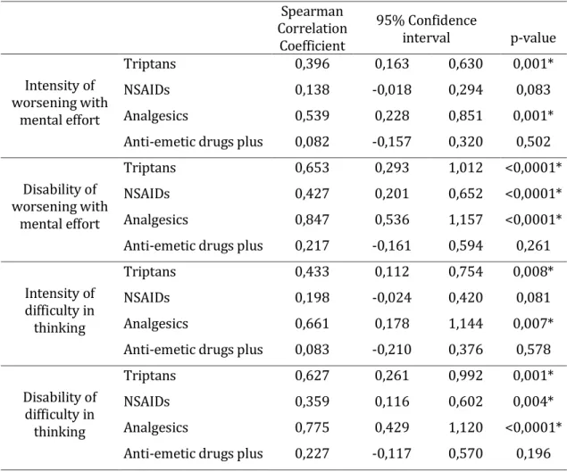
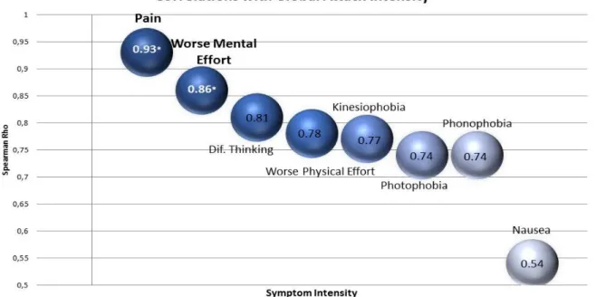
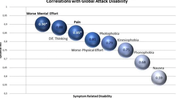
The migraine attacks of an independent clinical-based sample of 100 episodic migraine patients were prospectively recorded using paper headache diaries. Information collected included items of timing of the recorded attack (pain onset and relief, timing of rescue medication use) and scoring on a 0-10 Visual Analogue Scale the intensity and disability related to each attack and to each migraine symptom, including also two cognition related symptoms - pain worsening with mental effort and difficulty in thinking. Relationships between intensity and disability scores of the attacks an of each migraine symptom were explored.
Research question #4 – Can we identify and quantify attack-related cognitive symptoms
in migraine?
An extensive (43 items) cognitive symptoms checklist was assembled based on structured interviews of 37 migraine patients and with data from literature review in order to develop a questionnaire that allows identification and quantification of subjective cognitive symptoms in migraine attacks. The extensive checklist was applied prospectively to an independent sample of 93 migraine patients and factor analysis was conducted for item reduction. The reduced checklist retained 9 items that composed a multiple choice self-administered questionnaire – the Mig-SCog. Construct validity, internal consistency, temporal stability and external validity of the questionnaire were tested.
Research question #5 – Are cognitive complaints identified with the Mig-SCog specific
for migraine? How reliable is the Mig-SCog?
The Mig-SCog was prospectively applied in a clinical-based sample of headache patients in three different prospective studies with independent patient samples –one cross-sectional comparing migraine (N=98) and tension-type headache patients (N=51); the remaining included migraine patients using Mig-SCog for three different status (migraine, non-headache pain and pain-free, N=63) and in the last study the Mig-SCog was fulfilled within and in-between attacks, to screen for the recall bias (N=38). Scores obtained in each situation were calculated and compared with the appropriate statistic method. Validity analysis was used to determine the sensitivity and specificity of the Mig-SCog for the migraine diagnosis.
Research question #6 – What is the evidence of cognitive dysfunction occurrence during
migraine attacks?
A systematic review of medical databases (Medline and Cochrane Library) was performed to identify and collect available data about the existence and pattern of impaired neuropsychological performance during migraine attacks, compared to the headache-free status. Due to the high variability of the retrieved studies’ methodologies, data analysis was qualitative; Tables with summaries of relevant results were plotted.
Research question #7 - Do migraine patients have reversible cognitive impairment
during attacks?
A prospective two-period randomized cross-over study of neuropsychological performance of clinic-based independent sample of 39 migraine patients within a spontaneously occurring migraine without aura attack and in the headache-free period was conducted. Patients’ performance in an extensive neuropsychological battery was compared between both situations, while controlling for the most relevant potential bias.
Research question #8 – How can we measure attack-related cognitive impairment?
A battery composed of brief and practical routine neuropsychological tests focused on executive functions was assembled in order to be possible to sequentially test migraine patients in their ictal or/and inter-ictal status. A prospective cross-sectional controlled study of the performance of inter-ictal migraine patients in repeated short-term (6 weeks) applications of this battery was conducted. Cases’ performance was compared to that of matched controls, using a convenience sample of 48 volunteers from the hospital staff. The practice or learning effect of each test was quantified in order to determine the clinically meaningful predictable score change of repeated applications.
Research question #9 - Do brain perfusion changes exist during migraine without aura
27
A prospective longitudinal study of brain perfusion using Arterial Spin Labeling magnetic resonance imaging (ASL-MRI) was conducted in 13 female episodic migraine patients recruited among the hospital staff and in the acute care outpatient clinic during an untreated spontaneously occurring migraine without aura attack and repeated in a headache-free period. Cerebral global and regional brain perfusion was averaged for the total group and subtracted between the two sessions in order to identify perfusion differences.
Research question #10 – Are there neuronal network abnormalities underlying the
attack-related executive symptoms in migraine?
The previous study was complemented by the evaluation of cortical activation using Blood Oxygen Level Dependent functional magnetic resonance imaging (BOLD-fMRI) in response to an executive task (N-Back) and a brief neuropsychological evaluation focused on executive functions in the same conditions, during an untreated spontaneously occurring migraine without aura attack and repeated in a headache-free period. The cortical activation pattern in response to the N-Back task was averaged for the total group and subtracted between the two sessions in order to identify activation differences. The performance on the neuropsychological evaluation was compared between the sessions and differences found were paralleled to the predictable score change of repeated applications.
Research question #11 - Is ongoing migraine related to worse cognitive performance
late in life?
A prospective cross-sectional population based study of older adults (aged 50 or over) neuropsychological performance in an extensive neuropsychological battery was undertaken. The headache status of the sample was sought and classified into migraine, non-migraine headache and headache-free individuals, whom were used as controls. Cognitive performance was compared between groups.
Research question #12 - Is migraine associated with an increased risk of cognitive
The same sample of the previous study was revaluated after five years, to screen for cognitive decline, defined as a significant decline in memory and/or executive functions. The influence of persisting headache (migraine or non-migraine headache) on the risk of cognitive decline was sought.