2019
UNIVERSIDADE DE LISBOA
FACULDADE DE CIÊNCIAS
DEPARTAMENTO DE BIOLOGIA VEGETAL
Developing new molecular tools to study and visualize the
intermediate filament GFAP in living cells
Ricardo Jorge Pinheiro Quiteres
Mestrado em Biologia Molecular e Genética
Dissertação orientada por:
Professor Doutor Federico Herrera
ii
Acknowledgments
The completion of this thesis would not have been possible without the help, guidance and support of so, so many people:
I start by thanking my supervisor Dr. Federico Herrera for welcoming me in your group, for your guid-ance through each stage of this thesis and for all the patience and time you spent teaching me.
I would like to express my deepest gratitude to Dr. Zach Hensel for your tremendous help and advice, even when I was not technically your student.
I am also thankful to Dr. Alvaro Crevenna for all the advice and for the knowledge that he shared. I also wish to thank the Flow Cytometry and the Advanced Imaging Facilities of Instituto Gulbenkian de Ciência for their services and assistance.
Thank you to all the people from Fede’s lab, especially to three very talented PhD students. First to Cristina for all your help with that diabolic technique. To Fernanda thank you for all your help, encour-agement and support and for taking time off your experiments to teach me. Finally, to my mentor and friend Ricardo, thank you for your massive help, for teaching me basically everything that I know and for answering an enormous amount of questions and not answering a lot more, allowing me to think and grow as a scientist.
To three of the most kind, helpful and funny people I have ever met. Sara, Soraia and Teresa, thank you so, so much! Through the ups and downs of this thesis, you were always there for me, helping me and giving me advice. I want to specially thank you for the ice creams, for all the lunch hours we spend talking about random stuff and eating stolen chocolates, for getting us lost in the middle of nowhere, for the war games in the lab and for bringing up my goofiest side. Thank you for making this a year I will never forget! Ricciardi forever!
To my family, and especially to my mom, dad, sister and grandparents a massive thank you. Thank you for all your unconditional love and support, for being an inspiration, for always believing in me even when I did not and for always pushing me to do better and to be better. Everything that I accomplished is entirely thanks to you.
Lastly, a special thank you to my grandfather, for everything you taught me and for everything you did for me. Wherever you are I hope you are proud of me.
iii
Abstract
Intermediate filaments (IFs) are one of the three major filaments of the cytoskeleton. Due to their intrinsic physical properties, IFs provide mechanical support and give cells the elastic properties that allow them to resist deformation and maintain their shape. Different from actin filaments and microtu-bules, IFs have a cell type-specific expression pattern, and in mature astrocytes, the main IF protein expressed is the glial fibrillary acidic protein (GFAP). Although the exact function of GFAP remains unclear, GFAP is overexpressed in response to a variety of insults to the Central Nervous System (CNS) and the deletion of GFAP renders mice more susceptible to neurotoxicity. Furthermore, heterozygous gain-of-function mutations in the GFAP gene cause Alexander disease (AxD), a progressive and fatal neurodegenerative disorder that most frequently affects newborns and infants. IFs are the least charac-terized filaments of the three major cytoskeleton elements. Difficulties in studying the behavior of these filaments, and in particular of GFAP, arise from the fact that these proteins do not tolerate well the presence of protein tags in its termini. Therefore, we aimed to create new and more versatile molecular tools to study the behavior of GFAP in living cells, and that can easily be implemented in the study of other IFs. In this work, we developed a mouse GFAP-HaloTag fused system and the first tagged version of human GFAP. Both versions of tagged GFAP were able to incorporate into the IF network and form a normal filamentous network when transiently transfected in mammalian U251 cells. Opposingly, R236H/R239C AxD-causing mutations disrupted the endogenous filament network of GFAP and altered its diffusion properties. Furthermore, our results also show that GFAP dynamics is intimately linked to that of actin microfilaments and microtubules, as cytoskeleton-destabilizing drugs completely change the diffusion properties of GFAP molecules. Taken together, the results in this thesis demonstrate the enormous potential our two novel systems have for the study of GFAP oligomerization, aggregation, and dynamics in living cells.
iv
Resumo
A habilidade de uma célula de resistir a deformação, transportar carga intracelular ou de mudar a sua forma durante o movimento depende do citoesqueleto. O citoesqueleto é uma rede complexa de filamentos proteicos e proteínas motoras associadas que se estende pelo citoplasma de todas as células, incluindo bactérias e arquea. Em células eucarióticas, o citoesqueleto é composto por três tipos de polí-meros, distinguíveis entre si pelo tamanho: filamentos de actina ou microfilamentos (7 nm de diâmetro), microtúbulos (24 nm) e filamentos intermédios (10 nm). Contrariamente aos microfilamentos e microtú-bulos, cuja composição é muito semelhante entre todos os tipos celulares, a composição dos filamentos intermédios varia consideravelmente entre diferentes tipos de células, tecidos e órgãos, durante o desen-volvimento e em condições patológicas. Os filamentos intermédios são estruturas extremamente estáveis que fornecem suporte e protegem as células de danos mecânicos ao conferir-lhes propriedades elásticas para resistir à deformação e manter a sua forma. Além destas funções de suporte, estes filamentos de-sempenham ainda um papel na sobrevivência, divisão e migração celular. Devido à multitude de funções nas quais os filamentos intermédios participam, alterações nos níveis de expressão ou mutações em componentes específicos destes filamentos estão associadas ao aparecimento de várias patologias, a sua grande maioria raras. Apesar da sua enorme estabilidade, os filamentos intermédios são estruturas bas-tante dinâmicas e muitas vezes a participação dos microtúbulos e dos filamentos de actina é fundamental para a manutenção da sua rede.
A proteína ácida fibrilar glial (GFAP) é o filamento intermédio mais característico dos astrócitos, e é universalmente utilizada como o principal marcador para a sua identificação e classificação. Apesar da estrutura tridimensional desta proteína permanecer desconhecida, pensa-se que possua uma estrutura tripartida que é semelhante entre todos os filamentos intermédios. Esta estrutura consiste num domínio central alfa-helicoidal altamente conservado flanqueado por dois domínios N-terminal e C-terminal, sem uma estrutura secundária definida. Assim como os restantes filamentos intermédios, a GFAP tende a formar filamentos com 10 nm de diâmetro. Primeiramente, ocorre a formação de um dímero através da interação entre os domínios centrais de dois monómeros, que se enrolam um sobre o outro para formar uma estrutura superenrolada. De seguida, estes dímeros associam-se de forma antiparalela para formar tetrâmeros apolares. Posteriormente, com a contribuição dos domínios N-terminais, os tetrâmeros asso-ciam-se para formar protofilamentos que, por sua vez, se associam lateralmente para formar os filamen-tos maturos, com vários nanómetros de comprimento. Contrariamente aos restantes filamenfilamen-tos do cito-esqueleto, a construção dos filamentos intermédios é totalmente independente de nucleótidos. Para além de fornecer suporte mecânico aos astrócitos, pensa-se que a GFAP tenha também um papel importante na migração e motilidade celular, na mitose e ainda na manutenção da barreira hematoencefálica. A GFAP está também associada a várias patologias do sistema nervoso central, tais como a doença de Alexander. A doença de Alexander é uma doença neurodegenerativa rara, fatal e progressiva que afeta essencialmente recém-nascidos e crianças. Esta doença resulta do aparecimento de mutações pontuais no gene que codifica para a GFAP e é caracterizada pela acumulação massiva deste filamento intermé-dio, juntamente com proteínas de stress, em estruturas citoplasmáticas complexas, denominadas por fibras de Rosenthal.
Atualmente, os filamentos intermédios são os polímeros menos caracterizados do citoesqueleto e o nosso conhecimento atual relativamente à sua estrutura tridimensional, bem como dos processos dinâ-micos envolvidos na sua construção, está muito longe de estar completo. Esta escassez de conhecimento relativamente à organização e dinâmica dos filamentos intermédios deve-se em parte à dificuldade em visualizar estes filamentos in vivo. Isto acontece porque estes filamentos, e em particular a GFAP, não toleram bem a presença de marcadores proteicos nos seus terminais, como por exemplo as proteínas
v
fluorescentes, já que estes causam a rutura ou desorganização dos filamentos e levam à sua agregação. Assim, o principal objetivo deste trabalho é desenvolver novos modelos celulares para o estudo da oli-gomerização, função e dinâmica da GFAP em células vivas.
No presente estudo, começámos por construir um plasmídeo no qual a GFAP de ratinho foi ligada a uma HaloTag (mGFAP-HT), de forma a puder estudar a arquitetura e o comportamento da GFAP a um nível nanoscópico. A HaloTag é uma alternativa atrativa às proteínas fluorescentes, uma vez que combina a especificidade genética destas proteínas com as propriedades foto-físicas superiores de ligan-dos orgânicos, uma característica que a torna muito apelativa para microscopia de super-resolução e outras técnicas avançadas. Neste trabalho desenvolvemos também com sucesso a primeira versão mar-cada da GFAP de humano inserindo, de forma aleatória, a proteína fluorescente EGFP dentro da região codificante da GFAP. Quando expressos transientemente nas células de glioblastoma humano U251, ambos os sistemas foram capazes de se integrar na rede de filamentos intermédios endógena destas células e de formar estruturas filamentosas típicas da GFAP em aproximadamente 90% das células.
Por forma a estudar o efeito que as mutações relacionadas com a doença de Alexander têm na construção dos filamentos da GFAP, gerámos o mutante de ratinho R236H e o mutante de humano R239C, por mutagénese dirigida. Os nossos resultados mostram que na maioria das células transfetadas com os plasmídeos mutantes, a GFAP não é capaz de formar uma rede filamentosa. No entanto, o com-portamento da GFAP mutante não é o mesmo nos dois modelos. Enquanto que o mutante de humano R239C formou maioritariamente pequenos agregados, o mutante de ratinho R236H formou um padrão homogéneo disperso pelo citoplasma das células, sem nenhuma estrutura filamentosa aparente. Embora vários esforços continuem a ser feitos para tentar encontrar um composto que seja eficaz contra a doença de Alexander, a verdade é que esta doença permanece intratável e incurável. No entanto, estudos recen-tes demonstraram que a especiaria curcumina tem um efeito benéfico na redução da agregação da GFAP. Desta forma, decidimos testar o efeito neuroprotetor de dois compostos sintéticos e derivados da curcu-mina, J147 e CNB-001, na redução da agregação do mutante humano R239C. Observámos que, no nosso modelo celular, o composto CNB-001 é mais eficaz que o J147 e que apenas as células que cresceram na presença de CNB-001(10 µM) mostraram um aumento ligeiro, embora significativo, na formação de filamentos e uma respetiva diminuição da agregação da GFAP.
O plasmídeo mGFAP-HT foi ainda utilizado para estudar a dinâmica da GFAP de ratinho. Os nos-sos resultados mostram que a mutação R236H tem um forte efeito na dinâmica da GFAP, aumentando consideravelmente a sua velocidade de difusão, comparativamente à GFAP não mutada. Os filamentos intermédios estão em constante comunicação com os restantes elementos do citoesqueleto, e muitas vezes a sua dinâmica está dependente destes. Os nossos resultados são consistentes com estas observa-ções, e demonstram que a despolimerização dos microfilamentos de actina ou dos microtúbulos resulta numa diminuição significativa da velocidade de difusão das moléculas de GFAP, e num aumento da percentagem de moléculas com uma difusão mais lenta.
Em suma, os resultados apresentados neste trabalho demonstram o enorme potencial dos dois sis-temas que construímos no estudo da função, oligomerização e dinâmica da GFAP em células vivas. Em teoria, estas estratégias, podem também ser utilizadas para o estudo de qualquer outro filamento inter-médio. O desenvolvimento de novos e mais versáteis modelos celulares, continua, no entanto, a ser indispensáveis para uma melhor compreensão do comportamento da GFAP em células vivas e também dos mecanismos pelos quais mutações nesta proteína causam a doença de Alexander.
vi
General Contents
1. Introduction ... 1
1.1. Intermediate Filaments: a key component of the cytoskeleton ... 1
1.2. Glial Fibrillary Acidic Protein (GFAP) is a major intermediate filament typical of astrocytes ... 2
1.3. Alexander Disease is caused by point mutations in GFAP ... 5
2. Aims of the project ... 8
3. Material and Methods ... 9
3.1. Reagents ... 9
3.2. Plasmid construction and site-directed mutagenesis ... 9
3.2.1. Construction of a mouse GFAP-HaloTag fused plasmid ... 9
3.2.2. Construction of tagged human GFAP constructs ... 11
3.2.2. Site-directed mutagenesis ... 11
3.3. Cell culture ... 12
3.3.1. Cell growth, seeding and transfection ... 12
3.3.2. Drug treatments and cell labeling ... 12
3.4. Microscopy ... 12
3.5. Flow cytometry ... 13
3.6. Immunoblotting ... 13
3.7. Statistical analysis ... 14
4. Results ... 15
4.1. Mouse GFAP-HaloTag fused plasmid is successfully expressed in U251 cells ... 15
4.2. Alexander disease-causing mutation R236H is deleterious to the assembly and network formation of mouse GFAP ... 15
4.3. Generation of tagged human-GFAP constructs ... 19
4.4. Alexander disease-related R239C mutation induces human GFAP aggregation ... 21
5. Discussion ... 23
6. Main Conclusions ... 26
vii
Index of Figures
Figure 1.1: Schematic representation of IF protein structure ... 2
Figure 1.2: Different GFAP isoforms ... 3
Figure 1.3: Representative scheme of IFs assembly ... 4
Figure 1.4: Distribution of Alexander disease-related mutations in GFAP in relation to protein domain structure of IFs and clinical presentation ... 6
Figure 1.5: Rosenthal fibers (RFs) are the major histopathological feature of Alexander disease (AxD) ... 7
Figure 3.1: Cloning strategy for the construction of a mouse GFAP-HaloTag (mGFAP-HT) fused plasmid ... 10
Figure 4.1: Halo mouse GFAP is successfully expressed in U251 human glioblastoma cells. ... 15
Figure 4.2: Alexander disease-related R236H mutation causes filament disorganization of GFAP ... 16
Figure 4.3: GFAP molecules display different diffusion behaviors. ... 17
Figure 4.4: AxD-related R236H mutation alters the diffusion properties of mGFAP ... 18
Figure 4.5: Construction of tagged human GFAP plasmids ... 20
Figure 4.6: Transient expression of mutant R239C GFAP leads to aggregate formation in U251 cells ... 21
Figure 4.7: CNB-001 has a beneficial neuroprotective effect on GFAP filament organization and accumulation ………..………22
Index of Tables
Table 1.1: Classification of Intermediate filaments and associated pathologies. ... 1Table 3.1: Sequences of primers (5' ® 3') used to construct plasmids encoding tagged GFAP or in site-directed mutagenesis of these constructs ... 11
viii
List of Abbreviations
Aa Amp AxD BiFC bp BSA Ca2+ cm CNS CNTF CO2 CON DMEM DMSO DNA dNTPs E. coli EGFP EM EM-CCD ER FBS GAPDH GFAP GFP GLT-1 h hGFAP HRP HT IFs IgG IL-6 JAK JF549 JF646 JNK kan kb kDa kW LB LIF MAPK Mg2+ mGFAP Amino acids Ampicillin Alexander DiseaseBiomolecular Fluorescence Complementation Base Pairs
Bovine Serum Albumin Calcium Ion
Centimeter
Central Nervous System Ciliary Neurotrophic Factor Carbon Dioxide
Control
Dulbecco’s Modified Eagle Medium Dimethyl Sulfoxide
Deoxyribonucleic Acid Deoxynucleotides
Escherichia coli
Enhanced Green Fluorescent Protein Electron-Multiplying
Electron-Multiplying Charge-Coupled Device Endoplasmic Reticulum
Fetal Bovine Serum
Glyceraldehyde-3-Phosphate Dehydrogenase Glial Fibrillary Acidic Protein
Green Fluorescent Protein Glutamate Transporter 1 Hour
Human Glial Fibrillary Acidic Protein Horseradish Peroxidase HaloTag Intermediate Filaments Immunoglobulin G Interleukin-6 Janus Kinase Janelia Fluor 549 nm Janelia Fluor 646 nm c-Jun N-terminal Kinase Kanamycin
Kilobase Kilodalton Kilowatt Luria Broth
Leukemia Inhibitory Factor Mitogen-Activated Protein Kinase Magnesium Ion
ix
MHz min ml mM mRNA ms mTOR mW NaAz NaCl NF-kB ng nm nM Nrf2 p PA-JF549 PBS PCR PNS RFs sCMOS SD SDS SDS-PAGE s SRRF STAT3 TAE TBS TBST ULFs UTR V V1 V2 WT µg µl µm µM Megahertz Minute Milliliter Millimolar Messenger RNA MillisecondMammalian Target of Rapamycin Milliwatt
Sodium Azide Sodium Chloride
Nuclear Factor kappa-light-chain-enhancer of activated B cell Nanogram
Nanometer Nanomolar
Nuclear Factor Erythroid 2-Related Factor 2 p-Value
Photoactivatable Janelia Fluor 549 nm Phosphate Buffer Saline
Polymerase Chain Reaction Peripheral Nervous System Rosenthal Fibers
Scientific Complementary Metal-Oxide-Semiconductor Standard Deviation
Sodium Dodecyl Sulphate
Sodium Dodecyl Sulphate - Polyacrylamide Gel Electrophoresis Second
Super-Resolution Radial Fluctuations
Signal Transducer of Activators of Transcription Tris-Acetate-EDTA
Tris-HCl Buffer Saline
Tris-HCl Buffer Saline-Tween 20 Unit Length Filaments
Untranslated Region Volt Venus 1 Venus 2 Wild-Type Microgram Microliter Micrometer Micromolar
1
1. Introduction
1.1. Intermediate Filaments: a key component of the cytoskeleton
The structural framework of a cell is provided by the cytoskeleton, a complex network of protein-aceous filaments and motor proteins that extends throughout the cytoplasm of all cells, including
bacte-ria and archaea1. The cytoskeleton is an adaptive and dynamic structure that helps cells divide, move,
maintain their shape and regulate the traffic of intracellular cargo through the cytoplasm, among other
important functions2. Three major types of polymers, distinguishable by their size, make up the
cyto-skeleton of eukaryotic cells: actin filaments or microfilaments, microtubules, and a group of polymers collectively known as intermediate filaments (IFs). Microtubules are formed by a few isoforms of tubu-lin and are the thickest filaments with a typical diameter of 24 nm. These filaments are constantly as-sembling and disasas-sembling and play a crucial role in cell division and migration, and in the transport of intracellular cargo. Microfilaments are mainly formed by actin isoforms and reach 7 nm in diameter. They are involved in a variety of processes that require force, including muscle contraction and cell exploration and movement. Lastly, intermediate filaments, as the name suggests, have a diameter that falls between that of actin microfilaments and microtubules. These 10 nm-diameter filaments provide support for microfilaments and microtubules and give cells elastic properties that allow them to
with-stand tension1-4.
In contrast to actin filaments and microtubules, the composition of the proteins making up interme-diate filaments varies considerably between different tissues, organs, and cell types, during development
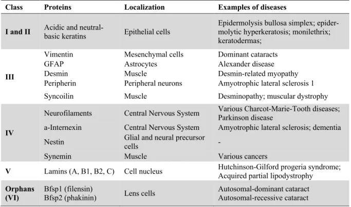
or in pathological conditions5. Intermediate filaments are classified into five major types based on
sim-ilarities in amino acid sequence, protein structure predictions, and assembly properties6. Because IFs
play such an important role in providing mechanical support to cells, changes in expression levels or mutations of specific IF components are associated with a multitude of tissue-specific degenerative
dis-orders7,8 (Table 1.1).
Table 1.1: Classification of Intermediate filaments and associated pathologies.
Class Proteins Localization Examples of diseases
I and II Acidic and neutral-basic keratins Epithelial cells Epidermolysis bullosa simplex; epider-molytic hyperkeratosis; monilethrix;
keratodermas;
III
Vimentin Mesenchymal cells Dominant cataracts
GFAP Astrocytes Alexander disease
Desmin Muscle Desmin-related myopathy
Peripherin Peripheral neurons Amyotrophic lateral sclerosis 1
Syncoilin Muscle Desminopathy; muscular dystrophy
IV
Neurofilaments Central Nervous System Various Charcot-Marie-Tooth diseases; Parkinson disease
a-Internexin Central Nervous System Amyotrophic lateral sclerosis; dementia
Nestin Glial and neural precursor cells -
Synemin Muscle Various cancers
V Lamins (A, B1, B2, C) Cell nucleus Hutchinson-Gilford progeria syndrome; Acquired partial lipodystrophy
Orphans
2
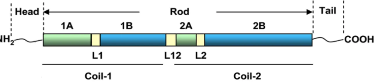
All IFs share a common tripartite structure consisting of a highly conserved a-helical central main (rod domain) flanked by an amino (N)-terminal head domain and a carboxy (C)-terminal tail do-main (Figure 1.1). The rod dodo-main is divided into four hydrophobic a-helical segments of conserved length: 1A, 1B, 2A and 2B. These four segments are interconnected by three short linkers (L1, L12, and L2). The beginning and end parts of the rod domain are particularly conserved in all IFs. Both of these conserved regions are involved in dimer-dimer interactions and mutations in these regions are deleteri-ous to IF organization and dynamics. On the contrary, the head and tail domains have very little
second-ary structure and their length and sequence vsecond-ary considerably among different IF proteins.9-11 The
three-dimensional structures of most IFs, including GFAP, remain incompletely described and this is a major barrier to our understanding of their assembly, protein-protein interactions, and other functional proper-ties.
Intermediate filaments are very stable structures and until recently they were thought to have very limited dynamics when compared to actin filaments and microtubules. This assumption originated from the observation that most IFs in a cell were not extracted and remained intact after cell lysis with
deter-gents and high concentrations of salts12. However, recent studies demonstrated that IFs are highly
dy-namic structures that can shorten, elongate and rearrange in response to a wide variety of extracellular
or intracellular stimuli13. IF dynamics often requires the participation of microtubules and actin
fila-ments. This contribution is particularly important in astrocytes, where microtubule- and actin-dependent
transport are essential for the maintenance of the IF network14.
Besides providing mechanical support to cells, IFs can also interact with cellular organelles either by forming cage-like structures around the nucleus, physically controlling their shape, position, and
movement15,16; or by serving as anchoring structures for mitochondria, positioning them near
high-en-ergy-demanding sites17. Furthermore, a large body of evidence points to a key role for IFs in cell
sur-vival18, division19,20 and migration21,22.
1.2. Glial Fibrillary Acidic Protein (GFAP) is a major intermediate filament typical of astrocytes
Astrocytes are the most numerous cells in the human brain, outnumbering neurons by over fivefold. These specialized glial cells that display an enormous heterogeneity in their morphology and function were originally thought to have only a passive role in the central nervous system (CNS) in the mainte-nance of neurons. However, over the past few decades, it has become clear that glial cells, particularly astrocytes, are involved in a wider variety of functions, vital for brain development, physiology and
pathology23. The most characteristic IF protein expressed in astrocytes is the glial fibrillary acidic
pro-tein (GFAP), being universally used as a molecular marker for their identification and classification.
This protein was initially discovered and isolated from patients with multiple sclerosis24 and its amino
acid composition was presented for the first time in 196925. GFAP, together with vimentin and nestin,
Figure 1.1: Schematic representation of IF protein structure. All IFs share a common tripartite structure with a conserved
3
makes up the intermediate filament system of astrocytes26. As astrocytes mature, typically GFAP
ex-pression increases, the nestin exex-pression stops and vimentin exex-pression decreases27,28. However, not all
populations of astrocytes express GFAP and different combinations of IF expression profiles are
possi-ble29.
The human GFAP gene is located on chromosome 17q21 and has nine exons and eight introns
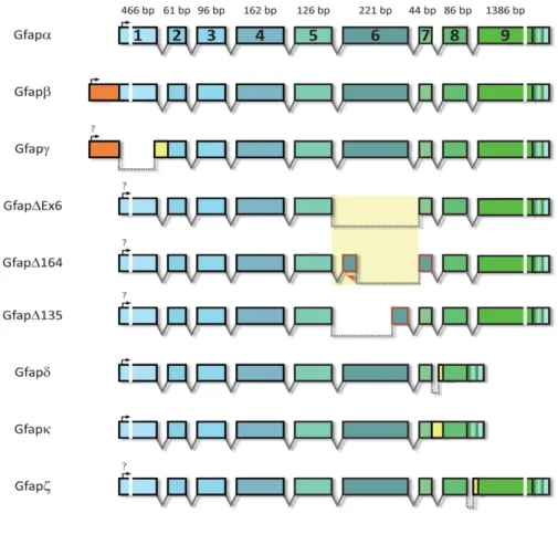
distributed over 10 kb in the human genome30. There are at least nine GFAP isoforms resulting from
alternative mRNA splicing31,32 (Figure 1.2). In humans, GFAP-a is the predominant isoform in the
CNS. It has 432 amino acids and a molecular weight of 50 kDa. GFAP-b is highly expressed in non-myelinating Schwann cells in the peripheral nervous system (PNS) and has an alternative upstream transcriptional start site in the 5’ UTR. The GFAP-d isoform is the second most studied isoform and is differentially expressed in neurogenic astrocytes in the subventricular zone and the olfactory bulb. This isoform has 431 amino acids and is generated by alternative mRNA splicing combined with alternative polyadenylation of the intron 7, which generates a new exon E7a. Due to differences in the tail region, this isoform is no longer capable of assembling homomeric GFAP-d filaments but can form heteromeric intermediate filaments with GFAP-a or vimentin. Other isoforms include the GFAP-g isoform which lacks exon 1 and includes the last 126 nucleotides of intron 1-2; the splice variants GFAPDEx6, GFAPD164 and GFAPD135 that skip sequences in exon 6; the GFAP-k isoform that lacks exons 8 and 9 like GFAP-d but the exon 7a is replaced by exon 7b, with the entire intron 7; and the GFAP-z isoform, a transcript that includes the last 284 nucleotides of intron 8. The amino acid sequence of human GFAP-a has a very high similarity (91% identity and 95% positivity) to that of mouse GFAP
(430 amino acids)33.
Figure 1.2: Different GFAP isoforms. At least nine GFAP transcript isoforms resulting from alternative mRNA splicing can
4
Although the three-dimensional structure is unknown, it is believed that GFAP shares with other intermediate filaments a common structure with three major domains: a head domain with 72 amino acids (1-72 Aa), a central helical rod domain with 305 amino acids (73-377 Aa) and a (C)-terminal tail
domain with 55 amino acids (378-432 Aa)34. Like other intermediate filament proteins, GFAP forms 10
nm diameter filaments. The first stage of filament assembly is the formation of a dimer through the interaction of the rod domain of two polypeptide chains that wind around each other in a coiled-coil structure. These dimers then associate in an anti-parallel manner to form apolar tetramers, which are the initial building blocks of all IFs. With the contribution of the nonhelical tail domains, these tetramers then associate to form protofilaments, and eight protofilaments wind around each other to form unit-length filaments (ULFs) that are 20 nm in diameter. Finally, the ULFs associate longitudinally to form
filaments with hundreds of nanometers in length11,35. The extended filaments undergo radial compaction
that reduces the diameter to 10 nm (Figure 1.3)36. Unlike microfilaments and microtubules, the
assem-bly of GFAP is totally nucleotide-independent 35.
IFs are often subject to multiple post-translational modifications and GFAP is no exception. Phos-phorylation, for example, plays a major role in the regulation of filament assembly/disassembly and in
the structural organization of GFAP and other IFs37-39. Site-specific phosphorylation of GFAP by protein
kinase A, calmodulin-dependent protein kinase II or protein kinase C is responsible for the disassembly of the filaments. The majority of phosphorylation sites are located in the head domain. Of the six phos-phorylation sites identified, five (Thr-7, Ser-8, Ser-13, Ser-17, and Ser-34) occur in the head domain;
the remaining one, Ser-389, is located in the tail domain38,39. Most of the amino acids that are subject to
phosphorylation in the head domain of GFAP are conserved among species and GFAP isoforms, which
reinforces the physiological importance of phosphorylation in the regulation of GFAP fibrillization31.
GFAP can also be citrullinated in its arginine residues, which creates epitopes that evoke autoimmune
responses40. Importantly, studies on post-translational modification were carried out in cell-free, in vitro
systems using protein extracts or in fixed cells. This limitation was due, in part, to the lack of chimeric
versions of GFAP that allowed their visualization in living cells until very recently14. IFs including
GFAP, rarely tolerate protein tags directly fused to their N- or C-termini, and this constitutes a major hurdle in our understanding of the physio- and pathological functions of this protein.
Similar to other IFs, GFAP is thought to provide mechanical support to astrocytes and help these cells to maintain their mechanical strength as well as their shape. However, the exact functions of GFAP remain elusive. Various studies using GFAP knockout or transgenic mice have provided some insights
Figure 1.3: Representative scheme of IFs assembly. In the first stage of filament assembly, two GFAP molecules interact to
form dimers. Two dimers associate in an anti-parallel manner to form apolar tetramers. Mature IFs are formed by the longitu-dinal association of unit length filaments (ULFs), that result from the association of apolar tetramers.Adapted from [35].
5
into the contribution of this protein in astrocyte physiology and pathology. GFAP plays very diverse
roles in cell migration and motility41, in mitosis by regulating filament assembly required for
cytokine-sis42,43 or in vesicle trafficking, including the movement of vesicles for exocytosis, vesicle recycling,
and lysosome-mediated autophagy44,45. Furthermore, GFAP is also suggested to be involved in the
mod-ulation of synaptic plasticity, by regulating the trafficking and the membrane anchoring of glutamate
transporters46,47, in the maintenance of the blood-brain barrier and normal CNS myelination48.
Studies using GFAP-null (GFAP-/-) mice have demonstrated that, despite all the cellular processes
that this protein has been implicated in, GFAP is not essential in mice. GFAP-null mice displayed nor-mal development, growth, fertility, and lifespan. When compared to wild-type mice, unchallenged GFAP-null mice showed no differences in brain architecture, and the blood-brain barrier was also in-tact49-51. However, mice deficient for GFAP, displayed an increased fragility of the CNS when subjected
to head injury, which suggests that GFAP plays an important role in neural protection. Some reports
suggest that the apparently innocuous phenotype displayed by GFAP-/- mice is due to a redundancy in
the function of intermediate filaments, as other intermediate filaments like vimentin or nestin could be
compensating for the absence of GFAP53.
In response to CNS injury and disease, astrocytes undergo a complex process called astrogliosis that is characterized by an increase in the number of astrocytes, profound morphological and
neuro-chemical changes in astrocytes, and an increase in the expression of GFAP54,55. The cellular and
molec-ular mechanisms that lead to astrogliosis are not completely understood, but neuroinflammatory path-ways like the Janus Kinase/Signal Transducer and Activator of Transcription 3 (JAK/STAT3) signaling pathway appear to trigger astrogliosis. The activation of astrocytes can be induced by several cytokines such as ciliary neurotrophic factor (CNTF), leukemia inhibitory factor (LIF) or interleukine-6 (IL-6),
produced and secreted by damaged cells and microglia in the CNS56,57. Mutations or overexpression of
GFAP over a toxic threshold can cause a lethal neurodegenerative disease known as Alexander disease58.
1.3. Alexander Disease is caused by point mutations in GFAP
Alexander Disease (AxD), described by Stewart Alexander in 1949, is a rare, fatal and progressive neurodegenerative disorder that most frequently affects newborns and infants. The first report of AxD described the case of an infant who died at 16 months of age after a several-month history of severe developmental delays, regressions in cognitive and motor skills, seizures and progressive
macroceph-aly59. According to the only population-based survey ever conducted, this disease has a prevalence of 1
in 2.7 million60,although Messing et al.58 believe this number is an underestimate because symptoms
are common to other disorders and often difficult to identify without previous clinical experience with this disorder. The disease occurs in both sexes, and there seems to be no predilection for race or ethnic-ity58,61.
The most common classification divides patients into three subtypes based on the age of onset:
infantile (0-2 years), juvenile (2-12 years) and adult (>12 years)62. More recently, different
classifica-tions that rely on the distribution of lesions and clinical presentaclassifica-tions rather than age of onset have been proposed. One of these alternative systems divides patients into two categories, type I and type II, with
all type I cases being early-onset, and type II cases occurring at all ages63. Early-onset patients normally
display a very different set of symptoms than late-onset patients. Early-onset patients have a prominent frontal-lobe involvement and typical symptoms include cognitive and motor developmental delays, megalencephaly, seizures, and hyperreflexia or spasticity. Patients with later onsets more often exhibit
6
hindbrain dysfunction and typically experience pseudobulbar symptoms such as ataxia, dysarthria and
dysphonia, sleep disturbances and very rarely have megalencephaly or seizures58,61-63. Lifespan is related
to age of onset with type I patients having a mean survival of 14 years and type II patients of 25
years58,61,63.
The genetic basis for AxD was defined in 2001 when Brenner and colleagues64 reported the
pres-ence of de novo mutations in 4 different GFAP residues in 10 unrelated AxD patients. At the moment, GFAP is the sole known cause of AxD, with approximately 95% of patients harboring mutations in the
GFAP gene65. In the majority of cases, GFAP mutations are not inherited but arise spontaneously. All
mutations detected so far are heterozygous and about 90% of these involve single amino acid changes,
though insertions and deletions have also been reported66. Disease-causing mutations are only known to
occur in the rod and tail domains of GFAP, with most of these mutations residing in the 1A, 2A and 2B segments of the conserved central rod domain of GFAP (Figure 1.4). Two mutation hotspots affecting amino acids R79 and R239 (equivalent to mouse GFAP R76 and R236, respectively) account for ~ 20% of all reported cases of AxD. These two missense mutations produce the greatest variety of substitutions
and cause a particular severe disease61,63-67.
AxD is genetically dominant and mutations appear to act in a gain-of-function manner, as
GFAP-null mice display a phenotype that does not resemble AxD49-51. However, it is still unclear if this
mech-anism is due to a gain of a normal function or gain of an abnormal function of GFAP. Analysis of various cases of AxD show that there is considerable phenotypic variability between affected individuals as (1) individuals with the same mutation display differences in severity of disease, (2) some patients with disease-causing mutations do not display symptoms, and (3) in rare cases of pathologically proven AxD, no mutation in GFAP has been found. The reasons for this variability are still unknown but some suggest
that genetic modifiers and environmental factors may influence the appearance of disease symptoms61,68.
Figure 1.4: Distribution of Alexander disease-related mutations in GFAP in relation to protein domain structure of IFs and clinical presentation. Aminoacids R76 and R239 are two mutation hotspots that account for approximately 20% of all
the AxD cases. These two missense mutations produce the greatest variety of substitutions and cause a particular severe disease. Adapted from [61].
7
An increase in GFAP levels beyond a toxic threshold, triggered by the presence of mutant GFAP, leads to the formation of eosinophilic protein inclusions known as Rosenthal fibers (RFs) (Figure 1.5A). These cytoplasmic inclusions mainly contain GFAP and vimentin intermediate filaments in association
with stress-related proteins like alpha-b-crystallin69 and the small heat shock protein hsp2770, as well as
other proteins in varying amounts71. The sequestration of both alpha-b-crystallin and hsp27 in GFAP
aggregates will most likely diminish the ability of astrocytes to cope with stress72,73 and prevent
apop-tosis74. RFs can be formed throughout the astrocyte and are thought to be generated by the continuous
incorporation of small aggregates into larger inclusions, a process that continues to happen over time in
parallel with the accumulation of GFAP (Figure 1.5B)75. Although RFs are characteristic of AxD, they
can sometimes be found in other pathological contexts like glial scars76 or pilocytic astrocytomas77.
However, their numbers and distribution in AxD are noticeably higher than in these other patholo-gies68,78. Overexpression of wild-type GFAP can also lead to the formation of RFs that are
indistinguish-able from the ones found in AxD patients79. Both these results indicate that GFAP mutations, although
not required for RF formation, may accelerate the process78.
Figure 1.5: Rosenthal fibers (RFs) are the major histopathological feature of Alexander disease (AxD). (A)
Morpholog-ical features of RFs. (B) Schematic representation of RF formation and growth. Adapted from [75].
Astrocytes in AxD display profound changes in cell shape and function, as AxD-causing mutations disturb the in vitro filament assembly and network formation of GFAP. When R239C GFAP was tran-siently expressed into Vimentin-negative SW13 cells, the protein did not form a filamentous network but instead formed small aggregates throughout the cytoplasm of cells. Although the R239C mutant also formed aggregates in primary rat astrocytes, in some of the cells it was also able to incorporate into the endogenous GFAP networks. These data suggest that GFAP mutations do not necessarily prevent GFAP
assembly but could affect the ability of ULFs to anneal and subsequently compact into filaments80,81.
Some authors propose that the effect of the mutations is to slow down the rate of normal polymer for-mation rather than to abolish it completely. To overcome the reduction in polymerization, astrocytes react in a self-destructive manner and increase the synthesis of mutant GFAP, resulting in a positive
feedback loop for disease progression67.
The accumulation of either wild-type or R239C GFAP in astrocytes impaired proteasome activity, but mutant GFAP produces a much stronger inhibitory effect. This proteasomal inhibition not only ag-gravates GFAP accumulation and aggregation, which in turn causes further proteasome inhibition but
may also prevent other proteins from being degraded81-83. GFAP aggregation also activates a number of
stress pathways such as the MAPK, p38 and JNK pathways. Constitutive activation of JNK in astrocytes leads to an increase in GFAP levels by further inhibiting proteasome activity. On the other hand, p38 activation produces the opposite effect as it negatively regulates mTOR activity, which in turn promotes
A)
A)
B)
B)
8
autophagy in an attempt to decrease GFAP levels84,85. Proteasome inhibition, and the consequent
accu-mulation of the short-lived transcription factor Nrf2, also triggers an oxidative stress response in
astro-cytes marked by activation of several oxidative stress-response genes86,87. Furthermore, aggregates of
GFAP can alter the morphology and cellular localization of some membranous organelles, like ER and Golgi, segregating these organelles to a perinuclear position. Such morphological changes may disrupt
vesicle formation and consequently intracellular and membrane trafficking75,88.
Since astrocytes play so many important roles, alterations in astrocyte function can impact other cells of the CNS. Actually, AxD astrocytes do not die, but patients show a massive white matter loss indicative of neuronal loss. One of the most characteristic changes in AxD astrocytes is the decrease of
its major glutamate transporter GLT-189. The loss of this transporter affects the ability of astrocytes to
uptake and buffer glutamate released from synapses into the extracellular medium, which in turn can lead to neuronal hyperexcitability and death. Recent studies using both mouse and human models of AxD show that AxD astrocytes induce a strong inflammatory environment characterized by an upregu-lation of inflammatory genes regulated by NF-kB-mediated gene transcription, a marked activation of
microglia and a modest influx of T cells into the CNS90.
At the moment, treatments for AxD can only address symptoms, but several strategies for therapy
have been proposed91. One approach is to screen libraries of drugs or compounds to reduce the
accumu-lation of GFAP, either by regulating the activity of the GFAP promotor or by increasing its
degrada-tion92. For example, the IL-6 family of cytokines induces GFAP expression by means of the JAK/STAT3
and/or MAPK pathways. Specific targeting of these or other cytokines or rate-limiting events in these pathways could be achieved by means of drugs, gene therapy or immunotherapy (with intrabodies, for example). Another possible approach is to minimize downstream effects of GFAP toxicity, such as the
deficit in the expression of the glutamate transporter GLT-193-95. Enhancing protective stress responses
such as those involving Nrf296,97 or alpha-b-crystallin98 could also be a promising target.
2. Aims of the project
Despite the knowledge we already have on GFAP, the spatio-temporal dynamics of this protein as well as the mechanisms by which GFAP mutations lead to astrocyte dysfunction remain poorly under-stood. This is due, at least in part, to the difficulties in visualizing this intermediate filament in vivo. These difficulties arise from the fact that GFAP does not tolerate well the presence of fluorescent tags in its N- or C- termini since they are deleterious for filament assembly and lead to aggregation of the protein.
With this in mind, the overall aim of this thesis is to develop new molecular tools to study GFAP location, oligomerization and dynamics in living cells without compromising its assembly and function. Our specific objectives are:
1) Creating a new, more versatile tool for the study of mouse GFAP behavior by super-resolution microscopy or other advanced methods; and
2) Developing the first tagged version of human GFAP for the study of its behavior in normal and AxD-related conditions.
9
3. Material and Methods
3.1. Reagents
Cloning enzymes were purchased from Thermo Scientific (Waltham, MA, USA) unless otherwise indicated. Exceptions include SrfI (New England Biolabs, Ipswich, MA, USA), PfuTurbo DNA poly-merase (Agilent, Santa Clara, CA, USA) and DpnI and alkaline phosphatase (NZYTech, Lisbon, Portu-gal). Cell culture media (Dulbecco’s Modified Eagle Medium - DMEM - and Leibovitz’s L-15 medium), Fetal Bovine Serum (FBS), penicillin-streptomycin commercial antibiotic mixture (Pen-Strep),
L-glu-tamine, TrypLE Express dissociation reagent, Ca2+- and Mg2+-free phosphate buffer saline (PBS) 1X
and Leukemia inhibitory factor (LIF) were obtained from Gibcoâ (Waltham, MA, USA). PCR primer
synthesis and DNA sequencing were performed by StabVida (Caparica, Portugal). Janelia Fluor 646 nm (JF646) and Janelia Fluor 549 nm (JF549) HaloTag ligands were obtained from Promega Corporation (Madison, WI, USA) and the photoactivatable Janelia Fluor 549 nm (PA-JF549) HaloTag ligand was a kind gift from Dr. Luke Lavis (HHMI’s Janelia research campus, Ashburn, VA, USA). Nocodazole was acquired from TargetMol (Wellesley Hills, MA, USA) and Latrunculin B from Focus Biomolecules (Plymouth Meeting, PA, USA). J147 and CNB-001 drugs were a kind gift from Dr. David Schubert (The Salk Institute for Biological Studies, LA Jolla, CA, United States). The following antibodies were used: rabbit anti-GFAP (1:1000, Sigma-Aldrich, St. Louis MO, USA), mouse anti-GADPH (1:2000, Santa Cruz Biotechnologies, Inc., Dallas, TX, USA), goat anti-rabbit IgG-Horseradish Peroxidase (HRP) conjugate (1:10000, Invitrogen, Carlsbad, CA, USA) and goat anti-mouse IgG-HRP conjugate (1:10000, Bio-Rad Laboratories, Hercules, CA, USA). Chemiluminescent HRP substrate was purchased from Bio-Rad Laboratories (Hercules, CA, USA). Human glioblastoma U251 cells were obtained from Public Health England (Salisbury, UK). The pEGFP-N3-mGFAP plasmid was a kind gift from Dr. Cécile Leduc (Institut Pasteur, Paris, France), the pZH504 Halo-Cro plasmid was kindly gifted by Dr. Zach Hensel (ITQB, Oeiras, Portugal) and the pcDNA3.1-EGFP-hGFAP plasmid encoding the human GFAP was developed at our laboratory by Ricardo Letra-Vilela.
3.2. Plasmid construction and site-directed mutagenesis
All plasmids were constructed using the conventional restriction digestion and ligation cloning technique. Vector and insert DNA fragments were obtained by restriction digestion of a host plasmid and through PCR amplification using Phusion polymerase, respectively.
3.2.1. Construction of a mouse GFAP-HaloTag fused plasmid
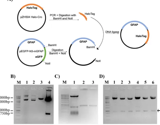
For the mGFAP-HaloTag construct, the host vector was obtained by substitution of the EGFP tag from a pEGFP-N3-mGFAP plasmid for a HaloTag using BamHI and NotI restriction sites (Figure
3.1A). We initially used the restriction enzymes BamHI and XbaI, but the double digestion failed. When
we separately single-digested the host plasmid with each restriction enzyme, BamHI was able to cleave its recognition site and produce a linear plasmid, but when we tried to digest with XbaI the digestion was not successful due to the methylation sensitivity of XbaI (Data not shown). To overcome this diffi-culty, we opted to change the pair of restriction enzymes to BamHI and NotI. Firstly, we single- and double-digested the host plasmid with BamHI and NotI and both enzymes were able to cleave the plas-mid and produce two bands, one which at 720 bp, the expected size for EGFP (Figure 3.1B). The Hal-oTag was obtained by PCR-amplification of the pZH504 Halo-Cro plasmid using specific primers
(Ta-ble 3.1) and carrying BamHI and NotI restriction sites (Figure 3.1C lanes 2 and 3). PCRs were done
10
µl containing 1 unit of Phusion DNA polymerase and the corresponding amplification buffer. The ther-mocycling conditions were the following: denaturation at 98ºC for 30 s followed by 30 cycles of 98ºC, 10 s; 65ºC, 30 s; 72ºC, 30 s (30 s/kb, as specified by the manufacturer) and a final extension at 72ºC for 10 min. Insert and vector fragments were digested for 2 h at 37ºC. Digested vectors were further incu-bated with 0.5 units of alkaline phosphatase for 1 h at 37ºC to prevent spontaneous religation of the vector. Digested vectors were then isolated by agarose gel electrophoresis (1% in Tris-acetate-EDTA – TAE - 1X) and purified with a NZYGelpure kit (NZYTech, Lisbon, Portugal). Digested vectors and inserts were ligated for 2 h at room temperature using 5 units T4 DNA ligase. Four different molar ratios of vector: insert were used – 1:0, 1:3, 1:6 and 1:9 – with the 1:0 ratio serving as a control for non-specific vector religation. Ligation products were transformed into NZY5a competent E. coli bacteria (NZYTech, Lisbon, Portugal) by heat shock and grown overnight at 37ºC on LB Agar 1X Petri dishes containing 50 µg/ml of kanamycin or 100 µg/ml of ampicillin for selection of transformed clones. Two different methods were used to determine if the cloning was successful. First, plasmids from randomly chosen colonies were isolated, digested with restriction enzymes and the resulting products analyzed by agarose gel electrophoresis. As shown in Figure 3.1D, in all of the tested colonies, plasmid digestion with BamHI and NotI generated 2 bands, one corresponding to the linearized plasmid and another in accordance with the size of the HaloTag (891 bp). To further confirm the cloning efficacy, 3 positive clones were sequenced, and in all the presence of the HaloTag was confirmed. No insertions or deletions were observed but all the clones had a single nucleotide mutation, that did not change the amino acid sequence. This mutation was probably present in the original template for HaloTag PCR amplification.
Figure 3.1: Cloning strategy for the construction of a mouse GFAP-HaloTag (mGFAP-HT) fused plasmid. (A) The
HaloTag protein was amplified as a 5’BamHI-3’NotI PCR fragment and cloned into a BamHI-NotI digested pEGFP-N3-GFAP plasmid. (B) Test digestion of the pEGFP-N3-GFAP plasmid; M – marker; lane 1- undigested plasmid; lane 2 - Single-digestion with BamHI; lane 3 – Single-digestion with NotI; lane 4 – Double-digestion with BamHI and NotI. (C) Vector and insert DNA fragments used for the construction of the mGFAP-HT plasmid. Vector fragments were prepared by double-digestion of the pEGFP-N3-GFAP plasmid with BamHI and NotI (lane 1) and insert fragments were obtained by PCR-amplification of a pZH504 Halo-Cro plasmid (lanes 2 and 3). (D) Confirmation of correct plasmid assembly by digestion with BamHI and NotI restriction enzymes. Positive clones are indicated by an arrow.
11
3.2.2. Construction of tagged human GFAP constructsAll cloning procedures were done as described in the previous subsection. Human GFAP constructs were obtained by substitution of the EGFP tag in our in-house pcDNA3.1-EGFP-hGFAP plasmid by Venus 1 (amino acids 1-157), Venus 2 (amino acids 158-238) or full-length HaloTag inserts. For these operations, we used the restriction enzymes AscI and SrfI. The Venus 1, Venus 2 and HaloTag inserts were amplified as 5’AscI – 3’SrfI PCR fragments using a pCS2-Venus or the pZH504 Halo-Cro plas-mids as templates and the primers described in Table 3.1.
3.2.2. Site-directed mutagenesis
Single p. Arg236His (R236H) and p. Arg239Cys (R239C) mutations were individually inserted by site-directed mutagenesis into the wild-type mGFAP and hGFAP constructs, respectively. PCRs were carried out using 25 ng of template DNA, 0.2 µM of mutagenesis primers (Table 3.1.) and 250 µM of dNTPs in a total volume of 50 µl containing 2.5 units of Pfu turbo DNA polymerase and the correspond-ing amplification buffer. The followcorrespond-ing thermocyclcorrespond-ing conditions were used: denaturation at 95ºC fol-lowed by 12 cycles of 95ºC, 30 s; 55ºC, 1 min; 68ºC, 8 min (1 min/kb, as specified by the manufacturer) and a final extension at 68ºC for 5 min. Mutagenesis reactions were then incubated with DpnI for 1 h at 37ºC to digest methylated parental plasmids and later transformed by heat shock into NZY5a competent bacteria. Transformed bacteria were grown overnight at 37ºC on LB Agar 1X Petri dishes with 50 µg/ml of kanamycin or 100 µg/ml of ampicillin for selection. Plasmids from randomly chosen colonies were isolated using a ZYMO Research Miniprep kit and sequenced to confirm the presence of the mutations.
Table 3.1: Sequences of primers (5' ® 3') used to construct plasmids encoding tagged GFAP or in site-directed muta-genesis of these constructs
Construct Primer sequence (5’ ® 3’)
mGFAP - HaloTag Forward: AATGGATCCATGGCAGAATCGGTACTGGC Reverse: TATGCGGCCGCTTAGCCGGAAATCTCGAG
hGFAP - Venus 1 Forward: AATGGCGCGCCATGGTGAGCAAGGGCG Reverse: AATGCCCGGGCTTGCTTGTCGGCGGTGAT
hGFAP – Venus 2 Forward: AATGGCGCGCCAAGAACGGCATCAAGGC Reverse: AATGCCCGGGCGTACAGCTCGTCCATGC
hGFAP - HaloTag Forward: AATGGCGCGCCATGGCAGAAATCGGTAC
Reverse: ATAGCCCGGGCGCCGGAAATCTCGAGC
Arg236His (R236H) Forward: CTGAGAGAGATTCACACTCAATACGAG
Reverse: CTCGTATTGAGTGTGAATCTCTCTCAG
Arg239Cys (R239C) Forward: CTGAAAGAGATCTGCACGCAGTATG
12
3.3. Cell culture3.3.1. Cell growth, seeding and transfection
Human glioblastoma U251 cells and rat glioma C6 cells were grown to confluence in 100 mm dishes (Thermo Scientific, Waltham, MA, USA) in DMEM, supplemented with 10% (v/v) fetal bovine serum (FBS), 1% (v/v) Penicillin-Streptomycin and 1% (v/v) L-glutamine and maintained at 37ºC in a
humidified atmosphere containing 5% CO2. Medium was changed every other day and cells were passed
once a week by trypsinization. Briefly, cells were incubated with TrypLE Express dissociation reagent for 5 min at 37ºC for cell detachment, followed by a centrifugation step at 300 g for 5 min at room temperature and sub-cultured according to the desired dilution. For fluorescent microscopy and
single-molecule tracking experiments, 3×10⁵ cells were grown on 35 mm glass-bottom µ- dishes (21 mm glass
surface diameter, ibidi GmbH, Gräfelfing, Germany). For flow cytometry, 3×10⁵ cells were seeded on
6-well plates (35 mm diameter, CorningÒ, Corning, NY, USA). For Western blotting, 8×10⁵ cells were
seeded on 60 mm dishes (Orange Scientific, Braine-L’Alleud, Belgium). Twenty-four hours after
seed-ing, cells were transiently transfected with 1 µg of the corresponding DNA constructs using jetPRIMEÒ
transfection reagent (Polyplus transfection, Illkirch, France) in a proportion of 3:1 (µL of transfection reagent: µg of DNA).
3.3.2. Drug treatments and cell labeling
All drugs and HaloTag ligands were diluted in DMSO (100% v/v). To test the neuroprotective activity of J147 and CNB-001, cells were treated with 100 nM, 1 µM or 10 µM of each drug 20 min before transfection and incubated for 24 h at 37ºC. For single-molecule experiments, 24 h after trans-fection, serum was removed for 2 h, and cells were treated with LIF (100 ng/ml), Nocodazole (10 µM) or Latrunculin B (10 µM) for 2 h at 37ºC in serum-free medium. Cells were then incubated with JF646 (100 nM), JF549 (100 nM) or PA-JF549 (1 nM) HaloTag ligands in serum-free medium for 20 min at 37ºC. Subsequently, cells were washed twice with sterile PBS. At the end of the final wash, the medium was changed to Leibovitz’s L-15 without phenol red.
3.4. Microscopy
Live-cell images of U251 cells were acquired at Instituto Gulbenkian de Ciência on a commercial widefield Nikon High Content Screening microscope, equipped with a 100x/1.45 plan-apo oil-immer-sion objective and an Andor Zyla 4.2 Scientific complementary metal-oxide-semiconductor (sCMOS) camera. To image eGFP/Venus–fused proteins, a 470 nm laser line, and GFP fluorescence filter sets were used and for the imaging of HaloTag-fused proteins, a 635 nm laser line and Cy5 fluorescent filter sets were used. An exposure time of 80 ms was used and the camera readout bandwidth was set to 200 MHz. The resulting pixel size was 65 nm using a 1024 x 1024 pixel as field of view. All acquisitions were done at room temperature. The microscope, cameras, and hardware were controlled through Nikon Elements software. Images were analyzed by means of the ImageJ free software.
Single-molecule imaging and SuResolution Radial Fluctuations (SRRF) imaging were per-formed on a Leica DMI6000 inverted microscope using a 100x/1.46 a-plan apochromat oil immersion objective. A 561 nm excitation laser (Coherent Sapphire) was set to 50 mW resulting in an effective
power density of ~ 2.3 kW/cm2. The laser beam was passed through a custom filter cube (Chroma
Tech-nology) with a zet405/561x excitation filter, a zt405/561/657rpc-uf2 dichroic beamsplitter, and an et610/75m emission filter. Fluorescent light was imaged on an Evolve 512 electron-multiplying charge-coupled device (EM-CCD) camera (Photometrics) after additional 1.6x magnification. An EM gain of
13
300 was used and the camera readout bandwidth was set to 10 MHz. The resulting pixel size was 100 nm using a 512 x 512 pixel as field of view. The incubation chamber was maintained at 37°C. The microscope, cameras, and hardware were controlled through MetaMorph software (Molecular Devices). For single-molecule experiments and for each cell, 1000 frames were acquired at 33 ms exposure time with no interval between frames. No photoactivation was necessary as spontaneous photoactivation of PA-JF549 combined with photobleaching by 561 nm illumination gave a reasonable density of single
molecules. The Trackmate ImageJ plugin99 was used to detect, fit and track individual GFAP molecules
in living cells. For the detection of GFAP molecules, we selected a LoG detector and an estimated blob
diameter of 0.4 µm and a threshold of 1500. For the tracking, we used a simple LAP tracker with a
linking max. distance of 0.5 µm, a gap-closing max. distance of 0.5 µm and a gap-closing max. frame gap of 0. The average diffusion of GFAP molecules and its fractions, per individual cell, were calculated
by fitting the SpotOn100 2-state (bound-free) kinetic model (0.05 µm2/s as maximum D
bound and 0.02
µm2/s as minimum D
free) to the distribution of translocations for individual molecules, obtained with
TrackMate. For SRRF imaging, 100 frames were acquired for each cell. SRRF images were generated
by running the NanoJ-SRRF ImageJ101 plugin on groups of 100 diffraction-limited images using the
default settings.
3.5. Flow cytometry
Twenty-four hours after transfection with the corresponding constructs, cells were washed once
with Ca2+-and Mg2+- free PBS and then detached with TrypLE Express dissociation reagent for 5 min
at 37ºC. Complete DMEM medium was added to the cells to neutralize TrypLE, and cells were collected to a microcentrifuge tube. Cells were centrifuged at 300 g for 5 min at room temperature, the supernatant was discarded, and pellets were resuspended in PBS. Fluorescent levels were determined using a 488 nm excitation laser and a 582/42 fluorescent filter. Flow cytometry was performed at Instituto Gulben-kian de Ciência on a FACSCalibur cytometer using Cell Quest software (Beckton Dickinson, Franklin Lakes, NJ, USA). Ten thousand events per experimental group were acquired and results were analyzed using FlowJo software (Tree Star Inc., Ashland, OR, USA).
3.6. Immunoblotting
Cells were washed once with PBS 1X and then incubated with a lysis buffer (50 mM Tris-HCl, 150 mM NaCl, 1% (v/v) NP-40 pH 7,5) supplemented with a protease inhibitor cocktail (Amresco, Fountain Parkway Solon, OH, USA). Cells were collected by scraping, transferred to microcentrifuge tubes, and incubated in ice for 10 min. To release intracellular proteins, cells were sonicated for 5 s using a Sonifier W - 450 D sonicator (Emerson, St. Louis, MO, USA). Proteins were collected after cell lysate centrifu-gation at 10000 g for 10 min at 4ºC and quantified by means of the Bradford method. Briefly, proteins were incubated with Bradford reagent (ApploChem Panreac, Cinisello Balsamo, Milan, Italy) for 10 min in the dark before measuring the absorbance at 595 nm. Protein concentrations were calculated by means of a standard curve of known bovine serum albumin (BSA) concentrations (0.125 to 2 µg/µL). For SDS-PAGE immunoblotting, 25 µg of total protein extracts were mixed with 4X loading buffer (4% (v/v) sodium dodecyl sulfate (SDS), 10% (v/v) 2- mercaptoethanol, 20% (v/v) glycerol, 0.004% (v/v) bromophenol blue, 0.125 mM Tris-HCl, at pH 6,8), boiled at 95ºC for 5 min to denature proteins, and incubated on ice for 5 min. Protein samples were separated by electrophoresis using a 10% (w/v) SDS-polyacrylamide gel and run at 125 V for 1 h. Proteins were transferred to a nitrocellulose membrane at 100 V for 1 h. Membranes were stained with 0.1% (w/v) Ponceau S to confirm sample quality and then washed with Milli Q water and Tris-HCl buffer saline (TBS) (150 mM NaCl, 50 mM Tris pH 7,4).
14
Membranes were subsequently blocked with 5% (w/v) non-fat dry milk in Tris-HCl buffer saline-Tween solution (TBS-T) (0.5% Tween-20) for 1 h at room temperature. After 3 washing steps with TBS-T, membranes were incubated with primary antibodies (diluted in 3% (w/v) BSA, 0.05% (v/v) sodium azide (NaAZ) in TBS-T) against GFAP (1:1000) and GAPDH (1:2000) overnight at 4ºC in a rotating wheel. To remove unbound antibody, membranes were rinsed 3 times with TBS-T for 10 min followed by an incubation with a secondary HRP-conjugated antibody (1:10000) in 5% (w/v) non-fat dry milk, for 2 h at room temperature with agitation. Membranes were rinsed 3 times for 10 min with TBS-T, incubated with chemiluminescent HRP substrate and imaged in a ChemiDoc XRS+ system (Bio-Rad, Hercules, CA, USA).
3.7. Statistical analysis
Statistical analysis and graphical representation of data were performed using Sigmaplot software (Systat Software, Inc., San Jose, CA, USA). Sample data are represented as mean ± standard deviation (SD) of at least 3 independent experiments. Statistical significance was evaluated by means of a one-way ANOVA followed by a Tukey’s test. Results were considered significant when p < 0.05, but in some cases, we also specified lower significance thresholds (p<0.01; p<0.001).
15
4. Results
Green fluorescent protein (GFP) and its spectral variants are versatile biological markers that have significantly changed our ability to study protein localization, dynamics, and interactions in living
cells102,103. However, these fluorescent proteins also have several disadvantages, including their low
photostability and brightness when compared to organic dyes, the potential formation of
homo-oligo-mers and limited choices in spectral wavelengths104,105. Due to these biophysical and photophysical
prop-erties, fluorescent proteins are often inadequate for super-resolution microscopy. An attractive
alterna-tive approach is the use of self-labeling protein tags, such as the HaloTag106, a hybrid approach that
combines the genetic specificity of fluorescent proteins with the superior photophysics of organic dyes. The HaloTag is a genetically engineered derivative of a hydrolase that, in physiological conditions, efficiently forms a highly specific and irreversible covalent bond with a multitude of synthetic chloro-alkane ligands, including a variety of spectrally distinct fluorescent tags, affinity tags, and attachments to solid supports.
4.1. Mouse GFAP-HaloTag fused plasmid is successfully expressed in U251 cells
We started by testing the effect of expressing a mGFAP-EGFP construct in U251 human glioblas-toma cells and C6 rat glioma cells by transient transfection. This construct was able to incorporate into the endogenous IF network and form a normal filamentous pattern when expressed in U251 cells
(Fig-ure 4.1A), but not in C6 cells (Fig(Fig-ure 4.1B), reflecting that the behavior of GFAP is not universal and
depends on the cellular context.
In order to study the architecture and behavior of GFAP at a nanoscopic level in living cells, we constructed a mouse-GFAP-HaloTag (mGFAP-HT) plasmid. This plasmid, as described in the Materials and Methods section, was generated by substitution of the EGFP tag from a host pEGFP-N3-mGFAP (mGFAP-EGFP) plasmid for a HaloTag using BamHI and NotI restriction sites and keeping the molec-ular linker between GFAP and HaloTag. As seen in Figure 4.1C, our mGFAP-HT construct was also able to form a normal filamentous network similar to that of mGFAP-EGFP, when transiently expressed in U251 cells. Furthermore, the application of the SRRF algorithm to the widefield images of mGFAP-HT was able to reconstruct these diffraction-limited low-resolution images into high-resolution images
and provide higher resolution details about the architecture of GFAP fibrils (Figure 4.1D).
Figure 4.1: Halo mouse GFAP is successfully ex-pressed in U251 human glioblastoma cells.
mGFAP-EGFP construct exhibited a normal GFAP filamentous network, when transiently expressed in U251 cells (A), but not in C6 cells (B). (C) U251 cells transfected with the mGFAP-HT construct and la-beled with the JF549 Halo ligand (100 nM) also dis-played a normal filamentous network characteristic of GFAP. (D) Widefield images of the mGFAP-HT construct were further analyzed with the NanoJ Su-per-Resolution Radial Fluctuations (SRRF) algo-rithm, which provided higher resolution details about the GFAP fibrils. Scale bars - 10 µm.
U251 C6
16
4.2. Alexander disease-causing mutation R236H is deleterious to the assembly and networkfor-mation of mouse GFAP
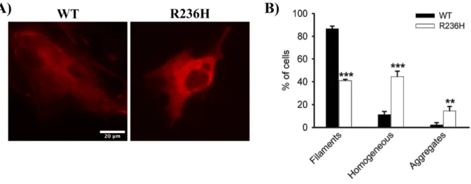
To understand the effect of AxD-causing mutations on the assembly properties of GFAP, a R236H AxD-mutant was generated by site-directed mutagenesis. Transient transfection of WT mGFAP into U251 cells resulted in its incorporation within the endogenous IF network and in the formation of fila-ment networks (Figure 4.2A, left panel) in approximately 87% of the transfected cells (Figure 4.2B). A diffuse homogeneous pattern or an aggregation pattern was occasionally observed in 11% and 2% of the transfected cells, respectively (Figure 4.2B). In contrast, transient expression of the R236H AxD-mutant disrupted the endogenous GFAP network, as the majority of the cells did not display an obvious filamentous structure (Figure 4.2A, right panel). Instead, GFAP either distributed homogeneously throughout the cytoplasm (45% of the cells) or formed cytoplasmatic inclusions (15% of the cells)
(Fig-ure 4.2B).
We next analyzed the dynamics of the mGFAP-HT construct using single-molecule tracking, a
powerful method to probe the mobility of molecules in living cells107. Images from living cells labeled
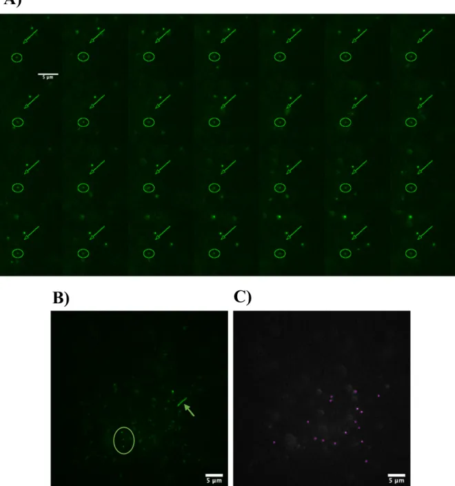
with the fluorescent HaloTag ligand PA-JF549 were acquired to generate 2D single-molecule tracks of the movement of each GFAP molecule. GFAP molecules do not have all the same diffusion motion, and we can clearly distinguish between molecules that have limited dynamics (Figure 4.3A and 4.3B,
cir-cle), and molecules exhibiting rapid diffusion or directional motion (Figure 4.3A and 4.3B, arrow). To
evaluate quantitatively the diffusive properties of GFAP we used the Trackmate ImageJ plugin to detect, localize and fit and track individual molecules. As observed in Figure 4.3C this plugin was able to detect with high accuracy the different GFAP molecules, in each frame. The individual tracks obtained were exported directly into the web-interface Spot-On and it was assumed that GFAP molecules could be distributed into 2 distinct subpopulations: a slow diffusion population and a rapid diffusion popula-tion. The Spot-On model does not account for directional motion or confined diffusion but was sufficient to provide a good fit for diffusion jump distributions for single-cell data.
Figure 4.2: Alexander disease-related R236H mutation causes filament disorganization of GFAP. U251 cells were
tran-siently transfected with either WT or mutant R236H GFAP, and 24 h later labeled with the JF646 Halo ligand (100 nM). (A) When expressed in this cell line, WT GFAP assembled into filaments (left panel), but mutant GFAP mostly formed a homoge-neous pattern throughout the cytoplasm without any apparent filament structures (right panel). (B) Quantification of the various patterns observed for transfected U251 cells with WT GFAP (black bars) and mutant GFAP (white bars). Results are shown as mean ± SD of triplicates. * significant versus WT GFAP. ** p<0.01, *** p<0.001. Scale bar – 20 µm.
17
B)
C)
A)
Figure 4.3: GFAP molecules display different diffusion behaviors. (A) Different types of diffusion motion of GFAP
mole-cules. arrow – fast-diffusing molecule; circle – slow-diffusing molecule. (B) Z-stack projection of the different diffusion mo-tions of GFAP molecules. arrow – fast-diffusing molecule; circle – slow-diffusing molecules. (C) Detection of GFAP mole-cules with the Trackmate ImageJ plugin. Scale bars – 5 µm.