Revista Brasileira de Odontologia Brazilian Journal of Dentistry
Associação Brasileira de Odontologia - Seção Rio de Janeiro
DOI: http://dx.doi.org/10.18363/rbo.v75.2018.e1200 Literature Review/Cariology
Current trends in the conservative treatment of
deep-caries lesion with risk of pulp exposure
Betina Paganini da Silva,1 Mayara Simão Gama,1 Apoena Aguiar Ribeiro21Department of Specific Department of Specific Formation, School of Dentistry, Universidade Federal Fluminense (UFF) Nova Friburgo, RJ, Brazil 2Department of Diagnostic Sciences, School of Dentistry, University of North Carolina, Chapel Hill, NC, USA
• Conflicts of interest: none declared.
AbstrAct
Objective: to present a review of the literature on the current state of the art of minimal intervention and conservation of hard dental tissues and pulp vitality in teeth
with deep caries lesion and risk of pulp exposure. Material and Methods: we searched the databases PubMed and SCOPUS, using the descriptors dental caries in dentin, deep caries lesion and conservative treatment, between 1960 and 2018. Results: of the 888 articles initially identified, 52 were selected following the inclusion and exclusion criteria. Conclusion: there is evidence that the current conservative techniques of stepwise excavation and selective caries removal prevents pulp exposure and maintains pulp vitality, thus improving the prognosis of these teeth. However, because they are innovative techniques, although presenting evidence-based scientific support in the English literature, no articles were found in Portuguese language, making it difficult to access and increase the use of these techniques as a routine by dentists in Brazil.
Keywords: Dental caries; Dentin; Dental pulp; Dental pulp capping; Dental pulp exposure.
Introduction
D
ental caries, even today, is the most prevalent dis-ease around the World1 and is regarded as a publichealth issue by the World Health Organization.2 It
can be defined as a localized dysbiosis or disease, resulting from bacterial activity in the biofilm3 and characterized
by a dissolution of the dental structure by acids formed from the fermentation of dietary carbohydrates, especially sucrose.3,4 Preventing the development of such disease, as
well as controlling existing lesions, shall essentially stem from the control of biofilm,5 associated with a less sucrose
consumption.6 Nonetheless, once the carious lesions have
already developed, their stages and presence of activity within the lesion must be assessed before planning the treatment, as each stage and feature of the carious lesion requires a different management.7
The conventional treatment of caries lesions is charac-terized by the removal of all infected (decomposed) and affected dentin, to eliminate all cariogenic activity and to leave a hardened mineral base for restoration.8 This is a
concern, particularly in the case of deep carious lesions, where the risk of pulp exposure is an aggravating aspect for the maintenance of pulp vitality. Thus, avoiding the pulp exposure has a great impact on the prognosis regard-ing the longevity of affected teeth and reduces the cost of dental treatment.9,10
As the contemporary dentistry is ultimately focused on minimal intervention, caries treatment have acquired a more conservative and preventive character.11 In cases
of deep carious lesions, with risk of pulp exposure,
alter-native treatments aimed at preserving hard tissues, and pulp vitality have been widely encouraged. This because much is known, currently, about the pathogenesis of car-ies in dental tissues and the curative potential of the den-tin-pulp complex. In this way, contemporary and con-servative techniques were developed and there is, today, scientific evidence of their success. Thus, the gradual or stepwise excavation and the selective caries removal are proposals, with scientific evidence, of ultraconservative treatment techniques for deep carious lesions.12
However, despite the vast scientific evidence of success, there is still lack of articles, in the national literature, that show these conservative techniques and the current state-of-art of minimal intervention and conservation of hard dental tissues and pulp vitality in teeth with risk of pulp exposure. Therefore, this article aims at reviewing the lit-erature and presenting current conservative techniques, to guide the clinician to minimize the risk of pulp expo-sure in deep carious lesions and to maintain the vitality and integrity of teeth affected by deep caries.
Material and Methods
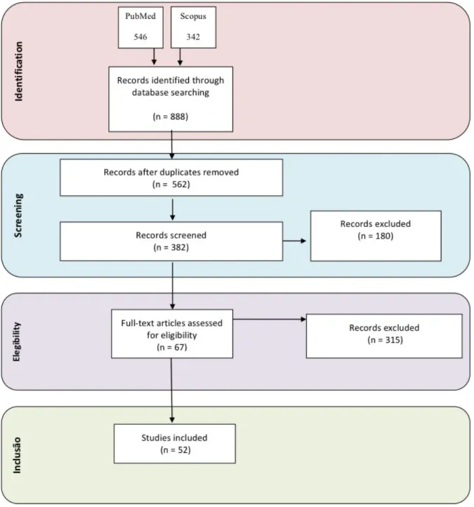
Searches were conducted in the databases Pubmed and SCOPUS, using the following descriptors: dental car-ies in dentin, deep dental carcar-ies, deep carcar-ies treatment, and conservative treatment, from 1960 to 2018. Figure 1 shows the diagram of selection and exclusion of the arti-cles included in this literature review.
Of the 888 articles initially identified, 326 were excluded because they were duplicates. Then, through reading the
ti-tles, 180 articles were excluded because they met the exclu-sion criteria, namely: articles on shallow or medium leexclu-sions in dentin, direct pulp capping, pulpotomy, pulpectomy, studies on animals, in vitro studies, and those that did not belong to the subject of interest. Of the 382 articles selected for reading the abstracts, 315 were excluded based on the same criteria. Therefore, of the 67 articles remaining, read in full, 52 articles in English and Portuguese were selected, fol-lowing the predetermined inclusion and exclusion criteria – including the record of study approval by the local ethics
committee –, to conduct this review.
Literature Review
The treatment planning of caries lesions shall aim at: (1) inactivating/controlling the carious process, (2) preserv-ing the hard dental tissue, (3) preventpreserv-ing the repetitive re-storative cycle, and (4) preserving the tooth for as long as possible.13 When in dentin, carious lesion is histologically
characterized by the formation of layers with different char-acteristics (Figure 2): (1) the most superficial layer, which is characterized by a necrotic zone, followed by a layer of in-fected, decomposed dentin (2), with high bacterial counts. The underlying layer (3) is made of affected, demineralized dentin, and is followed by the last layer of reaction or scle-rotic dentin (4).14 Layers of necrotic and infected dentin are
Current trends in the conservative treatment of deep-caries lesion with risk of pulp exposure
characterized as a superficial layer with extensive deminer-alization, degenerated collagen fibers and negative odonto-blastic processes, non-physiologically remineralizable and strongly infected. Affected dentin layer is characterized by intermediate demineralization, healthy collagen fibers and live odontoblastic process, in addition to being physiologi-cally remineralizable.15
Traditionally, the indirect pulp capping technique is commonly indicated for treating deep carious lesions. Such therapeutic procedure is defined by the total removal of ne-crotic, infected and affected tissues, until one finds a hard-ened dentin that offers resistance to be removed by manual or low-rotation instruments (Figure 3). Then, a calcium hy-droxide coating is usually added to induce the formation of tertiary dentin, thus avoiding pulp aggression when mak-ing the definitive restoration in the same session.16
Figure 2. Clinical picture of carious lesion affecting dentin, with the
forma-tion of 4 distinct layers: (1) necrotic dentin; (2) infected (or decomposed) dentin; (3) demineralized (or affected) dentin; (4) reactional (or sclerotic) dentin. Image kindly provided by Prof. Dr. Aline de Almeida Neves
Figure 3. Clinical sequence of indirect pulp capping technique in single
session. a) initial clinical aspect; b) initial X-ray; c) tooth after rubber dam placed for restoration; d) total removal of the infected dentin. Source: Apoena de Aguiar Ribeiro. Treatment of deep carious lesions in decidu-ous teeth. PRO-odonto prevenção. 2012;6(2):9-37. Rights for the use of images grantes by Atmed Panamericana Editora Ltda.
The advantage of the indirect pulp capping technique is the fact that it can be conducted in a single session, being indi-cated mainly to child care due to decreasing the amount of dental consultations and treatment costs.17 However, its main
downside is that, due to being a less conservative technique, due to the need to leave only the dentin tissue of leathery, hardened consistency, it presents greater risk of pulp expo-sure, leading to a less favorable treatment prognosis due to possible pulp exposure and loss of pulp vitality.18
Currently, this technique is regarded as over-treatment because, with the increased knowledge on the histopatho-genesis of dentin caries and on the healing potential of the dentin-pulp complex, the treatment of deep caries should only be applied to stop the lesion progression and to stim-ulate the dentin reaction through sclerosis and repair.13,19
Currently, this may be achieved by the ultra-conservative treatment of deep carious lesions.
The conservative treatment of deep carious lesions at risk of pulp exposure aims at removing the superficial necrotic dentin and only part of the infected dentin during the acute phase of the process, followed by placement of a coating material that fosters the bactericidal or bacteriostatic action and the sealing of the cavity.20 In the literature, two different
types of ultra-conservative treatment are described for deep carious lesions: (1) stepwise excavation, (2) selective caries removal. The two treatment options are described below:
• Stepwise Excavation
Stepwise excavation is defined as a conservative treat-ment for deep carious lesion, where only part of the infect-ed dentin is removinfect-ed, throughout two or more sessions in a time interval – hence the term “stepwise”.20 Such interval is
for inducing the formation of tertiary dentin (through phys-iological processes of the dentin-pulp complex) and for re-ducing the amount of bacteria within the lesion, in addition to enabling the complete removal of the carious tissue, thus minimizing the chances of pulp exposure.21
The clinical protocol of the stepwise excavation consists of the total removal of the necrotic and infected (decomposed) dentin of the surrounding walls (layers 1 and 2, presented in Figure 2) and only partial removal of this same dentin of the bottom of the cavity, followed by coating with a calcium hydroxide paste base and sealing of the cavity with Zinc Ox-ide and Eugenol Cement or Glass Ionomer Cement. After 4 to 6 months, this cavity must be reopened, with the total removal of the sealing material, so that a new excavation of the remaining infected dentin may be performed (Figure 4). If it is not possible to remove all infected dentin and leave the bottom of the cavity only with affected dentin (layer 3, shown in Figure 1), a new cavity sealing should be placed and subsequent appointments for reopening may be sched-uled every 4 to 6 months.22
One of the advantages of this technique is the lower pos-sibility of pulp exposure in deep caries, mainly when com-pared with the indirect pulp capping technique. In addition, it allows the reduction of bacterial load between sessions and the physiological recovery of the dentin-pulp complex, through the production of tertiary dentin.8,23-25 Leksellet al.26
verified that, by using the stepwise excavation technique, there was a lower rate of pulp exposure (17.5%) in compari-son to the conventional removal of carious dentin (indirect pulp capping), which exposure rate was 40%.
However, the stepwise excavation technique has some disadvantages, such as an additional step for reopening the cavity, which can cause problems in the pulp due to the in-creased risk of exposure, thus damaging its vitality. Anoth-er disadvantage is due to the risk of loss of the temporary sealing material between appointments, compromising the formation of tertiary dentin and, consequently, the lesion inactivation. In addition, since it requires an extra-visit by the patient, dentist should be aware to indicate it only to those patients who would collaborate with subsequent con-sultations for review and cavity reopening.27
• Selective Removal of Carious Tissue
The selective removal of carious tissue consists of the to-tal removal of the infected and affected dentin of the sur-rounding walls of the cavity and the partial removal of the softened central dentin (infected dentin), leaving part of it in the bottom of the pulp wall (similar to the first step of the stepwise excavation technique). Thus, after the selective removal of part of the infected dentin from the pulp wall, a base with Calcium Hydroxide paste-paste might be applied (albeit not essential) and part of the cavity is filled with a
layer of resin-modified glass ionomer cement. Finally, the cavity is restored with direct composite resin, in the same session, i.e., without re-entrance in the cavity (Figure 5).28-31
Such technique became feasible due to the evolution of
adhesive materials, which ensure satisfactory clinical results of marginal sealing, even with the infected dentin remnant in the bottom of the cavity.32 This is because the axial walls
are free of carious dentin, so sealing is effective in preventing the remaining bacteria (in the affected dentin at the bottom of the cavity) to have acces to nutrients, not favoring bacteri-al survivbacteri-al, thus significantly reducing the bacteribacteri-al popula-tion within the carious lesion.33 Thus, for the success of this
technique, all the softened carious tissue must be removed (necrotic, infected and affected dentin) from the axial walls of the cavity, to ensure proper sealing of it with an adhesive material, aiming the inactivation of the carious lesion.33
The reduced cost and the benefit of the decreased risk of pulp exposure are its main advantages of this technique34
since it does not need additional visits to the dentist, being less costly for the patient and even facilitating, mainly, the treatment of children.35 However, as this is a recent
tech-nique, its disadvantage is that it requires more longitudinal
Figure 4. Clinical sequence of the treatment through stepwise excavation technique. a) initial clinical aspect; b) initial X-ray; c) cavity after the first
excavation. Six months after: d) tooth after rubber dam placed for restoration; e) cavity after removal of temporary filling; f) total removal of the infected dentin. Source: Apoena de Aguiar Ribeiro. Treatment of deep carious lesions in deciduous teeth. PRO-odonto prevenção. 2012;6(2):9-37. Rights for the use of images grantes by Atmed Panamericana Editora Ltda.
Figure 5. Clinical sequence of the selective caries removal technique a)
initial clinical aspect; b) partial removal of the infected dentin; c) direct composite restoration
studies, even though it already shows positive results in re-search performed with permanent and primary teeth.12,36-40
Discussion
The discussion on the amount of carious tissue to be re-moved in deep cavity lesions is not new and it aims to eval-uate the success of the treatment and, mainly, the mainte-nance of the tooth longevity through the reduction of the pulp risk of exposure.40-42 In 1859, Tomes wrote that “it is
best to maintain a layer of carious dentin to protect the pulp than have the risk of sacrificing the teeth.” Nonetheless, in 1908, Black registered that “it is best to expose the pulp than leaving it covered with softened dentin.”43
Since then, the complete removal of the softened, affect-ed carious dentin (or indirect pulp capping) has been con-sidered an essential step in the treatment of caries, and it was assumed that the success of the restorative treatment depends on the complete elimination of bacteria.44 However,
even today, there is no definitive clinical diagnosis equip-ment to define when to stop the removal of carious dentin tissue, therefore, this criterion shall be defined by the dentist. Since the traditional knowlege advocates that the removal of carious dentin should be performed until the total removal of the softened dentin, this procedure ends up extending the cavity preparation not only horizontally but mainly verti-cally, towards the dental pulp.20,45
However, there is current scientific evidence that indirect pulp capping often leads to pulp exposure26,46 and,
there-fore, conservative approaches have been proposed to man-age deep carious lesions in permanent and primary teeth, aiming at repairing the dentin by the sclerosis of dentinal tubules and production of tertiary dentin.3
Reactional changes due to the treatment with partial re-moval of infected dentin and stepwise excavation were in-vestigated by several authors.30,38-42,48 Maltz et al.30 evaluated
32 permanent teeth at risk of pulp exposure, in patients aged from 12 to 23 years. The stepwise excavation technique was carried out, with total removal of carious tissue from the surrounding walls and partial removal of the caries in the pulp wall, leaving the softened, infected dentin at the bot-tom of the cavity. The teeth were sealed with calcium hy-droxide as base and modified zinc oxide and eugenol cement (IRM). After 6 to 7 months, the teeth were reopened, with total removal of the temporary material, and then restored with direct composite resin. They observed that there was a change in the physical characteristics of the dentin after the time interval, as it was dry and, in 80% of the teeth, the den-tin became hard. In 16.7% of the teeth the denden-tin was leath-er-looking and in only 3.3% it remained soft, thus showing that, even before the incomplete removal of infected dentin, it is possible to stop the lesion progression by the formation of tertiary or sclerotic dentin.
Regarding the effectiveness of these techniques in pre-venting pulp exposure, comparative studies between total removal (indirect pulp capping technique) and partial re-moval (stepwise excavation technique) of carious dentin have shown that the stepwise excavation resulted in less pulp exposures in comparison with complete direct excava-tion (a difference ranging from 11.4% to 22.5%).10,18,26,51
Af-ter years of follow-up, there is a significantly higher success rate with stepwise excavation – being success defined as a non-exposed pulp with tooth vitality and without apical ra-diolucency.24,52
However, among more conservative professionals, there is always the question about microorganisms present in the infected dentin and that were left at the base of the lesion, near the pulp. The lesion will not continue to progress? The knowledge that the lesion stops progressing when the cavi-ty is closed, even if it still presents viable microorganisms, is scientifically established since the 1940s.49 Clinical and
microbiological studies, when analyzing the selective caries removal and the stepwise excavation treatments highlight-ed that the reopening step the reopening step, for the total removal of the remaining carious tissue left in the cavity, can be dismissed. This because the proper sealing of the cavity may promote beneficial changes in deep carious le-sions, even in the presence of infected dentin. This reinforc-es the current scientific approach that the re-entry phase can be excluded, and the treatment may be held in a single session.8,41,42,50 The recent microbiological study of Singhal
et al.41 compared the microbial counts among cavities
sub-jected to total or partial removal of caries, showing that the progression of the caries lesion is paralyzed when the cavity is closed, even if living microorganisms are left within the cavity. Other clinical and microbiological trials also came to the same conclusion, strengthening the approach of con-servative techniques for deep caries at risk of pulp exposure, without the need of a step for reopening the cavity.18,30,41,43
These studies were the basis for the design of the selective caries removal technique, i.e, without the cavity reopening step. However, for this technique to be successful, it is es-sential that all carious tissue must be removed from the ax-ial walls of the cavity, ensuring its proper sealing with an adhesive material, to control the progression and promote inactivation of the carious lesion.30,41 Such technique is
feasi-ble due to the evolution of capabilities of adhesive materials, which ensure satisfactory clinical results of marginal seal-ing, even with the infected dentin remnant at the bottom of the cavity.20,32,33 This because the satisfactory sealing of
the cavity prevents the entry of nutrients for the bacteria re-maining in the carious tissue, not favoring bacterial survival and significantly reducing the population within the carious lesion.32 Thus, the selective caries removal, albeit an
inno-vative approach, presents high success rates in maintaining
FDI task group. Int Dent J. 2012; 62(5):223-43.
12. Jardim JJ, Simoneti DNM, Maltz M. Partial caries removal in permanent teeth: six-year follow-up. RFO UPF. 2001;(20):1.
13. Schwendicke F, Frencken JE, Bjørndal L, Maltz M, Manton DJ, Ricketts D, et al. Managing Carious Lesions: Consensus Recommendations on Carious Tissue Removal. Adv Dente Res. 2016;28(2):58-67.
14. Browning W.D. Critical appraisal. 2015 Update: Approaches to Caries Re-moval. J Esthet Restor Dent. 2015;27(6):383-96.
15. Ogushi K, Fusayama T. Electron microscopic structure of the two layers of carious dentin. J Dent Res.1975;54(5):1019-26.
16. Straffon LH, Feigal RJ, Welch KB. Indirect pulp treatment of primary posteri-or teeth: a retrospective study. Pediatr Dent. 2003;25(1):29-36.
17. Mosele GT, Imparato JCP, Parizotto SPCOL. Avaliação do capeamento pul-par indireto e tratamento expectante em molares decíduos. Rev. Assoc. Paul. Cir. Dent. 2012;66(3):214-9.
18. Phonghanyudh U, Phantumvanit P, Songpaisan Y, Petersen PE. Clinical eval-uation of three caries removal approaches in primary teeth: a randomised con-trolled trial. Community Dent Health. 2012;29(2):173-8
19. Ribeiro AA. Tratamento de lesões cariosas profundas em dentes decíduos. PRO-ODONTO PREVENÇÃO. 2012;6(2):9-37.
20. Almeida NA, Coutinho E, Cardoso VM, Lambrechtsd P, Meerbeeke VB. Cur-rent Concepts and Techniques for Caries Excavation and Adhesion to Residual Dentin. J Adhes Dent. 2011; 13(1):7-22.
21. Ricketts D. Management of the deep carious lesion and the vital pulp dentine complex. British Dental Journal. 2001;191(11):606-10.
22. Bjorndal L, Kidd EA. The treatment of deep dentine caries lesions. Dent Up-date. 2005;32(7):402-4.
23. Ricketts D, Lamont T, Innes NPT, Kidd E, Clarkson JE. Operative caries man-agement in adults and children. Cochrane Database Syst Rev. 2013;(3):CD003808.
References
1. Bourgeois DM, Llodra JC. Global burden of dental condition among children in nine countries participating in an international oral health promotion pro-gramme. Int Dent J. 2014;64(2):27-34.
2. Hashim R, Thonson WM, Ayer KM, Lewsey JD, Awad M. Dental caries experi-ence and use of dental services among preschool children in Ajman. Int J Paediatr Dent. 2006;16(4):257-62.
3. Fejerskov O, Nyvad B, Kidd EAM. Dental Caries: The Disease And Its Clinical Management. 3rd Ed. Oxford (UK):Wiley Blackwell;2015.
4. Bjørnda L, Thylstrup A. A practice-based study on stepwise excavation of deep carious lesions in permanent teeth: a 1-year follow-up study. Community Dent Oral Epidemiol. 1998;26(2):122-8.
5. Lima OEJ. Cárie dentária: um novo conceito. R Dental Press Ortodon Ortop Facial. 2007;12(6):119-0.
6. Arcella D, Ottolenghi L, Polimeni A, Leclercq C. The relationship between frequency of carbohydrates intake and dental caries: a cross-sectional study in Italian teenagers. 2002;5(4):553-60.
7. Ribeiro AA. Biological Features of Dental Caries. JSM Dent. 2016;(3):1065-70. 8. Ricketts DN, Kidd EA, Iness NPT, Clarkson J. Complete or ultraconser-vative removal of decayed tissue in unfilled teeth. Dados Cochrane Syst Rev. 2006;(3):CD003808.
9. Whitworth JM, Myers PM, Smith J , Paredes AW, McCabe JF. Endodon-tic complications after plasEndodon-tic restorations in general pracEndodon-tice. Int Endod J. 2005;38(6):409-16.
10. Bjørndal L, Claes R, Gitte B, Merete M, Marianne K, Peggy N et al. Treatment of deep caries lesions in adults: randomized clinical trials comparing stepwise vs. direct complete excavation, and direct pulp capping vs. partial pulpotomy. Eur J Oral Sci. 2010;118(3):290-7.
11. Frencken JE, Peters MC, Manton DJ, Leal SC , Gordan VV , Eden E. Minimal Intervention Dentistry (MID) for managing dental caries – a review: Report of a
the viability in permanent and deciduous teeth.12,38-41
Recent studies also compared the stepwise excavation with selective caries removal and reported high rates of suc-cess, specially in the selective removal technique.12,38-41 In
the study of Maltz et al.38, the success rates were 95.45% for
the selective caries removal and 80.85% for stepwise exca-vation after one year of follow-up. Maltz et al.,39 in 2012, also
showed that, after two years, the success rates were 91% for the selective caries removal and 69% for stepwise excavation technique, suggesting that the selective caries removal treat-ment promotes higher longevity . Also, in a 6-year longitu-dinal follow-up study, Jardim et al.12 showed that the success
rate of the treatments through selective caries removal was much higher than the stepwise excavation technique (60% and 32%, respectively).
Another important point of discussion is regarding the longevity of restorations performed on the remaining cari-ous dentin tissue. Franzon et al.52 monitored, for 2 years, 120
primary teeth with deep caries lesions, submitted to selec-tive removal or indirect pulp capping, to compare the lon-gevity of restorations performed and the rate of prevention of pulp exposure. Pulp exposure occurred in 1 and 15 of the teeth treated with selective caries removal and indirect pulp capping, respectively. However, the survival rate of resto-rations after 24 months was 66% for selective caries removal and 86% for indirect pulp capping. Selective caries removal carried out in occlusal-proximal restorations showed a sig-nificantly lower rate of success. Thus, the implementation
of selective caries removal prevented the pulp exposure in primary teeth and, consequently, the need for more invasive treatments. However, this technique showed lower longevity for the direct composite resin restoration in comparison to the indirect pulp capping, suggesting that restorations after selective caries removal must be monitored over time, spe-cially when they involve two or more surfaces of the tooth. Ribeiro et al.32 showed, through scanning electron
micros-copy, the presence of a altered hybrid zone formed on the carious dentin; however, limited information is available about the impact of this over time.
Conclusion
Considering the literature consulted, we concluded that the conservative techniques of stepwise excavation and se-lective caries removal features a lower pulp exposure rate. Among these, the selective caries removal showed better clinical performance and greater longevity. Thus, these cur-rent conservative techniques should be used to minimize the risk of pulp exposure and to maintain the vitality and integ-rity of teeth with deep carious lesions. However, as these are innovative techniques, no articles on the topic were found in the scientific literature in Portuguese – even though there is plenty scientific support in the literature in English –, which may justify the lack of information about the use of these techniques in the routine practice among dentists in Brazil.
Submitted: 05/22/2018 / Accepted for publication: 08/06/2018
Corresponding Author Apoena Aguiar Ribeiro
E-mail: apoena@email.unc.edu
Mini Curriculum and Author’s Contribution
1. Betina Paganini da Silva – DDS. Contribution: literature search, article reading, initial writing of the manuscript. ORCID: 0000-0002-5466-2304 2. Mayara Simão Gama – DDS. Contribution: literature search, article reading, initial writing of the manuscript. ORCID: 0000-0003-4696-1512 3. Apoena Aguiar Ribeiro – DDS, MS and PhD. Contribution: conception, guidance and final writing of the manuscript. ORCID: 0000-0001-7702-6178 24. Uribe S. Partial caries removal in symptomless teeth reduces the risk of pulp
exposure. Evid Based Dent. 2006;7(4):94.
25. Fagundes TC, Barata TJE, Prakki A, Bresciani E, Pereira JC. Indirect pulp treatment in a permanent molar: case report of 4-year follow-up. J Appl Oral Sci. 2009;17(1):70-4.
26. Leksell E, Ridell K, Cvek H, Mejáre EU. Pulp exposure after stepwise versus direct complete excavation of deep carious lesions in young posterior permanent teeth. Endod Dent Traumatol.1996;12(4):192-6.
27. Oliveira EF, Carminatti G, Fontanella V, Maltz M. The monitoring of deep caries lesions after incomplete dentine caries removal: results after 14-18 months. Clin Oral Investig. 2006;10(2):134-9.
28. Orhan AI, Oz FT, Ozcelik B, Orhan K. A clinical and microbiological com-parative study of deep carious lesion treatment in deciduous and young perma-nent molars. Clin Oral Investig. 2008;12:369-78.
29. Orhan AI, Oz FT, Orhan K. Pulp exposure occurrence and outcomes after 1- or 2-visit indirect pulp therapy vs. complete caries removal in primary and permanent molars. Pediatr Dent. 2010;32:347-55.
30. Maltz M, de Oliveira EF, Fontanella V, Bianchi R. A clinical, microbiologic, and radiographic study of deep caries lesions after incomplete caries removal. Quintessence Int. 2002;33(2):151-9.
31. Maltz M, Oliveira EF, Fontanella V, Carminatti G. Deep caries lesions af-ter incomplete dentine caries removal: 40-month follow-up study. Caries Res. 2007;41(6):493-6.
32. Ribeiro CC, Baratieri LN, Perdigão J, Baratieri NM, Rittler AV. A clinical, radiographic and scanning electron microscopic evoluation of adhesive res-torations on carious dentin in primary teeth. Quintessence International. 1999;30(9):591-9.
33. Ribeiro CC, Lula OCE, Da Costa NCR, Nunes MMA. Rationale of partial removal of carious tissue in primary teeth. Pediatric Dentistry. 2012; 34(1):39-41. 34. Carvalho JC, Dige I, Machiulskiene V, Qvist V, Bakhshandeh A, Fatturi-Pa-rolo C, et al. Occlusal Caries: Biological Approach for Its Diagnosis and Manage-ment. Caries Res. 2016;50(6):527-42.
35. Schwendicke F, Paris S, Stolpe M. Cost-effectiveness of caries excavations in different risk groups - a micro-simulation study. BMC Oral Health. 2014;14:153. 36. Ferreira SMJ, Pinheiro SL, Sampaio FC, de Menezes VA. Caries removal in primary teeth--a systematic review. Quintessence Int. 2012;43(1):9-15.
37. Schwendicke F, Dorfer CE, Paris S. Incomplete caries removal: a systematic review and metaanalysis. J Dent Res. 2013;92(4):306-14.
38. Maltz M, Mauricio M, Jardim JJ, Marques C, De Paula LM, Mestrinho DH. Partial Caries Removal in Deep Lesions: 19-30 months follow-up study Par-tial Caries Removal in Deep Lesions: 19-30 months follow-up study. Rev Fac
Odontol. 2010;1(51):20-3.
39. Maltz M, Garcia R, Jardim JJ, de Paula LM, Yamaguti PM, Moura MS, et al. Randomized trial of partial vs. stepwise caries removal: 3-year follow-up. J Dent Res. 2012; 91(11):1026-31.
40. Schwendicke F, Schweigel H , Petrou MA , Santamaria R, Hopfenmüller W, Fink C et al. Selective or stepwise removal of deep caries in deciduous molars: study protocol for a randomized controlled trial. Trials. 2015;16:11.
41. Singhal DK, Acharya S, Thakur AS. Microbiological analysis after complete or partial removal of carious dentin using two different techniques in primary teeth: A randomized clinical trial. Dent Res J (Isfahan). 2016;13(1):30-7. 42. Bjørndal L, Thylstrup A. A practice-based study on stepwise excavation of deep carious lesions in permanent teeth: a 1-year follow-up study. Community dentistry and oral epidemiology. 1998;26(2):122-8.
43. Kidd EAM. How “clean” must a cavity be before restoration? Caries Res. 2004;38:305-13.
44. Weerheijm KL, Kreulen CM, de Soet JJ, Groen HJ, van Amerongen WE. Bac-terial counts in carious dentine under restorations: 2-year in vivo effects. Caries Res. 1999;33(2):130-4.
45. Bjørndal L. Indirect Pulp Therapy and Stepwise Excavation. Pediatric Den-tistry. 2008;30(3):225-9.
46. Magnusson BO, Sundell SO. Stepwise excavation of deep carious lesions in primary molars. J Int Assoc Dent Child. 1977;8(2):36-40.
47. Chibinski, RCA, Wambier L, Reis A, Wambier DS. Clinical, mineral and ul-trastructural changes in carious dentin of primary molars after restoration. Int Dent J. 2016;66(3):150-7.
48. Besic FC. The Fate of Bacteria Sealed in Dental Cavities. J Dent Res. 1943;22(5):349-59.
49. Thompson V, Craig RG, Curro FA, Green WS, Ship JA. Treatment of deep carious lesions by complete excavation or partial removal. J Am Dent Assoc. 2008;139(6):705-12.
50. Villat C, Attal JP, Brulat N, Decup F, Doméjean S,Dursun E, Fron-Chabouis H et al. One-step partial or complete caries removal and bonding with antibacte-rial or traditional self-etch adhesives: study protocol for a randomized controlled trial. Trials. 2016;17(1):404.
51. Hayashi M, Fujitani M, Yamaki C,Momoi Y. Ways of Enhancing Pulp Pres-ervation by Stepwise Excavation - A Systematic Review. Journal of Dentistry. 2011;(39):95-107.
52. Franzon R, Opdam NJ, Guimarães LF, Demarco FF, Casagrande L, Haas AN, et al. Randomized controlled clinical trial of the 24-months survival of compos-ite resin restorations after one-step incomplete and complete excavation on pri-mary teeth. J Dent. 2015;43(10):1235-41.