Universidade de Lisboa
Faculdade de Medicina da Universidade de Lisboa
Role of SNARE-dependant gliotransmitter
release by astrocytes on the modulation of
synaptic plasticity by BDNF
João Pedro de Almeida Jesus
Supervisor: Sandra Henriques Vaz, PhD
Thesis elaborated for the obtainment of the Master’s degree in Neurocience
Lisbon, 2017
Universidade de Lisboa
Faculdade de Medicina da Universidade de Lisboa
Role of SNARE-dependant gliotransmitter
release by astrocytes on the modulation of
synaptic plasticity by BDNF
João Pedro de Almeida Jesus
Supervisor: Sandra Henriques Vaz, PhD
Thesis elaborated for the obtainment of the Master’s degree in Neurocience
Everything that is implied in the present document is of the responsibility of its author, with no responsibility falling to the Faculty of Medicine of the University of Lisbon for the content that is here presented.
The printing of this thesis was approved by the scientific counsel of the Faculty of Medicine of the University of Lisbon in a meeting on the 18th of
The experimental work contained in this thesis was performed at the
Instituto de Farmacologia e Neurociências, Faculdade de Medicina e Unidade de Neurociências, Instituto de Medicina Molecular da Universidade de Lisboa, under the supervision of Sandra Henriques Vaz, PhD.
O trabalho experimental constante da presente tese foi realizado no Instituto de Farmacologia e Neurociências, Faculdade de Medicina e Unidade de Neurociências, Instituto de Medicina Molecular da Universidade de Lisboa, sob orientação da Doutora Sandra Henriques Vaz
.
V
RESUMO
Os astrócitos são um dos quatro tipos de células da glia que podemos encontrar no Sistema Nervoso Central (SNC), sendo os outros três a microglia, os oligodendrócitos e as células NG2 positivas. Os astrócitos são o tipo de célula da glia mais abundante no SNC, sendo responsáveis por numerosas e complexas funções essenciais para bom funcionamento neuronal, através da sua ação sobre a transmissão sináptica e excitabilidade neuronal e sobre o processamento da informação transmitida pelos circuitos neuronais. Estas células têm também a capacidade de executar numerosas funções de suporte neuronal, colaborando no suporte trófico dos neurónios, nos processos de sobrevivência e diferenciação neuronal, no crescimento neurítico, em processos de manutenção da eficiência sináptica, e regulação das concentrações extracelulares de iões, contribuindo assim para a homeostasia do cérebro.
Os astrócitos desempenham grande parte das suas funções através da libertação de mensageiros neuroactivos, denominados gliotransmissores. Os principais gliotransmissores são o glutamato, a adenosina trifosfato(ATP), a D-Serina, o
brain-derived neurotrophic factor (BDNF) e também o tumor necrosis factor alpha (TNF-α).
A libertação destas moléculas para a fenda sináptica e as interações resultantes entre estes gliotransmissores com os seus recetores, localizados tanto a nível pré como pós-sináptico, levam à modulação da atividade sináptica.
O modelo que descreve o mecanismo de comunicação bidirecional entre astrócitos e neurónios denomina-se de sinapse tripartida. Este modelo propõe que após a libertação de neurotransmissores/neuromoduladores para a fenda sináptica pelo neurónio pré-sináptico, estes mesmos neurotransmissores/neuromoduladores irão ligar-se não só aos seus recetores a nível do neurónio pós-sináptico mas também ao nível dos seus recetores específicos que se encontram ao nível da membrana plasmática do astrócito que envolve a sinapse. Os astrócitos possuem inúmeros tipos distintos de recetores para os vários neurotransmissores/neuromoduladores ao nível da sua membrana plasmática, pelo que podem assim desencadear respostas consoante os neurotransmissores/neuromoduladores que são libertados. A ligação destas moléculas aos respetivos recetores astrocitários podem desencadear um aumento da concentração intracelular de Ca2+. O aumento da concentração intracelular deste ião leva à libertação
VI
de gliotransmissores para a fenda sináptica, permitindo fenómenos de modulação da atividade sináptica.
As sinapses possuem plasticidade que varia consoante a sua atividade, sendo que este mecanismo tem um papel vital no desenho das conexões sinápticas, particularmente durante o período de desenvolvimento. Esta plasticidade, num entanto, também se encontra presente no cérebro adulto, sendo aceite que a formação de memórias se baseia em alterações da eficiência sináptica que fortalecem as associações entre neurónios comunicantes, o que por sua vez permite o armazenamento de informação. A este fortalecimento dá-se o nome de Long Term Potentiation (LTP). A indução de LTP envolve numerosos recetores, nomeadamente a família dos recetores NMDA de glutamato. Durante fenómenos de estimulação, onde existe uma intensa despolarização da membrana do neurónio pós-sináptico, verifica-se um desbloqueio dos canais de cálcio destes recetores, que normalmente se encontram bloqueados pela presença de iões Mg2+. A abertura destes canais leva à entrada de Ca2+ para o interior do neurónio pós-sináptico, levando a um aumento da concentração intracelular deste mesmo ião, que leva à ativação das vias metabólicas da proteína cinase dependente de calmodulina II (CaMKII) e Proteína Cinase A (PKA) que têm um papel fundamental na potenciação da sinapse. A ativação da proteína CaMKII leva a alterações morfológicas menores, como o aumento das espinhas dendríticas e o aumento da condutividade dos recetores AMPA; por outro lado, a ativação da PKA leva à ativação dos fatores de transcrição CREB e ERK, que desencadeiam também alterações que potenciam a sinapse.
Nos últimos anos vários grupos demonstraram que os astrócitos modulam a LTP através da libertação de gliotransmissores, nomeadamente o glutamato, a D-serina e o ATP. A libertação de ATP e a sua consequente metabolização a adenosina ao nível da fenda sináptica tem especial importância, uma vez que a adenosina extracelular formada exerce a sua acção sobre a transmissão sináptica. A adenosina actua através da ligação a quatro tipos diferentes de recetores acoplados a proteínas G: os recetores A1 e A3, que
estão acoplados a subunidades Gi/o, levando a respostas inibitórias quando ativados; e recetores A2A e A2B, que estão acoplados a subunidades Gs, levando a respostas
excitatórias quando ativados.
O BDNF é uma neurotrofina com um papel neurofisiológico bastante importante por estar envolvido na regulação do desenvolvimento dos circuitos nervosos, na diferenciação e crescimento de axónios e dendrites, na formação e maturação de sinapses. Para além disso, o BDNF tem também um papel na regulação dos circuitos neuronais
VII
maduros e na regulação da LTP e long term depression (LTD), podendo potenciar a magnitude da LTP. O efeito facilitatório do BDNF sobre a magnitude da LTP acontece devido à ativação dos recetores TrkB, aos quais se liga com grande afinidade. A ativação dos recetores TrkB leva a alterações tanto a um nível pré-sináptico como pós-sináptico, que têm como consequência o aumento da excitabilidade do neurónio pós-sináptico. Existe também evidências de que este efeito potenciador do BDNF sobre a magnitude da LTP é dependente da ativação dos recetores A2A de adenosina.
Uma vez que os astrócitos têm a capacidade de controlar a LTP por libertação de gliotransmissores, e adicionalmente o BDNF também modula a magnitude da LTP de um modo dependente da activação dos receptores A2A de adenosina, o objectivo do presente
trabalho foi avaliar se o efeito do BDNF na LTP depende da libertação de gliotransmissores, nomeadamente ATP/adenosina.
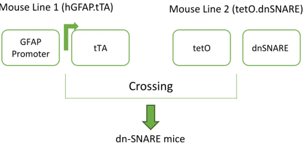
Para tal, foram realizados estudos electrofisiológicos, nomeadamente indução de LTP, utilizando fatias agudas de hipocampo obtidas a partir de ratinhos da estirpe dn-SNARE. Estes animais expressam um transgene (porção citosólica do domínio SNARE da sinaptobrevina 2) selectivamente em astrócitos, que pode ser manipulado, por administração de doxiciclina, de modo a bloquear ou ativar a gliotransmissão.
Neste trabalho observou-se que um estimulo de indução de LTP θ-burst aumentou o declive dos fEPSP em 22±10% nos ratinhos WT (+DOX), e que nas mesmas fatias mas em presença de BDNF (20ng/ml), o mesmo paradigma de estimulação aumentou o declive dos fEPSP em 55±6.8% (p<0.05, n=5). O que corresponde a um efeito estatisticamente significativo do BDNF sobre a magnitude da LTP. Em ratinhos dn-SNARE (-DOX) a magnitude da LTP foi de 24±4% em condições controlo (sem BDNF) e de 29±3% em fatias tratadas com BDNF (p>0.05, n=4). Observou-te também que o efeito do BDNF é dependente da activação dos receptores de adenosina, uma vez que o efeito potenciador desta neurotrofina sobre a LTP foi perdido na presença do antagonista selectivo dos receptores A2A (SCH 58261) em animais dn-SNARE (+DOX). Estes
resultados sugerem que a libertação de gliotransmissores pelos astrócitos controla o efeito potenciador do BDNF sobre a LTP.
Uma vez que a activação dos receptores A2A da adenosina é fundamental para os
efeitos mediados pelo BND na LTP, colocou-se a hipótese que os astrócitos seriam a possível fonte de adenosina envolvida da neste processo. Para testar esta hipótese fatias de hipocampo provenientes de ratinhos dn-SNARE (-DOX) foram perfundidas com o agonista selectivo dos receptores A2A de adenosina (CGS 21680 (30nM)), previamente
VIII
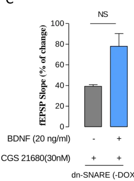
ao tratamento com BDNF (20ng/ml). Na presença de CGS 21680 (30ng/ml), o estimulo θ-burst induziu um aumento do declive de 39±2%, e na presença de CGS 21680 (30ng/ml) e BDNF (20ng/ml) o declive dos fEPSP foi de 78±12% (p<0.05, n=3), o que corresponde a um efeito estatisticamente significativo do BDNF sobre a magnitude da LTP de 100% .
Estes resultados mostram que os astrócitos têm um papel ativo na ação facilitadora do BDNF sobre a LTP e sugerem também que a principal fonte de adenosina envolvida no efeito do BDNF serão os astrócitos, através da sua libertação de ATP para a fenda sináptica e a posterior transformação deste ATP em adenosina. A adenosina assim formada leva à ativação de recetores A2A, permitindo a ação facilitadora do BDNF sobre a LTP.
É importante de notar que outros gliotransmissores, nomeadamente o glutamato e a D-serina poderão ter também um papel ativo sobre esta ação do BDNF, juntamente com a adenosina. Assim sendo, o estudo do papel destes gliotransmissores sobre o mecanismo de potenciação da LTP pelo BDNF seria bastante interessante. Por outro lado, a replicação dos resultados obtidos neste trabalho usando um maior número de animais seria de elevado interesse, de modo a confirmar a viabilidade destes mesmos resultados.
Palavras-chave: Astrócitos, LTP, BDNF, dn-SNARE, gliotransmissão, recetores A2A de
IX
ABSTRACT
Astrocytes are one of the four types of glial cells that we can find in the Central Nervous System (CNS), with the remaining three being the microglia, oligodendrocytes and NG2 positive cells. Astrocytes are the type of nervous cell more abundant in brain, being responsible for numerous and complex functions that are essential for its correct functioning, through their role over the modulation synaptic transmission and neuronal excitability, as well as their role over the processing of information transmitted by neuronal circuits. These cells are also able of doing numerous functions of neuronal support, helping with the trophic support of neurons, in processes of neuronal survival and growth, in the process of neurite growth and in processes of the maintenance of synaptic efficiency. Besides this, these glial cells also contribute to the maintenance of the homeostasis of the brain, through their regulation of the concentrations of certain ions and neuroactive substances.
Most of the astrocytes functions are executed through the release of neuroactive messengers, called gliotransmitters. The main Gliotransmitters are glutamate, adenosine triphosphate (ATP), D-serine, brain-derived neurotrophic factor (BDNF) and also tumor necrosis factor alpha (TNF- α). The release of these molecules to the synaptic cleft and the interactions between these gliotransmitters and their receptors, localized both at a pre-synaptic and a post-pre-synaptic level, lead to the modulation of the pre-synaptic activity.
The model that describes this mechanism of bidirectional communication between astrocytes and neurons is called the tripartite synapse model. This model describes that after the release of neurotransmitters by the pre-synaptic neuron to the synaptic cleft, these same neurotransmitters will bind not only to their receptors located on the
post-X
synaptic neuron but also to their specific receptors present in the membrane of the astrocyte that encircles the synapse. Astrocytes possess numerous distinct types of receptors for the various neurotransmitters on their membrane, and because of this they can respond in different ways based on the kind of neurotransmitter that is released. The binding of these neurotransmitters to their receptors in the membrane of the astrocytes leads to an increase in the excitability if these cells due to an increase of the astrocytic intracellular calcium (Ca2+) concentration. This increase in Ca2+ concentration leads to
the release of gliotransmitters to the synaptic cleft, which allows the modulation of the synaptic activity.
Synapses possess plasticity that varies depending on their activity, which plays a role in the sculpting of synaptic connexions, especially during development. This plasticity, however, is also present in the adult brain, with the formation of memories being based around alterations of synaptic efficiency that strengthen the connexions between communicating neurons, which leads to the storage of information. To this strengthening we give the name Long Term Potentiation (LTP). The induction of LTP involves various receptors, namely the NMDA glutamate receptor family. During stimulation events, when there is an intense depolarization of the membrane of the post-synaptic neuron, it’s possible to observe the unblocking of the NMDA receptor calcium channels that are normally blocked by magnesium ions. The opening of these channels leads to the entry of Ca2+ ions into the post-synaptic neuron, which leads to an increase in the intracellular concentration of this same ion, which in turn culminates in the activation of the calmodulin-dependent protein kinase II (CaMKII) and Protein Kinase A (PKA) pathways that have a very important role in the potentiation of the synapse. The activation of CaMKII leads to morphological changes, like the increase in dendritic spines and the increase in the conductivity of AMPA receptors; on another hand, the activation of PKA leads to the activation of the ERK and CREB transcription factors, which also lead to changes that potentiate the synapse.
An important aspect to take into account for this work is the fact that astrocytes can modulate LTP through their release of gliotransmitters, like glutamate, D-serine and ATP, in response to synaptic activity. The release of ATP and its consequent possible metabolization into adenosine in the synaptic cleft is of special importance, due to the fact that extracellular adenosine that is formed this way will be then capable of modulating synaptic transmission. Adenosine exerts its effect through its binding to four different types of receptors, which are coupled to G-proteins: A1 and A3 receptors, which
XI
are coupled to Gi/o subunits, leading to inhibitory responses when activated; and A2A and
A2B receptors, that are coupled to Gs receptors, leading to excitatory responses when
activated.
BDNF is a neurotrophin that possesses a very important neurophysiologic role due to being involved in the regulation of the development of nervous circuits, in the differentiation and growth of axons and dendrites, in the formation and maturation of synapses. Besides that, BDNF also plays a role in the regulation of mature neuronal circuits and in the regulation of LTP and long-term depression (LTD), being capable of potentiating the magnitude of the invoked LTPs. The facilitating effect of BDNF over the magnitude of LTP is possible due to the activation of TrkB receptors, to which it binds with high affinity. The activation of TrkB receptors leads to changes at both a pre-synaptic and a post-synaptic level, and all of these changes lead to an increase in the excitability of the post-synaptic neuron, which explains the potentiating effect of BDNF over the magnitude of LTP. There is also evidence that this potentiating effect of BDNF over the magnitude of LTP is dependent of the activation of adenosine A2A receptors.
Since astrocytes can control LTP through the release of gliotransmitters and, on the other hand, LTP can be enhanced by BDNF, the main aim of this work was to investigate the role of astrocytes upon the potentiation of hippocampal LTP by BDNF, and to identify gliotransmitters involved in this crosstalk between astrocytes, BDNF and LTP.
fEPSP were recorded from the CA1 area of hippocampal slices prepared from WT and transgenic mice in which the SNARE-dependent release of gliotransmitters was selectively impaired in astrocytes (dn-SNARE). LTP was induced by theta-burst protocol in the Schaffer collaterals/CA1, by 3 trains separated by 200 ms, 3 pulses each, of 100Hz. In dn-SNARE mice the cytosolic portion of the SNARE domain of synaptobrevin 2 expression is suppressed by the presence of doxycycline (Dox) administration in their drinking water (25 µl/ml).
The θ-burst stimulation increased the slope of fEPSP by 22±10% in WT (+DOX) mice, whereas in the same slices but in the presence of BDNF (20 ng/ml) the same induction paradigm enhanced fEPSP slope by 55±6.8% (p<0.05, n=5). In dn-SNARE (-DOX) mice the LTP magnitude was 24±4% in control condition (absence of BDNF (20ng/mL)) and 29±3% in slices superfused with BDNF (p>0.05, n=4).
Since activation of adenosine A2A receptor is crucial for BDNF mediated effects
XII
processes. To test this hypotheses hippocampal slices from dn-SNARE (-DOX) mice were superfused with the selective A2AR agonist, CGS21680 (30nM), before the
treatment with BDNF (20ng/ml). In the presence of CGS 21680 alone, θ-burst stimulation increased the slope of the fEPSP by 39±2%. In the presence of CGS 21680 and BDNF the LTP magnitude that was obtained was of 78±12% (p<0.05, n=3). This corresponds to a statistically significant effect of BDNF over LTP of 100%.
The results obtained in this thesis show that astrocytes play an active role in the facilitating action of BDNF upon LTP, and suggest that they do so by being a source of the gliotransmitter adenosine and/or its precursor ATP, seeing as the facilitating action of BDNF over the magnitude of LTP is dependent on the activation of A2A receptors.
It is important to note that other gliotransmitters, namely glutamate and D-serine might also have a role over this potentiating effect, together with adenosine, which makes them an interesting target for future studies of this particular mechanism. On another hand, the replication of the results obtained in this study with a larger amount of animals would also be of interest so as to increase the certainty of these findings.
Keywords: Astrocytes, LTP, BDNF, dn-SNARE, gliotransmission, A2A adenosine
XIII
Index
RESUMO ... V ABSTRACT ... IX ABBREVIATIONS ... XV 1. INTRODUCTION ... 1Astrocytes and their role in brain function: ... 1
The Tripartite Synapse: Astrocyte-neuronal communication ... 3
Long-Term Potentiation (LTP) ... 4
Mechanisms Underlying LTP ... 6
Brain-derived neurotrophic factor (BDNF) ... 10
Adenosine ... 15
Role of astrocytes over the potentiation of LTP triggered by BDNF ... 17
2. AIMS ... 17
3. TECHNIQUES ... 18
4. METHODOLOGY ... 23
4.1. Animals ... 23
4.2. Animal Genotyping and Dn-SNARE Animal Identification... 24
4.3. Preparation of Acute Hippocampal Slices ... 26
4.4. fEPSP Recordings ... 27
4.5. LTP induction ... 27
4.6. Quantification and Result Analysis ... 28
4.7. Input–output curve ... 29
4.8. Drugs ... 29
5. RESULTS ... 30
5.1. INPUT/OUTPUT curves ... 30
5.2 Effect of BDNF on LTP is impaired in dn-SNARE mice ... 32
5.3 Facilitation of the action of BDNF upon LTP in WT mice is dependent of adenosine A2A receptor activation ... 34
5.4 BDNF EFFECT upon LTP in dn-SNARE mice (-DOX) is re-established by activation of adenosine A2A receptors ... 36
XIV
CONCLUSION ... 43 REFERENCES ... 44
XV ABBREVIATIONS ACh aCSF Akt AMPA ATP BDNF CA1 CA2 CA3 Ca2+ CaCl2 CaMKII cAMP CBP CGS 21680 CNS CNT CREB DNA -DOX +DOX DMSO dn-SNARE EDTA EGFP Elk-1 Acetylcholine
Artificial Cerebrospinal Fluid Protein kinase B
α- Amino 3-hydroxy-5-methyl-4 isoxazolepropionic acid Adenosine 5’-triphosphate
Brain-derived neurotrophic factor Cornu Ammonis Area 1
Cornu Ammonis Area 2 Cornu Ammonis Area 1 Calcium Ion
Calcium chloride
Ca2+/calmodulin-dependent protein kinase II
Cyclic Adenosine Monophosphate CREB binding protein
4-[2-[[6-amino-9-(N-ethyl-β-D-ribofuranuronamidosyl)- 9H-purinyl] amino]ethyl]benzenepropanoic acid hydro-chloride
Central Nervous System
Concentrative nucleoside transporter cAMP response element-binding protein Deoxyribonucleic acid
Not treated with doxycycline Treated with doxycycline Dimethyl sulfoxide
Dominant-negative SNARE Ethylenediaminetetraacetic acid Enhanced Green Fluorescent Protein ETS domain-containing protein Elk-1
XVI E-LTP ENT ERK fEPSP GluR1 GPCR HSF-1 IEG IP3 JNK KCl LTD LTP L-LTP MAP2 MAP/ERK MEK Mg2+ MgSO4 NaCl NaH2PO4 NaHCO3 NF-κB NGF NMDA NMDAR NT-3 NT-4 p75 NTR PCR
Early Long-term Potentiation Equilibrative nucleoside transporter Extracellular signal–regulated kinases Field Excitatory Post-Synaptic Potential Glutamate Receptor 1
G protein–coupled receptor Heat shock factor protein 1 Immediate Early Gene Inositol Triphosphate Jun kinase
Potassium Chloride Long-Term Depression Long-Term Potentiation Late Long-Term Depression Microtubule-associated protein 2 Mitogen-activated protein kinase Mitogen-activated protein kinase kinase Magnesium Ion Magnesium Sulfate Sodium Chloride Monosodium phosphate Sodium bicarbonate Nuclear factor κB Nerve Growth Factor N-Methyl-D-aspartate
N-Methyl-D-aspartate Receptor Neurotrophin-3
Neurotrophin-4
Low-affinity nerve growth factor receptor Polymerase Chain Reaction
XVII PI3k PLC PLC-γ PKA PKC PS PSFV RSK2 SCH 58261 SNAP-25 SNARE TCA tetO TrisHCL Trk TrkA TrkB TrkC TRPC TNF-α tTA WT Phosphatidylinositol-4,5-bisphosphate 3-kinase Phospholipase C Phospholipase C Gamma Protein Kinase A Protein Kinase C Population Spike
Post-synaptic fiber volley Ribosomal protein S6 kinase
2-(2-furanyl)-7-(2-phenylethyl)- 7H-pyrazolo[4,3-e][1,2,4]triazolo[1,5-c]pyrimidin-5-amine)
Synaptosomal-associated protein 25
SNAP Soluble NSF Attachment Protein Receptor Tricarboxylic acid cycle
tet Operator
Tris(hydroxymethyl)aminomethane Tyrosine Kinase Receptor
Tyrosine Kinase Receptor A Tyrosine Kinase Receptor B Tyrosine Kinase Receptor C
Transient receptor potential cation channels Tumor necrosis factor alpha
Tetracycline transactivator Wildtype
1
1. INTRODUCTION
Astrocytes and their role in brain function:
The concept of Neuroglia was introduced for the first time in 1856, by Rudolf Virchow, being characterized as the group of cells belonging to the Nervous System that participate in the functional maintenance of neurons, exerting a support role, and with whom these same neurons are intimately related to (Kettenmann & Verkhratsky 2008). Throughout the 19th century, numerous hypotheses were raised over the function that
astrocytes could possibly have, contrary to the popular belief that these cells functioned only as neural support elements: in 1870, Camillo Golgi suggested that astrocytes would be responsible for the metabolic communication between neurons and blood vessels (Verkhratsky & Butt 2013); later on, Santiago Ramón y Cajal defended that astrocytes exerted direct control over the diameter of the blood vessels of the brain (Verkhratsky & Butt 2013); in 1989, Carl Ludwig Schleich proposed that astrocytes would be involved in the control of neural communication through inhibitory mechanisms (Kettenmann & Verkhratsky 2008).
The idea that there are various types of cells that belong to the glial cell family was presented by Michael von Lenhossek, in 1893, in which the astrocytes characteristic star-shaped morphology was used to differentiate them from microglia and oligodendrocytes (Matyash & Kettenmann 2010). Presently, it is considered that glial cells are subdivided in four main groups of cells: microglia, oligodendrocytes, NG2 positive cells and astrocytes (Maldonado et al. 2011), and it is also considered that all of these execute fundamental roles throughout the entirety of the nervous system (Sofroniew & Vinters 2010).
Microglia are known to have an immunological role in the Central Nervous System (CNS), playing an important part during brain infections and inflammation (Wake et al. 2011). Oligodendrocytes, on the other hand, are involved in the myelination of the dendrites and its repair when the myelin sheets are damaged (Bradl & Lassmann 2010). They are also known to secrete certain neurotrophic factors like brain-derived neurotrophic factor (BDNF) and glial cell line-derived neurotrophic factor (GDNF) in order to provide support to neurons (Bradl & Lassmann 2010). Finally, NG2 positive cells
2
are known to be precursor cells to oligodendrocytes, being able to differentiate into this type of cell in response to various stimulus, like myelin damage (Gensert & Goldman 1997; Zawadzka et al. 2010). There is also evidence that NG2 positive cells form synapses with neurons (Bergles et al. 2000; De Biase et al. 2010). Astrocytes comprise the most abundant cellular population of the CNS, encompassing 20-40% of the entirety of the cerebral cells (Khakh & Sofroniew 2015), and are responsible for a large diversity of complex and essential functions for the functioning of an healthy CNS, through their action over synaptic transmission and neuronal excitability, and also over the processing of information of the neuronal circuits (Sofroniew & Vinters 2010) (Perea & Araque 2010) (Khakh & Sofroniew 2015).
Indeed, there is evidence that astrocytes play a role in various aspects of neuronal function, like in trophic support, neuronal survival and differentiation, neuronal guidance, neurite outgrowth and synaptic efficacy, while also contributing to the homeostasis of the brain, through regulation of local concentrations of ions and neuroactive substances (Perea & Araque 2002). Astrocytes play critical roles in the development and physiology of the CNS, being involved in key aspects of neuronal function, such as trophic support (Cajal 1911) (Tsacopoulos & Magistretti 1996), through their uptake and subsequent glycolysis of glucose, culminating in the release of lactate as a metabolic substract to be used by neurons; neuronal survival (Raff et al. 1993) and differentiation (Takeshima et al. 1994) through the secretion of neurotrophic factors; neuronal guidance in development stages of the brain, through direct neuron-glial cell-cell interactions (Kuwada 1986; Rakic 1990); neurite outgrowth (through the secretion of soluble astrocyte-derived substances) (Le Roux & Reh 1994; Smith et al. 1990) and synaptic efficacy, by potentiating synaptogenesis and synaptic activity (Mauch et al. 2001; Pfrieger & Barres 1997). Furthermore, astrocytes contribute to brain homeostasis, regulating the local concentrations of ions (due to the presence of several types of ion pumps and channels in their membranes) (Largo et al. 1996 ; Orkand et al. 1966) and neuroactive substances like neurotransmitters and neuromodulators, which they can secrete and reuptake selectively in different situations (Largo et al. 1996; Bergles & Jahr 1997; Mennerick & Zorumski 1994).
3
The Tripartite Synapse: Astrocyte-neuronal communication
During the last decades it has been observed that astrocytes conduct most of their functions through the release of chemical, neuroactive messengers, called gliotransmitters. The release of these molecules into the synaptic cleft and the resulting interactions between gliotransmitters and their specific receptors, which are located at a pre-synaptic and a post-synaptic level, lead to the modulation/regulation of synaptic events, with this mechanism being known as the tripartite synapse model.
Thus, the term ‘tripartite synapse’ refers to a concept in synaptic physiology in which bidirectional communication between astrocytes and neurons exists. Taking such concept into account, there seems to be proof that astrocytes exchange information with the synaptic neuronal elements (the pre-synaptic and post-synaptic neurons) in response to synaptic activity, which leads to a regulation and modulation of synaptic transmission (Perea et al. 2009). This, however, doesn’t happen through an electrical stimulus, since astrocytes are not electrically excitable and as such are not capable of generating action potentials (Orkand et al. 1966; Sontheimer 1994; Verkhratsky & Steinhäuser 2000; Seifert & Steinhäuser 2001). However, astrocytes do possess excitability through variations of their intracellular calcium concentrations. This has been shown in several fluorescent imaging studies conducted in the 1990s, in which it was shown that these intracellular calcium concentration variations mainly manifested due to a mobilization of the calcium ions stored in the endoplasmic reticulum. This rise in cytosolic calcium concentration then serves as an intracellular signal, being the main agent through which the vast majority of the astrocytes’ cellular responses are triggered.
Astrocyte Ca2+ elevations can occur spontaneously as intrinsic oscillations even when in the absence of neuronal activity (Aguado et al. 2003;Nett et al. 2002; Parri et al. 2001), and they can also be triggered by the release of neurotransmitters from the pre-synaptic terminal during pre-synaptic activity (Perea & Araque 2005). This is very important, seeing as it demonstrates that neuron-to-astrocyte communication exists.
Astrocytes possess a wide variety of membrane receptors for neurotransmitters, including glutamate, adenosine, norepinephrine, GABA, histamine, and acetylcholine (ACh), that are capable of sensing neuronal activity. So, when the presynaptic terminal releases a neurotransmitter, or a neuromodulator, into the synaptic cleft, these molecules
4
will then bind either to their postsynaptic or astrocytic membrane receptors (Halassa et al. 2007), triggering cellular responses (Porter & McCarthy 1997). Most of the neurotransmitter receptors present in the astrocytes’ membrane are G-protein coupled receptors (GPCRs), whose activation leads to the stimulation of the phospholipase C pathway, where the formation of inositol (1,4,5)-triphosphate (IP3) and its subsequent binding to IP3R2 receptors in the endoplasmic reticulum culminates in an increase of the intracellular calcium concentration, through the release of the cell’s intracellular storages of this ion (Santello et al. 2012). This is important, since in physiological conditions there is a tight control of the intracellular calcium concentration of each cell, seeing as a dysregulation in the intracellular homeostasis of this ion can lead to cellular dysfunction and even death (Ronco et al. 2014). However, in the case of astrocytes, these brief phenomena of intracellular calcium concentration increase, in response to external stimuli, enables the cell to respond to neuronal activity through the release of specific gliotransmitters.
Gliotransmitters are a group of a wide variety of neuroactive substances that are released by astrocytes to the synaptic cleft, including glutamate (Angulo et al. 2004), adenosine triphosphate (ATP) (Coco et al. 2003), D-serine (Oliet & Mothet 2009), GABA (Yoon & Lee 2014), brain-derived neurotrophic factor (BDNF) (Bergami et al. 2008) and even tumor necrosis factor alpha (TNF-α) (E. C. Beattie et al. 2002), where they can act both at a presynaptic or a postsynaptic level, leading to phenomena of neuromodulation (Allen & Barres 2009) that depend on the type of gliotransmitter that is released.
Long-Term Potentiation (LTP)
The activity-dependent plasticity of the synapses play a vital role in the sculpting of synaptic connections, particularly during the critical periods of early development. This plasticity, however, is also present in the adult brain and it is accepted that the formation of memory is based on changes in synaptic efficiency that will strengthen the associations between communicating neurons, which in turn permits the storage of information.
5
Cajal originally postulated in 1911 that the strength of synaptic connections between active neurons was the basis for the storage of information. Hebb went further with this notion in 1949, and proposed that the synaptic efficiency of the synapse between two neurons would be strengthened if both neurons were active at the same time. In 1966, Lomo reported that a short, single test shock following an initial period of conditioning test shocks to the perforant path would elicit a potentiated response from these neurons to the dentate gyrus. This was further explored by Bliss and Lomo, whom in 1973 wrote the first full description of Long-Term Potentiation (LTP), where they reported that the application of trains of high-frequency stimulation to a rabbit hippocampal perforant path caused a sustained increase in the efficiency of the synaptic transmission of granule cells of the dentate gyrus. This report set the basis for the recognition that changes in synaptic plasticity in certain forms of learning and memory could be similar to those in which LTP relied on.
There are three well-described characteristics of LTP: cooperativity, associativity and input specificity (Bliss & Collingridge 1993). There is cooperativity in LTP due to the existence of an intensity threshold for induction, which explains why weak tetanic stimulation does not trigger LTP, despite the fact that they activate some afferent fibres (McNaughton et al. 1978). This is caused by the blockage of NMDA channels in the post-synaptic neuron by Mg2+ (Bliss & Collingridge 1993). Associativity, on the other hand, is the capability of a weak input being potentiated if it is active at the same time as a strong tetanic stimulus to a different but convergent input (McNaughton et al. 1978; Levy & Steward 1979). Finally, LTP is input-specific due to the fact that only active inputs at the time of the tetanus will share the potentiation induced in the tetanized pathway, with inactive inputs not sharing in this potentiation (Lynch et al. 1977). These three properties can be explained by the fact that a synapse will only be potentiated if it is active at the time that the region of the dendrite on which it terminates is sufficiently depolarized (Bliss & Collingridge 1993).
It is also important to note that LTP consists of two distinct phases involving different molecular mechanisms: the early phase long-term potentiation (E-LTP), which lasts two to three hours, is independent of protein synthesis (Lynch 2004), and the persistent long-lasting long-term potentiation (L-LTP), that lasts several hours in vitro and weeks in vivo, requires synthesis of new proteins (Lynch 2004). Thus, blocking
6
protein synthesis prevents LTP measured several hours after a stimulus but does not affect LTP measured at earlier times (Lynch 2004).
Mechanisms Underlying LTP
The induction of LTP relies on the involvement of various receptors, namely the NMDA family of glutamate receptors. The NMDA receptor channel is permeable to Ca2+, but it is normally blocked by physiological concentrations of Mg2+ (Purves et al. 2001). This makes it so that during low-frequency synaptic transmission, the glutamate that is released by the pre-synaptic neurons (in the case of the hippocampus, the Schaffer collaterals) will bind both to the AMPA/kainate-type and NMDA-type receptors present in the post-synaptic neuron, but only the AMPA/kainate-type receptors will elicit a response (Purves et al. 2001). This happens due to the voltage-dependent gating of NMDA receptors by Mg2+, which means that these receptors can only elicit a response when a state of cellular depolarization is achieved (Purves et al. 2001). As such, during situations of high-frequency stimulation (or when the cell is directly depolarized), the Mg2+ that blocks the NMDA channel will be expelled from it, leading to the opening of
the Ca2+ channel of the receptor, and culminating in the entrance of Ca2+ to the inside of
the post-synaptic neuron (Purves et al. 2001). This increase of intracellular Ca2+ is thus a
critical trigger for the induction of LTP, leading to the activation of various metabolic pathways that will lead to the potentiation of the synapse. In this way, NMDA receptors will function as a gate for the induction of LTP, due to the fact that its channel will only open when there is the release (and subsequent binding to the NMDA receptor) of glutamate by the pre-synaptic neuron and, at the same time, the depolarization of the post-synaptic cell (Purves et al. 2001). These properties of the NMDA receptor explain both the property of specificity and associativity of LTP: LTP has specificity due to the fact that glutamate only opens NMDA channels in active synapses, leading to a confinement of LTP to these particular synapses (Purves et al. 2001). On another hand, associativity in LTP is also explained by its dependence on the NMDA channel opening, due to the fact that even if a weakly stimulated input cannot sufficiently depolarize the post-synaptic neuron so as to relieve the Mg2+ blockage (even if it is sufficient for the release of glutamate to the synaptic cleft), if there are neighbouring inputs that are strongly
7
stimulated, these will provide the “associative” depolarization that is needed to relieve the blockage (Purves et al. 2001).
The importance of the rise in intracellular Ca2+ concentration in the post-synaptic
neuron for the induction of phenomena of LTP has been observed through various experiments: the injection of Ca2+ chelators blocks the induction of LTP (Purves et al.
2001), for example; on another hand the rise of intracellular Ca2+ levels leads to a potentiated synaptic transmission (Purves et al. 2001). What is known is that the increase of intracellular Ca2+ will lead to the activation of two main protein kinases,
Ca2+/calmodulin-dependent protein kinase (CaMKII) and protein kinase A (PKA), which lead to the induction of changes that help potentiate the synapse (Purves et al. 2001).
Role of CaMKII in LTP
CaMKII is one of the most abundant proteins in neurons, being expressed both pre-synaptically and post-synaptically. In the post-synaptic neuron, this protein is known to cause the phosphorylation of GluR1 on Ser-831, leading to an increase in AMPA receptor conductance (Derkach et al. 1999). There is evidence that the delivery of extra AMPA receptors to the dendritic spine after the induction of LTP may also be dependent on CaMKII activation (Liao et al. 2001; Shi et al. 2001; Shi et al. 1999) through the phosphorylation of PDZ domain proteins (type II PDZ domain proteins) (Shi et al. 2001; Piccini & Malinow 2002). Besides that, there is also evidence that CaMKII activation may lead to morphological changes (like an increase of the number of large spines (Desmond & Levy 1988) and axospinous perforated synapses (Geinisman et al. 1991; Geinisman et al. 1993; Schuster et al. 1990) and of perforated synaptic densities with larger apposition zones between pre- and postsynaptic structures (Buchs & Muller 1996). Other changes have also been reported, like in the distribution and the number of synaptic vesicles (Meshul & Hopkins 1990) (Arvanov et al. 2000) and changes in synaptic morphology (Desmond & Levy 1990), which seem to accompany more persistent components of LTP. This is due to CaMKII binding to several molecules like actinin, PSD95 and densin-180, and also due to the fact that it also leads to the phosphorylation of microtubule-associated protein 2 (MAP2) and neurofilament L, which have a role as cytoskeletal regulation molecules (Lynch 2004). Synapsin, synaptotagmin and
8
synaptophysin, which are present in the pre-synaptic terminal, play a role over neurotransmitter release, meaning that their phosphorylation by CaMKII can be attributed, at least in part, to the enhanced transmitter release that is observed during LTP (Bliss & Collingridge 1993; Lynch 1998). This is indicative that CaMKII has an important role over the effects of LTP on the pre-synaptic side of the synapse.
The induction of LTP also triggers an increase in intracellular cAMP concentration, which leads to the activation of PKA and ultimately culminates in the activation of certain transcription factors such as CREB and ERK, as well as increased translation of proteins. The activation of PKA and its signalling cascade appears to be important especially in the case of L-LTP, and this is evidenced by the fact that the induction of high-duration LTP (6-10h) is blocked by the administration of PKA inhibitors (Huang & Kandel 1994), while such an effect is not observed when a lower intensity LTP was triggered and the same drug was administered. Despite this, there is some evidence that PKA may actually have a role over early LTP, due to that fact that it was shown to be activated transiently in the time period of 10 minutes after the induction of LTP in the hippocampus (Roberson & Sweatt 1996) There is also evidence that this might be due to the activation of calmodulin-dependent adenylyl cyclase that is a consequence of NMDA receptor activation (Wong et al. 1999). It is also important to note that there seems to be some evidence that cAMP can possibly have a role over BDNF potentiating effect over LTP, through its rapid stimulation of TrkB receptors (Patterson et al. 2001)
ERK
One of the proteins that is activated due to an increase of cAMP concentration during LTP induction is mitogen-activated protein kinase (MAP/ERK). The activation of this kinase has been associated with the phosphorylation of synapsin I (Jovanovic et al. 1996; Matsubara et al. 1996) (a substrate for cAMP-dependent kinase) and CaMKII (Greengard et al. 1993), which leads to an increase of vesicle movement to the active zone and, consequently, to an increased likelihood of vesicle fusion, due to a reduction of synapsin-actin bundling (Greengard et al. 1993).
9
On the long-term, the activation of the MAP/ERK pathway increases the translation and transcription (Frödin & Gammeltoft 1999, Thomson et al. 1999), that requires ERK translocation to the nucleus (Boglari et al. 1998). The activation of ERK, CREB and Elk-1 by LTP induction is accompanied by the upregulation of the zif268 gene (Davis et al. 2000). On another hand, ERK activation leads to the indirect activation of CREB by coupling with the RSK2 kinase that in turn recruits the CREB binding protein (CBP) together with other kinases, thus beginning gene transcription of immediate early genes.
The activation of CREB is critically important for memory formation, being associated with long-term memory in Drosophila, Aplysia, mice and rats (Casadio et al. 1999; Silva et al. 1998). Besides that, CREB phosphorylation has been associated with protein synthesis, since the activation of CREB has been regarded as essential in the cascade that originates new dendritic spines (which are the primary targets for excitatory synaptic inputs that are associated with the long-term morphological changes that are seen in LTP) (Murphy & Segal 1997). There is also evidence that the activation of CREB plays a role in the potentiation of LTP by BDNF and is essential for BDNF-induced transcription (Finkbeiner et al. 1997).
Activation of Immediate early genes (IEGs) and Late-Response Genes in LTP
IEGs are described as early-response genes, whose translation products will act as transcription factors to induce the transcription of late-response genes, by binding to regulatory sites on DNA in the nucleus of the neurons. After translation in the cytoplasm, early-response gene products bind to regulatory sites on DNA in the nucleus, stimulating transcription of late-response genes. . The protein products of the late-response genes can have a wide variety of functions which are involved in neuronal growth and neuronal plasticity, since these products can be structural proteins, enzymes, ion channels, or neurotransmitters (Lynch 2004).Receptors are another possible protein product of late-response genes, and there is already evidence that L-LTP is associated with the synthesis of AMPA receptors (Nayak et al. 1998)
As described above, LTP can be divided into two primary phases: E-LTP and L-LTP. L-LTP however can also be divided into two distinct phases: the LTP2 and the LTP3.
10
LTP2 is the component of L-LTP that is protein synthesis dependent, which has a decay time constant of 4 days. LTP3 has a decay time constant of 23 days and is dependent on new transcription and translation. Studies conducted in various laboratories have demonstrated that there is a requirement of gene expression and protein synthesis for the switch from early-phase LTP to late-phase LTP to occur. This has been proven by the fact that the administration of protein synthesis inhibitors in animal models leads to a short-lived tetanus-induced potentiation of the synaptic response in the hippocampus, which translates to a lack of L-LTP but a maintenance of E-LTP (Krug et al. 1984; Mullany & Lynch 1997; Otani et al. 1989; Otani et al. 1992; Stanton & Sarvey 1984).
Among other functions, astrocytes modulate LTP through the release and regulation of different gliotransmitters, namely glutamate, D-serine and ATP: on one hand, the Ca2+-dependent release of glutamate by astrocytes has a role in the triggering of LTP, when it occurs simultaneously with postsynaptic neuronal activity in the hippocampal CA1 area (Perea & Araque 2007); on another hand, Ca2+-dependent release of D-serine from CA1 astrocytes is involved in the control of NMDAR-dependent plasticity in the excitatory synapses of nearby neurons (Henneberger et al. 2010). Extracellular adenosine (that is derived from the metabolization of astrocytic ATP) is also known to have a role in the regulation of synaptic transmission and modulation of LTP (Pascual et al. 2005). There is also evidence that the blockage of the intracellular calcium concentration variations in astrocytes (Henneberger et al. 2010), the inhibition of gliotransmission (Pascual et al. 2005) as well as the selective decrease in the astrocytes metabolism by the use of fluorocitrate (Vaz et al. Unpublished data) all lead to a decrease or even a complete inhibition of the induction of LTP, in the mouse’s hippocampus. Taking all of this into account, we can safely conclude that astrocytes play a very important role in the modulation of synaptic plasticity.
Brain-derived neurotrophic factor (BDNF)
Neurotrophins are a family of neurotrophic factors that are essential for the development and maintenance of the nervous system of vertebrates. These neurotrophic factors are synthesized by peripheral tissues or neurons (non-neuronal cells in the periphery and neurons in the CNS) (Thoenen 1995) that are contacted by axons of
11
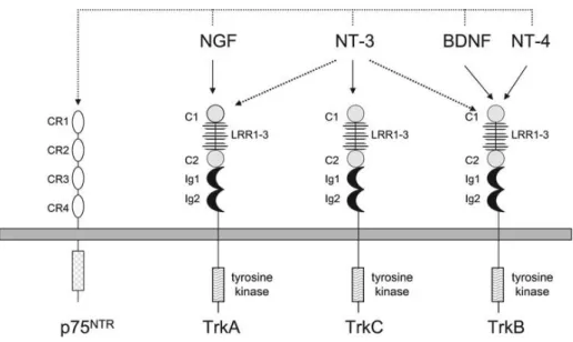
neurotrophin-sensitive neurons. During development, neurotrophins are transported in a retrograde manner from their place of origin into the nerve terminal that innervates it and then up through the axon and into the cell body (Barde et al. 1989). This ensures the survival of the neurons that establish this retrograde flow of neurotrophins through the period of selective cell death. The establishment of the retrograde neurotrophin flow is continued through the lifetime of the neuron, therefore maintaining the functional and differentiated state of the cell (Barde et al. 1989). There are four main types of neurotrophins: Nerve Growth Factor (NGF), Brain-derived Neurotrophic Factor (BDNF), Neurotrophin-3 (NT-3) and Neurotrophin-4 (NT-4). Each neurotrophin can signalize through two main types of receptors: Tropomyosin-receptor-kinase (Trk) and p75 NTR receptors, leading to the activation of various metabolic pathways that can promote the survival or the death of the cells, according to the circumstances in which these pathways are activated (Chao 2003).
In mammals, the Trk family of tyrosine kinase receptors consists of an extracellular domain of a cysteine-rich cluster followed by three leucine-rich repeats, another cysteine-rich cluster and two Ig-like domains (Skaper 2012). Each Trk receptor possesses a single transdomain region that ends in a cytoplasmic domain containing a tyrosine-kinase domain surrounded by several residues of tyrosine, which serve as phosphorylation-dependent docking sites for cytoplasmic adaptors and enzymes (Skaper 2012). The binding of a neurotrophin to its receptor triggers the latters dimerization, which results in the activation of the receptor by transphosphorylation of the cytoplasmic domain kinases. Each neurotrophin possesses specificity of its action due to their selective binding to specific receptors: NGF binds to TrkA (Kaplan et al. 1991) (Rüdiger Klein et al. 1991), TrkB binds BDNF and NT-4 with high affinity (R Klein et al. 1991; Squinto et al. 1991), and TrkC binds NT-3 (Lamballe et al. 1991). NT-3 can also interact, albeit with less efficiency, with TrkA and TrkB (Squinto et al. 1991; Ip et al. 1993) It is important to note that pro-neurotrophins (the immature forms of neurotrophins) are more selective ligands for the p75 receptor than their mature forms (Lee et al. 2001) and are more effective at inducing p75-dependent apoptosis (Lee et al. 2001; M. S. Beattie et al. 2002) This is indicative that pro-neurotrophins preferentially activate p75 to mediate apoptosis, while on the other hand mature neurotrophins selectively activate Trk receptors to promote cell survival (Chao 2003).
12
The cytoplasmic domains of Trk receptors contain several additional tyrosine residues that also serve as substrates for phosphorylation by each receptor’s tyrosine kinase. When phosphorylated, these tyrosine residues form the core of the binding sites that serve as a scaffolding for the recruitment of various proteins and enzymes, which leads to the propagation of the neurotrophin signal (Segal & Greenberg 1996). The phosphotyrosines and their surrounding aminoacid residues present within the activated Trk receptor create binding sites for proteins with phosphotyrosine-binding or Src-homology 2 domains (Skaper 2012), which enables the activation of two separate intracellular signalling pathways. The binding of Shc to the Trk receptor leads to neuronal survival, through the increase of phosphatidylinositol 3-kinase (PI3K) and Akt (protein kinase B) activities (Skaper 2012). The phosphorylation of Shc by the activated Trk tyrosine kinase domain also lead to an increase of Ras and ERK, which in turn induces transcriptional events, like the activation of CREB. CREB will in turn have effects over the cell cycle, neurite outgrowth and synaptic plasticity (Lonze & Ginty 2002). In addition, phospholipase Cγ (PLC-γ) also binds to the activated Trk receptors, initiating an intracellular signalling cascade that leads to the release of inositol phosphates and ends up activating protein kinase C (PKC) (Chao 2003).
p75 NTR receptors do not contain a catalytic domain. However, these receptors still interact with several proteins that relay important signals for the regulation of neuronal cell survival, differentiation and synaptic plasticity (Chao 2003). The activation of the p75 NTR receptor results in the activation of nuclear factor κB (NF-κB) and Jun kinase (JNK), as well as other signalling pathways, with p75 activation being correlated directly with the promotion of programmed cell death (Hempstead 2002). This might provide a means for the selection of neurons during development, and also to refine correct target innervation. Apoptosis by p75 activation is also manifested after seizure or inflammation; and also in injuries to the spinal cord, leading to oligodendrocyte death (Dowling et al. 1999; Roux et al. 1999; M. S. Beattie et al. 2002). p75 NTR’s apoptotic function is accompanied by an increase in Rac and JNK activities, which are essential for NGF-dependent death (Harrington et al. 2002). Despite this, there is also some evidence that p75 might mediate non-apoptotic or survival responses like other tumour necrosis factor receptors (Khursigara et al. 2001; DeFreitas et al. 2001).
The function of the Trk receptor can be modulated by the p75 receptor on several levels, through its actions over ligand binding (in which there is a promotion of axonal
13
growth and target innervation that culminates in the promotion of the receptor’s accessibility to neurotrophins) and endocytosis and retrograde transport to membrane compartments where the engagement of neurotrophins to their Trk receptors can eventually promote signalling (Skaper 2012). One example of this is the p75 NTR inhibiting action over the activation of Trk receptors by non-preferred neurotrophins both in vivo and in vitro (Benedetti et al. 1993; Bibel et al. 1999). Another interesting thing to note is that there is evidence that neurons can respond to a wide variety of extracellular stimuli through the transactivation of the receptor’s tyrosine kinases by G-protein coupled receptors (GPCRs) in situations where their ligand is absent (Daub et al. 1996; Luttrell et al. 1999), like in the case of TrkA and TrkB (Lee & Chao 2001; Lee et al. 2002). An example of this is the capability of adenosine and pituitary adenylate cyclase-activating peptide to trigger Trk receptor activity by their binding with their GPCRs, leading to a stimulation of protein kinase B (Akt) activity that in turn leads to an increase in neuronal survival.
Fig. 1 – Neurotrophins and their respective receptors. Adapted from: Stephen D. Skaper (ed.), Neurotrophic Factors: Methods and
Protocols, Methods in Molecular Biology, vol. 846
BDNF is a neurotrophin that has a very important neurophysiological role, due to its involvement in the regulation of the development of the neuronal circuitry (leading to the survival and differentiation of neural stem cells (Bibel & Barde 2000) and of differentiated mature neurons (Huang & Reichardt 2003). It is also involved in the differentiation and growth of axons and dendrites (Park & Poo 2012) and also in the
14
formation and maturation of synapses (Vicario-Abejón et al. 2002). Besides that, BDNF has a role in the regulation of mature neuronal circuits (acting by modulation of the synaptic efficacy (Lu & Figurov n.d.) and of LTP and LTD (Figurov et al. 1996)).
BDNF has a definitive role in enhancing synaptic efficacy and plasticity, being able to produce rapid increases in synaptic strength in nerve-muscle synapses, increases in excitatory post-synaptic currents in hippocampal neurons and also rapid and long-lasting enhancements of synaptic strength by LTP in the hippocampal neurons (Lohof et al. 1993; Kang & Schuman 1995; Levine et al. 1995). This is proven through the fact that BDNF-deficient mice show an impairment of LTP in hippocampal slices (Korte et al. 1995), but also through the evidence that the exogenous administration of BDNF can rescue normal LTP activity in these cases (Patterson et al. 1996).
Figurov et al. 1996 were the first to demonstrate that there was a facilitating action of LTP when BDNF was administered to hippocampal slices, due to its activation of TrkB receptors, which elicits cellular responses and changes both at a presynaptic and at a postsynaptic level. At a presynaptic level, it is observed that the activation of TrkB receptors by BDNF leads to a modification in the release of neurotransmitters by the neuron, which culminates in an increase in the quantity of neurotransmitters present in the synaptic cleft, therefor potentiating neuronal transmission (Poo 2001). On another hand, at a postsynaptic level, the activation of TrkB receptors by BDNF acts in three different aspects: there is an activation of the Fyn protein, which will lead to an increase in the probability of the opening of the NMDA receptor ionic channels (Levine et al. 1998); there is also an increase of the influx of calcium and sodium ions into the intracellular space of the postsynaptic neuron, through the TRPC proteins (transient receptor potential channel) (Li et al. 1999); and finally, there is an increase of the modulation of the expression and trafficking of AMPA receptors to the active site of the postsynaptic neuron (Caldeira et al. 2007). All of these mechanisms will trigger an increase of the excitability of the postsynaptic neuron, which explains the potentiating effect of BDNF over the magnitude of LTP.
It has been shown that BDNF-induced potentiation of LTP is dependent on the activation of the NMDA receptors and of the ERK and CREB pathways. This was observed by Messaoudi E et al in 2002, where their group described that although the potentiation of LTP by BDNF was associated with an increase in ERK and CREB phosphorylation, it was inhibited by local infusion of MEK inhibitors, and that in such a
15
situation there was little to no evidence of the presence of activated ERK or CREB. (Messaoudi et al. 2002). As such, there is evidence to believe that the binding of BDNF to TrkB receptors triggers the phosphorylation of the receptor’s tyrosine kinase domain and also of ERK, which in turn will trigger the various modification in the neurons that culminate in an increase of the synapse’s strength.
Adenosine
Adenosine and ATP are two other substances that play a critical role over signalling pathways in the nervous system, both at an intracellular and extracellular level. ATP can be released by neurons and astrocytes in several brain areas, serving as a regular neurotransmitter. Adenosine, on the other hand, is neither stored nor released like other classical neurotransmitters (Burnstock 2007), reaching the extracellular space through the use of several non-exocytotic mechanisms, like the conversion of ATP to adenosine in the synaptic cleft by action of ectonucleotidase pathway (which is the predominant mechanism related with high frequency neuronal firing and astrocytic stimulation (Fredholm et al. 2005); and also through the action of the adenosine transporter proteins from the equilibrative nucleoside transporter (ENT) and concentrative nucleoside transporter (CNT) families (Zhong et al. 2017).
Adenosine exerts its action through the modulation of neuromodulators and neurotransmitters released during synaptic transmission, thus leading to the fine tuning of neuronal communication. Because of this, adenosine plays a key role in several physiological and pathological events, like sleep and epilepsy, respectively (Sebastiao & Ribeiro 2009). It is important to note that the actions of adenosine are mediated by the activation of G-protein-coupled seven transmembrane domain receptors, which are expressed by both neurons and glial cells (Fields & Burnstock 2006). Up to this point, four different adenosine receptors have been identified: A1 and A3 receptors, which are negatively coupled to adenylyl cyclase through their Gi/o protein α-subunits, leading to inhibitory responses; and A2A and A2B receptors, which are positively coupled to adenylyl cyclase through their Gs protein subunits and lead to excitatory responses. (Fredholm et al. 2005).
16
There is evidence that all four kinds of adenosine receptors are expressed in astrocytes (Björklund et al. 2008). There is also some evidence of some actions that are triggered due to the activation of these receptors in these cells: on one hand, the activation of A1 receptors leads to a reduction of the proliferation rate of astrocytes, in culture (Boison et al. 2010). The activation of these receptors is also linked to the protection of astrocytes from damage and death (Björklund et al. 2008) as well as to the inhibition of GABA uptake (Cristóvão-Ferreira et al. 2013). On another hand, the activation of astrocytic A2A receptors is associated with important functions such as the increase of extracellular levels of glutamate (Matos et al. 2012), the increase of the activation and proliferation of astrocytes (Boison et al. 2010) and the enhancement of GABA uptake (Cristóvão-Ferreira et al. 2013).
There is also evidence that this potentiating effect of BDNF over the magnitude of LTP is dependent of the activation of A2A adenosine receptors, through the activation
of the PKA (protein kinase A) signal transduction pathway, by the stimulation of the cAMP production by adenililcyclase (Fontinha et al. 2008).
Another thing to note is the capability of adenosine to actually activate Trk receptors while in the absence of neurotrophins, through its activation of GPCRs. This has been described previously in the literature, where it was seen that the administration of adenosine led to an increase in Trk receptor autophosphorylation in hippocampal neurons, due to the activation of A2A receptors (Lee & Chao 2001). It has also been
described that this increase of Trk activity could be inhibited by the administration of PP1or K-252a (protein kinase inhibitors), which further indicates the presence of a cross-talk between adenosine receptors and Trk receptors (Chao 2003).
These GPCR transactivation events are unique: The activation of Trk receptors by adenosine requires a long period of time of about 1-2 hours, and this leads to an activation of PI3K and Akt, which in turn culminates in enhanced cell survival, even in the absence of neurotrophins (Chao 2003). Because of this, it has been hypothesized that the transactivation of Trk receptors by GPCRs might be the reason why neuronal survival in the CNS is not adversely affected by the absence of neurotrophins, since the ligands for the GPCRs end up compensating the lack of neurotrophins by providing a survival function through a neurotrophin-receptors signalling pathway (Chao 2003). There is also
17
some indication that transactivated receptor signalling might have a role in the regulation of ion channels (Chao 2003).
Finally, it is important to note that the presence of mutations in components of the adenosine signalling pathway give rise to behavioural problems in learning and memory (Kotecha et al. 2002; Otto et al. 2001), as well as increased aggression (Ledent et al. 1997), similar to what is observed when there are mutations in the BDNF and TrkB receptor genes (Lyons et al. 1999; Kernie et al. 2000; Patterson et al. 1996; Minichiello et al. 1999; Xu et al. 2000). This seems to indicate that adenosine can work in parallel with neurotrophin action and also that Trk receptors might act as convergence points for signals that originate from other receptor systems.
Role of astrocytes over the potentiation of LTP triggered by BDNF
Taking all of the above into account, both astrocytes and BDNF seem to have an active role over the procedures of induction and potentiation of LTP. However, the possible role of astrocytes over the potentiating effect of BDNF over the magnitude of LTP has only been recently verified. Based on results obtained by Vaz and collaborators, still not published, it is possible to conclude that the reduction of the astrocytic metabolism by application of fluorocitrate (a drug that selectively reduces the metabolism of astrocytes, by inhibition of the aconitase enzyme of the tricarboxylic acid cycle, TCA), results in a loss of the excitatory effect of BDNF over LTP, in the hippocampus. This is indicative that the entirety of the process of LTP potentiation by BDNF will also be under the control of astrocytes.
2. AIMS
Even though there are several studies about the potentiation of LTP magnitude by BDNF and despite the fact that there is already evidence of the underlying mechanisms of this phenomenon, little is still known about the relationship that seems to exist between the effect of BDNF over LTP and the neuromodulation performed by astrocytes.
18
- to study the role of SNARE-dependant gliotransmitter release done by astrocytes in the modulation of synaptic plasticity by BDNF
- to investigate the influence of specific gliotransmitters, namely ATP/adenosine, over this potentiating effect of BDNF over the magnitude of LTP.
3. TECHNIQUES
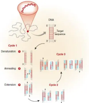
3.1. Polymerase Chain Reaction (PCR)
Biological science suffered an advent with the discovery of the PCR technique, since it offered the possibility to detect and produce large amounts of DNA (Mullis 1990). Nowadays, the PCR technique is widely used by clinicians and researchers to diagnose diseases and pathogens, to clone and sequence genes and to carry out sophisticated quantitative and genomic studies in a rapid and sensitive manner (Garibyan & Avashia 2013). The PCR technique is a simple but elegant enzymatic essay that enables the amplification of a specific DNA fragment from a complex pool of DNA (Garibyan & Avashia 2013). PCR can be performed by obtaining DNA from a wide variety of tissues such as peripheral blood, skin, hair and saliva. Only trace amounts of DNA are needed to generate a sufficient number of copies by using this technique, which can then be analysed using conventional laboratory methods (Garibyan & Avashia 2013).
Each PCR assay requires the presence of template DNA, primers, nucleotides (the four bases: adenine, thymine, cytosine, and guanine (A, T, C, G) that are found in DNA.) and DNA polymerase. The DNA polymerase is the key enzyme that will lead to the linking of the nucleotides to form the finished PCR product. The primers on the other hand are short DNA fragments, with a defined sequence that is complementary to the target DNA, that end up being detected by the DNA polymerase and are then amplified (Garibyan & Avashia 2013). The primers are therefore the agents through which the DNA product that is to be amplified is specified c.
Each of the previous mentioned reagents are mixed in a test tube and then placed in a machine the enables repeated cycles of DNA amplification to occur. This machine is
19
normally a thermocycler, possessing a thermal block with holes into which the test tubes holding the PCR mixture (including the sample DNA) can be inserted. The thermocycler can then be programmed to raise and lower the temperature of the thermal block in discrete and precise steps (Weier HU; Gray JW. 1988): this happens in a predetermined way, with the reaction solution first being heated above the melting point of the complementary DNA strands of the target DNA (which allows the strands to separate) so that there is denaturation (Garibyan & Avashia 2013). Afterwards, the temperature is lowered, which allows the specific primers to bind to the DNA segments (hybridization or annealing) that they complement (Garibyan & Avashia 2013). Finally, the temperature is raised again, which permits the DNA polymerase to extend the primers by adding nucleotides to the developing DNA strand (Garibyan & Avashia 2013). With each repetition of these three steps, the number of copied DNA molecules doubles (Garibyan & Avashia 2013).