This article was published in International Journal of Experimental Pathology, 95, 191-198, 2014 http://dx.doi.org/10.1111/iep.12082
Cytokeratin 7/19 expression in N-Diethylnitrosamine-induced mouse hepatocellular lesions: implications for histogenesis
Nuno P. Santos1,2, Paula A. Oliveira1,3, Regina Arantes-Rodrigues1,2, Ana I. Faustino-Rocha1, Aura Colaço1,2, Carlos Lopes4,5, Rui M. Gil da Costa4,6
1Veterinary Sciences Department, University of Trás-os-Montes and Alto Douro, UTAD, www.utad.pt, 5001-801, Vila Real, Portugal
2Veterinary and Animal Science Research Centre (CECAV), Veterinary Science Department, University of Trás-os-Montes and Alto Douro, UTAD, www.utad.pt, 5001-801, Vila Real, Portugal 3Centre for the Research and Technology of Agro-Environmental and Biological Sciences (CITAB), University of Trás-os-Montes and Alto Douro, UTAD, www.utad.pt, 5001-911, Vila Real, Portugal 4Experimental Pathology and Therapeutics Group, CI-IPOP, Portuguese Institute of Oncology, Rua Dr. António Bernardino de Almeida, 4200-072 Porto, Portugal
5Pathology and Molecular Immunology Department, Abel Salazar Institute for Biomedical Sciences -University of Porto (ICBAS-UP), Rua de Jorge Viterbo Ferreira n.º 228, 4050-313 Porto, Portugal 6Laboratory for Process Engineering, Environment, Biotechnology and Energy (LEPABE),
Chemical Engineering Department, Faculty of Engineering, University of Porto (FEUP), Rua Dr. Roberto Frias, 4200-465 Porto, Portugal
Corresponding author: Paula A. Oliveira, Veterinary Sciences Department, University of Trás-os- Montes and Alto Douro, UTAD, www.utad.pt,5001-801, Vila Real, Portugal.
Abstract
Hepatocellular carcinoma (HCC) is a common malignancy with poor clinical outcome, whose histogenesis is the subject of intense debate. Specifically, expression of cytokeratins (CKs) 7 and 19, associated with aggressive biological behaviour, is proposed to reflect a possible progenitor-cell origin or tumour de-differentiation towards a primitive phenotype. This work addresses that problem by studying CKs 7 and 19 expression in N-diethylnitrosamine (DEN)-induced mouse HCCs. ICR mice were divided into 6 DEN-exposed and 6 matched control groups. Samples were taken from each group at consecutive time points. Hyperplastic foci (13 lesions) occurred at 29 to
40 weeks (groups 8, 10 and 12) with diffuse dysplastic areas (19 lesions) and with one hepatocellular adenoma (HCA) (at 29 weeks). HCCs (4 lesions) were observed 40 weeks after the first DEN administration (group 12). CKs 7 and 19 showed identical expression patterns and located to large, mature hepatocytes, isolated or in small clusters. Hyperplastic foci and the single HCA were consistently negative for both markers while dysplastic areas and HCCs were positive. These results support the hypothesis that CKs 7 and 19 expression in hepatocellular malignancies result from a de-differentiation process rather than from a possible progenitor cell origin.
Introduction
Liver cancer is a major public health problem, with approximately 748 000 new cases diagnosed yearly (Ferlay et al. 2010). The majority of these cases are histologically classified as hepatocellular carcinomas (HCC) and occur mostly in older males in developing countries, due to the distribution of the risk factors (Motola-Kuba et al. 2006). HCCs are believed to emerge through a process of multi-step carcinogenesis, but their histogenesis is still a matter of discussion. One long-established view is that normal mature hepatocytes undergo neoplastic transformation to originate pre- malignant lesions and that early-stage HCCs arise within such lesions, giving then place to high- grade carcinomas, originating a characteristic nodule-within-nodule structure (Sakamoto et al. 1991). However, it has also been hypothesized that a set of HCCs may originate from hepatic stem cells (Durnez et al. 2006; Lee et al. 2006). This hypothesis is supported by the expression patterns of several hepatic differentiation markers. Cytokeratins (CKs) 8 and 18 are equally expressed by hepatocytes and bile duct cells, while cytokeratin (CK) 7 and CK19 are considered as markers of cholangiocytic differentiation (Strnad et al. 2008). Nevertheless, it has been observed that some HCCs express CKs 7 and 19, as well as c-kit that is a hepatic stem cell marker (Kim et al. 2004), displaying an intermediate hepatocellular-cholangiocytic progenitor cell-like phenotype. Furthermore, expression of CK19 has been associated with poor prognosis (Uenishi et al. 2003; Ding et al. 2004; Durnez et al.
2006). However, Shibuya and collaborators (Shibuya et al. 2011) have recently put forward another
contribution to this debate by proposing that malignant hepatocytes may trans-differentiate (or de-differentiate) into immature stem cell-like cells. An experimental carcinogenesis experiment may be useful to address this problem, by studying the expression of differentiation markers over time and thus determining whether tumours result from the clonal expansion of progenitor cells or de-differentiate to acquire a progenitor cell-like phenotype. Over the years, several experimental rodent models have been used to study the pathogenesis of HCC, particularly mice and rats (Heindryckx et al. 2009). Chemically-induced
models using N-diethylnitrosamine (DEN) are long-established and among the most frequently used (Goldfarb et al. 1983). DEN belongs to the N-nitroso group and is widely known for its ability to induce tumours in a multiplicity of organs, including the liver, the stomach, the lung and the haematopoietic organs. DEN is a genotoxic, DNA-alkylating carcinogen that requires metabolic activation (Hecht et al. 1989). The first biotransformation step is hydroxylation to - hydroxylnitrosamine, mediated by cytochrome P450, which shows its highest activity in centrilobular hepatocytes (Kang et al. 2007). Upon cleavage of acetaldehyde an ethyldiazonium ion is formed, reacting with DNA and other nucleophiles, including DNA repair enzymes. DEN- induced mouse tumours often harbour H-ras activating mutations (Stahl et al. 2005). While activation of the Ras pathway is a common event in human hepatocarcinogenesis (Calvisi et al. 2006), mutations of the H-ras proto-oncogene itself are less frequent and associated with a more aggressive biological behaviour (Wang et al. 2001). Accordingly, DEN-induced tumours are thought to closely mimic the more aggressive human HCCs.
The necessary development time after a single DEN administration for HCC depends on several factors, namely administered dose, administration route, age, sex and strain of the animals (Vesselinovitch & Mihailovich,1983; Vesselinovitch et al. 1984; Heindryckx et al. 2009). The present work employs a multiple dose DEN-induced mouse HCC model to study the expression of CKs 7 and 19 in pre-neoplastic and neoplastic hepatic lesions.
Materials and Methods
Animals and experimental conditions
One hundred and twenty male ICR mice with five weeks of age (Harlan, Barcelona) were housed at the bioterium of the University of Trás-os-Montes and Alto Douro, according to National (Portaria
submitted to one week of quarantine Mice were identified with ear cuts and kept in ventilated facilities, under controlled conditions of temperature (23±2ºC), light-dark cycle (12h light/12h dark) and relative humidity (50±10%), using hardwood bedding. Standard diet (Global Diet 2014, Harlan, Barcelona) and water were provided ad libitum.
Experimental procedures
At six weeks of age, all mice were randomly divided into twelve groups. Animals from groups 1, 3, 5, 7, 9 and 11 (controls, CTL) were intraperitoneally injected with a saline solution weekly for eight consecutive weeks (administration route - i.p. once a week), whereas animals from groups 2, 4, 6, 8, 10 and 12 (exposed groups) received DEN (Sigma-Aldrich) at a dose of 35mg per Kg of body weight (Heindryckx et al., 2012). Throughout the experimental protocol (Figure 1), animals were daily monitored for signs of distress. Food and water intake, as well as animal weights, were weekly recorded. Animals from groups 1 and 2, 3 and 4, 5 and 6, 7 and 8, 9 and 10 were euthanized at 8, 15, 22, 29 and 36 weeks (W) after the first administration of DEN, respectively, by means of a lethal dose of pentobarbital sodium, injected intraperitoneally. Euthanasia of animals from groups
11 (40W CTL) and 12 (40W exposed) was performed at 40 weeks after the DEN first administration, because some mice of the DEN-exposed group exhibited external signs of distress.
Sample collection and histological processing
A complete necropsy was carried out in all animals in order to check for tumoral masses. The liver, brain, heart, lungs, spleen, stomach, intestine, pancreas, kidneys, adrenal glands, testes, gastrocnemius muscles and a dorsal skin sample were collected into 10% neutral buffered formalin and fixated for 48 hours. Posteriorly the samples were routinely processed and paraffin-embedded.
Histological sections (2µm-thick) were obtained and stained with haematoxylin and eosin (H&E) for examination on light microscopy. The presence of haemorrhage and other vascular disorders, bile cysts, necrosis, apoptosis, nuclear and cytoplasmic changes/accumulations were noted. Proliferative hepatic lesions were classified as hyperplastic foci, hepatocellular adenoma (HCA) or HCC (Deschl et al. 2001). Furthermore, multifocal to regionally extensive, poorly-delimited dysplastic areas, showing loss or distortion of lobular architecture, irregular hepatocyte plates, moderate cell atypia and mitotic activity were classified as diffuse dysplasia. Immunohistochemical detection of CK7 and CK19 was performed on 2µm-thick sections using a standard peroxidase protocol. Heat-induced antigen retrieval was performed employing citrate buffer in a microwave for 15 minutes. Monoclonal anti-CK7 (RCK105) and anti-CK19 (A-3) were acquired from Santa Cruz Biotechnology, diluted in phosphate-buffered saline (1:500) and incubated with the tissue sections for one hour at room temperature. Immunoreactivity was detected using diaminobenzidine. Negative controls were obtained by omitting the primary antibody. Bile duct epithelial cells were used as positive internal controls.
Statistical analysis
Data were expressed as mean±standard deviation (SD) and compared by one - way analysis of variance (ANOVA) followed by Tukey’s multiple comparison test at the 5% significant level (p<0.05). All tests were performed using the GraphPad Prism, version 5.01 (GraphPad Software, Inc., La Jolla, CA, USA).
Results
The mortality rate in the DEN group during the experimental protocol was 6.67% (4 out of 60 animals, one in group 2, another in group 10 and two in group 12). Despite the enrichment of cage
environment, male competitive behaviour was noticed, resulting in punctual injuries and focal loss of hair (barbering behavior).
Regarding animal growth, the ponderal homogeneity index (iPH) and ponderal gain (PG) for control and DEN-exposed groups were calculated (Table 1). Drinking-water consumption was lower in DEN-treated animals compared with control groups (not significant). Food intake was also lower in DEN-exposed groups. Statistically significant differences between groups were noticed during the assay at 29 and 36 weeks after the first DEN exposure, regarding final body weight (p<0.05).
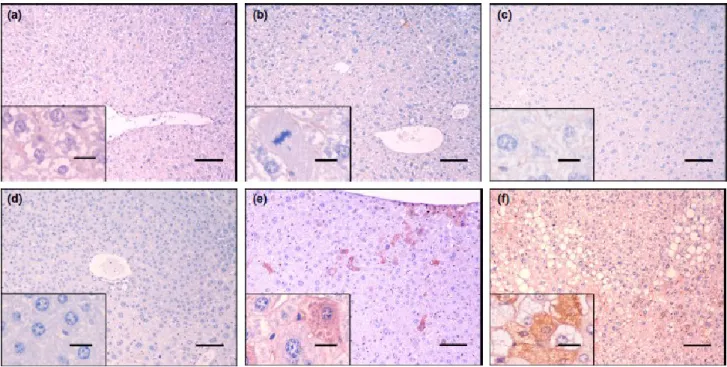
Control animals (Figure 3) did not show any changes to the normal hepatic architecture at any time point of the assay. Animals from group 2 (8W exposed) revealed acute toxic lesions, such as hepatocellular hydropic degeneration (Figure 4a) and necrosis. Animals from groups 4 and 6 (15W and 22W, exposed) revealed the same kind of lesions (Figures 4b and c, respectively). No alterations to normal liver architecture were present. At the same time points, occasional cytoplasmic immunostaining for CKs 7 (Figure 5a, b and c, respectively) and 19 (Figure 6a, b and c, respectively) was observed in the biliary epithelium and hepatocytes surrounding necrotic areas, in both exposed and control animals.
Nodular hepatic lesions occurred from twenty-nine weeks after the first DEN administration and were often visible macroscopically (Figure 2). The first hyperplastic foci were observed in group 8 (29W exposed). Such foci were clearly delimited from the adjacent parenchyma, were composed of tight hepatocellular plates which compressed the hepatic sinusoids and showed distinct, basophilic or mixed type, cytoplasmic coloration (Figure 4d). Four out of 10 animals (40%) showed hepatic hyperplastic foci (six nodules, Table 2). One animal showing 2 hyperplastic foci also exhibited dysplastic changes in a distant and poorly-delimited area classified as diffuse dysplasia (10%) as well as a larger, well-defined nodule classified as a HCA (10). Dysplastic lesions (Figure 4e) were
composed of hepatocytes arranged in irregular and variably-oriented plates which disrupted normal liver architecture lacking the formation of distinguishable nodules. Hepatocytes were moderately pleomorphic showing significant variations in size and occasional cytoplasmic vacuoles. Pseudonucleoli and mitotic figures were present (1 per high-power field). The HCA was a large (1.0 cm in diameter) nodule which compressed the adjacent parenchyma, composed of regularly- oriented hepatocellular trabeculae. Hepatocytes were smaller than adjacent cells, displaying basophilic cytoplasm and large, round nuclei with coarse chromatin and occasional mitotic figures (up to 1 mitotic figure per high power field). Both dysplastic and adenomatous areas showed scattered immunopositive cells for CK7 and CK19, isolated or in groups of two to ten large mature hepatocytes (Figures 5e and 6e, respectively), while hyperplastic foci (Figures 5d and 6d) were consistently negative. Immunopositive cells in dysplastic areas were often located in the subcapsular region or in centrilobular areas, around the central vein. Immunostaining was diffusely distributed in the cytoplasm and was moderate to strong. Antibodies against CK7 and CK19 consistently stained the same areas, with similar distribution and intensity. All animals from group 10 (36W exposed) showed extensive areas of diffuse hepatic dysplasia (10 out of 10 animals, 100%). Hyperplastic foci were recorded less frequently (2 out of 10 animals, 20%, a total of 3 nodules). As observed in group 8 (29W exposed), dysplastic areas showed scattered cells exhibiting moderate to strong immunostaining for CK7 and CK19, while hyperplastic foci remained consistently negative. Dysplastic areas also showed a sinusoidal accumulation of erythroblasts distributed in small foci of 2 to 20 cells. Occasionally, myeloid precursor cells were found in association with such foci.
Four out of 8 (50%) animals from group 12 (40W exposed) exhibited large hepatic, soft, grey to light brown or occasionally haemorrhagic nodules, measuring up to 1.0 cm in diameter. Histologically, these nodules were classified as HCCs. These lesions arose within diffuse dysplastic areas and showed invasive growth and a multifocal distribution. HCCs (Figure 4f) were composed
of moderately to highly pleomorphic cells disposed in solid nests, trabeculae or, multifocally, pseudo-acinar structures, supported by a loose fibrovascular stroma. Neoplastic cells showed moderate nuclear pleomorphism, a prominent nucleolus and up to four mitotic figures per high power field. Cells varied in size, were often vacuolated and frequently assumed a signet-ring morphology. Multifocally, variably sized intracytoplasmic hyaline bodies were present. Scattered cells or cell groups exhibiting moderate to strong cytoplasmic immunostaining for CK7 and CK19 were present (Figures 5f and 6f). Immunopositive hepatocytes were mostly large, little- or non- vacuolated cells, with vacuolated cells showing a fainter immunostaining. Immunostaining for both CKs was consistently present in the same areas. Groups of erythroblasts were present in all HCCs and were larger than those observed in dysplastic areas. In one instance, fully-differentiated bone marrow developed inside a carcinomatous nodule, with myeloid, erythroid, lymphoid and platelet precursors distributed between bone lamellae and adipocytes (Figure 3f). All animals from group 12 (40W exposed) showed diffuse dysplastic areas (8 out of 8 animals, 100%) and 3 out 8 (37.5%) showed hyperplastic foci (4 lesions in total). The immunostaining patterns observed in these lesions were identical to those observed in previous groups.
Discussion
The histogenesis of HCC is the subject of an intense debate. In fact, since some tumour subtypes show different biological behaviour, refining their histopathological classification has important consequences for patient treatment and prognosis (Ding et al. 2004; Durnez et al. 2006). HCCs with a progenitor cell-like phenotype express certain characteristic markers of cholangiocytes (Ding et al. 2004; Durnez et al. 2006). While CKs 8 and 18 are expressed by both hepatocytes and epithelial bile duct cells, CK7 and CK19 are used as markers of cholangiocytic differentiation (Strnad et al. 2008). In the present work, the expression of CKs 7 and 19 was studied in a murine model of chemical hepatocarcinogenesis, in non-neoplastic, pre-neoplastic and neoplastic tissues.
Immunopositive cells were not observed in control tissues in the early exposed groups, apart from bile duct epithelial cells and hepatocytes bordering necrotic foci. These were not small, scattered cells resembling hepatic progenitor cells but a continuous lining of large, mature hepatocytes. It seems likely that these hepatocytes are responding to liver damage and have suffered corresponding cytoskeletal rearrangements involving CKs 7 and 19 expression - a similar phenomenon was previously described in alcoholic steatohepatitis and cholestatic liver diseases (Vaneyken et al. 1989; Zatloukal et al. 2004). Activation of hepatic progenitor cells in the early stages following DEN exposure was not detected immunohistochemically. Hyperplastic foci are considered early pre-neoplastic changes in the process of multi-step hepatocarcinogenesis. Overexpression of CKs 8 and 18 has been described in hyperplastic foci in DEN-exposed RasH2, B6C3F1 and C57BL/6 mice (Kakehashi et al. 2010; Kawai et al. 2010; Kushida et al. 2011). These authors agree that CKs 8 and 18-positive foci seem to be reliable markers for pre-malignant lesions in those mouse strains. Expression of CK19 has also been recently described in DEN-induced hyperplastic foci (especially in eosinophilic foci) and HCAs in C57BL/6 mice (Kushida et al. 2011). Andersen et al. (Anderson et al. 2010) also associated CK19 expression with malignancy and with malignant progression of pre-neoplastic liver lesions in a rat model.
In the present study, immunopostivity for CKs 7 and 19 was always coincident and distributed in small cell clusters or isolated cells, in a pattern resembling that described by Shibuya et al. (Shibuya et al. 2011) and not forming nodules as described by Kushida et al. (Kushida et al. 2011). However, in contrast with these last author’s findings, hyperplastic nodules were consistently negative for both CK7 and CK 19. This discrepancy must be regarded with caution due to the low number of hyperplastic foci detected in the present study and the fact that these were basophilic or mixed instead of eosinophilic. On the other hand, immunopositive hepatocytes were found outside hyperplastic foci, scattered through diffuse dysplastic areas, and especially in subcapsular and centrilobular areas
hepatocytes did not resemble hepatic progenitor cells, but were large, mature hepatocytes with abundant cytoplasm, a coarse nuclear chromatin pattern and, frequently, a prominent nucleolus, in agreement with the results of Shibuya et al. (Shibuya et al. 2011). A similar pattern was observed in all HCCs (although there were only four of these). The only diagnosed HCA showed an identical CK7/CK19 expression pattern as well. These results support the association of CK7 and CK19 expression with hepatocellular malignancy in the mouse DEN-induced hepatocarcinogenesis model. Interestingly, immunopositivity for CKs 7 and 19 coincides with the presence of extramedullary haematopoiesis, consisting first of scattered groups of erythroblasts in dysplastic areas and then, of bone marrow foci in HCCs. Considering that the immature mouse liver is, physiologically, a haematopoietic organ, this finding provides yet another indication of the primitive or immature phenotype of these malignancies. Furthermore, bearing in mind that: a) all observed hyperplastic foci were CK7/CK19-negative, b) positive cells occurred isolated or in small clusters, rather than in nodules and c) positive cells showed hepatocellular rather than progenitor cell morphology, the present results give additional support to the hypothesis that CKs 7 and 19 expression in HCCs may result from de-differentiation of mature hepatocytes This is in line with the findings of Shibuya et al. (2011), but contrasts with results presented by other autors. In fact, Libbrecht et al. (2000) presented results suggesting that hepatic progenitor cells give rise to HCCs via small-cell dysplastic foci. Using an experimental appraoch, Dumble et al. (2002) showed that p53-null hepatic progenitor cells implanted in nude mice did give rise to tumors resembling HCCs. The discrepancy between these findings and our present results appears to lie in the different models used by each research group. Most human HCCs arise from a background of chronic liver disease, with hepatic progenitor cell activation. In our model, DEN preferentially targets centrilobular hepatocytes (not progenitor cells) over a 8-weeks period, with no evidence of early-stage progenitor cell activation. These results thus provide new perspectives on the mechanisms of hepatocarcinogenesis, especially on the contribution of the stem-cell phenotype in the context of DEN-induced carcinogenesis.
Acknowledgements
The authors express their deepest appreciation to J.H. Teixeira and Lígia Lourenço for technical support in preparing some histological and immunohistochemical slides. The Authors gratefully acknowledge the financial support provided to N. Paula-Santos (BD/60280/2009) by the Foundation for Science and Technology (FCT) and Research Unit in Vila Real (POCTI-SFA-3- 616).
References
Anderson J, Loi R., Perra A., Factor V., Ledda-Columbano G., Columbano A. & Thorgeirsson S. (2010) Progenitor-derived hepatocelular carcinoma model in the rat. Hepatology 511401-1409.
Calvisi D F, Ladu S., Gorden A., Farina M., Conner E. A., Lee J. S., Factor V. M. & Thorgeirsson S. S. (2006) Ubiquitous activation of Ras and Jak/Stat pathways in human HCC. Gastroenterology 130(4), 1117-1128.
Deschl U., Cattley R., Harada T., Küttler K., Hailey J., Hartig F., Leblanc B., Marsman D. & Shirai T. International Classification of Rodent Tumors: The Mouse, In: International Classification of Rodent Tumors: The mouse. Mohr, U, Eds., Springer-Verlag, Heidelberg. 2001.
Ding S J, Li Y., Tan Y. X., Jiang M. R., Tian B., Liu Y. K., Shao X. X., Ye S. L., Wu J. R., Zeng R., Wang H. Y., Tang Z. Y. & Xia Q. C. (2004) From proteomic analysis to clinical significance - Overexpression of cytokeratin 19 correlates with hepatocellular carcinoma metastasis. Molecular & Cellular Proteomics 3(1), 73-81.
Dumble M. L., Croager E. J., Yeoh G. C. T., Quail E. A. (2002) Generation and characterization of p53 null transformed hepatic progenitor cells: oval cells give rise to hepaticellular carcinoma. Carcinogenesis 23, 435-445.
Durnez A, Verslype C., Nevens F., Fevery J., Aerts R., Pirenne J., Lesaffre E., Libbrecht L., Desmet V. & Roskams T. (2006) The clinicopathological and prognostic relevance of cytokeratin 7 and 19 expression in hepatocellular carcinoma. A possible progenitor cell origin. Histopathology 49(2), 138-151.
Ferlay J, Shin H. R., Bray F., Forman D., Mathers C. & Parkin D. M. (2010) Estimates of worldwide burden of cancer in 2008: GLOBOCAN 2008. International Journal of Cancer 127(12), 2893-2917.
Goldfarb S, Pugh T. D., Koen H. & He Y. Z. (1983) Preneoplastic and Neoplastic Progression During Hepatocarcinogenesis in Mice Injected with Diethylnitrosamine in Infancy. Environmental Health Perspectives 50(APR), 149-161.
Hecht S S, Mirvish S. S., Gold B., Nagel D. & Magee P. N. (1989) Conference on Advances in the Biology and Chemistry of N-Nitroso and Related-Compounds. Cancer Research 49(5), 1327-1329.
Heindryckx F, Colle I. & Van Vlierberghe H. (2009) Experimental mouse models for hepatocellular carcinoma research. International Journal of Experimental Pathology 90(4), 367-386.
Heindryckx F, Kuchnio A, Casteleyn C, Coulon S, Olievier K, Colle I, Geerts A, Libbrecht L, Carmeliet P, Van Vlierberghe H (2012) Effect of prolyl hydroxylase domain-2 haplodeficiency on the hepatocarcinogenesis in mice. Journal of Hepatology 57, 61-68.
Kakehashi A, Kato A., Inoue M., Ishii N., Okazaki E., Wei M., Tachibana T. & Wanibuchi H. (2010) Cytokeratin 8/18 as a new marker of mouse liver preneoplastic lesions. Toxicology and Applied Pharmacology 242(1), 47-55.
Kang J S, Wanibuchi H., Morimura K., Gonzalez F. J. & Fukushima S. (2007) Role of CYP2E1 in diethyinitrosamine-induced hepatocarcinogenesis in vivo. Cancer Research 67(23), 11141-11146.
Kawai M, Saegusa Y., Kemmochi S., Harada T., Shimamoto K., Shibutani M. & Mitsumori K. (2010) Cytokeratin 8/18 is a Useful Immunohistochemical Marker for Hepatocellular Proliferative Lesions in Mice. Journal of Veterinary Medical Science 72(3), 263-269.
Kim H, Park C., Han K. H., Choi J. S., Kim Y. B., Kim J. K. & Park Y. N. (2004) Primary liver carcinoma of intermediate (hepatocyte-cholangiocyte) phenotype. Journal of Hepatology 40(2), 298-304.
Kushida M, Kamendulis L. M., Peat T. J. & Klaunig J. E. (2011) Dose-Related Induction of Hepatic Preneoplastic Lesions by Diethylnitrosamine in C57BL/6 Mice. Toxicologic Pathology 39(5), 776-786.
Lee J S, Heo J., Libbrecht L., Chu I. S., Kaposi-Novak P., Calvisi D. F., Mikaelyan A., Roberts L. R., Demetris A. J., Sun Z. T., Nevens F., Roskams T. & Thorgeirsson S. S. (2006) A novel prognostic subtype of human hepatocellular carcinoma derived from hepatic progenitor cells. Nature Medicine 12(4), 410-416.
Libbrecht L., Desmet V., Van Damme B., Roskams T. (2000) The imunohistochemical phenotype of dysplastic foci in human liver: correlation with putative progenitor cells. Journal of Hepatology 33, 76-84.
Motola-Kuba D, Zamora-Valdés D., Uribe M. & Méndez-Sánchez N. (2006) Hepatocellular carcinoma. An overview. Annals of Hepatology 5(1), 16-24.
Sakamoto M, Hirohashi S. & Shimosato Y. (1991) Early Stages of Multistep Hepatocarcinogenesis - Adenomatous Hyperplasia and Early Hepatocellular-Carcinoma. Human Pathology 22(2), 172- 178.
Shibuya M, Kondo F., Sano K., Takada T. & Asano T. (2011) Immunohistochemical study of hepatocyte, cholangiocyte and stem cell markers of hepatocellular carcinoma. Journal of Hepato- Biliary-Pancreatic Sciences 18(4), 537-543.
Stahl S, Ittrich C., Marx-Stoelting P., Kohle C., Altug-Teber O., Riess O., Bonin M., Jobst J., Kaiser S., Buchmann A. & Schwarz M. (2005) Genotype-phenotype relationships in hepatocellular tumors from mice and man. Hepatology 42(2), 353-361.
Strnad P, Stumptner C., Zatloukal K. & Denk H. (2008) Intermediate filament cytoskeleton of the liver in health and disease. Histochemistry and Cell Biology 129(6), 735-749.
Uenishi T, Kubo S., Yamamoto T., Shuto T., Ogawa M., Tanaka H., Tanaka S., Kaneda K. & Hirohashi K. (2003) Cytokeratin 19 expression in hepatocellular carcinoma predicts early postoperative recurrence. Cancer Science 94(10), 851-857.
Vaneyken P, Sciot R. & Desmet V. J. (1989) A Cytokeratin Immunohistochemical Study of Cholestatic Liver-Disease - Evidence That Hepatocytes Can Express Bile Duct-Type Cytokeratins. Histopathology 15(2), 125-135.
Vesselinovitch S D, Koka M., Mihailovich N. & Rao K. V. N. (1984) Carcinogenicity of Diethylnitrosamine in Newborn, Infant, and Adult Mice. Journal of Cancer Research and Clinical Oncology 108(1), 60-65.
Vesselinovitch S D & Mihailovich N. (1983) Kinetics of Diethylnitrosamine Hepatocarcinogenesis in the Infant Mouse. Cancer Research 43(9), 4253-4259.
Wang Q, Lin Z. Y. & Feng X. L. (2001) Alterations in metastatic properties of hepatocellular carcinoma cell following H-ras oncogene transfection. World Journal of Gastroenterology 7(3), 335-339.
Zatloukal K, Stumptner C., Fuchsbichler A., Fickert P., Lackner C., Trauner M. & Denk H. (2004) The keratin cytoskeleton in liver diseases. Journal of Pathology 204(4), 367-376.
Figure 3 N-diethylnitrosamine-exposed mouse liver. H&E, 100x, bars = 100 lm. Inserts 400x, bars = 20 lm. (a) 8W animal. Hepatocellular hydropic degeneration. Note intense cytoplasmic vacuolization. (b) 15W animal. Note coagulative necrosis focus. (c) 22W animal. Note nuclear changes with condensed, fuzzy chromatin. (d) 29W animal. Note small, mixed type hyperplastic nodule.(e) 36W animal. Note pleomorphic nuclei with pseudonucleoli over a dysplastic area. (f) 40W animal. Note a pseudo-acinar structure amidst a hepatocellular
Figure 4 N-diethylnitrosamine-exposed mouse liver. Immunostaining for CK7-Mayer’s haematoxylin, 100x, bars = 100 m. Inserts 400x, bars = 20 m. (a) 8W animal. (b) 15W animal. Note mitotic figure. (c) 22W animal. (d) 29W animal. Note hyperplastic nodule on the upper half of the picture. (e) 36W animal. Note scattered, large, mature, immunopositive hepatocytes over a dysplastic area. (f) 40W animal. Note variably stained, mature, immunopositive hepatocytes scattered amidst a hepatocellular carcinoma.
Figure 5 N-diethylnitrosamine-exposed mouse liver. Immunostaining for CK19-Mayer’s haematoxylin, 100x, bars = 100 m. Inserts 400x,bars = 20 m. (a) 8W animal. (b) 15W animal. Note immunopositive biliary epithelium. (c) 22W animal. (d) 29W animal. Note hyperplastic nodule on the lower half of the picture. (e) 36W animal. Note scattered, large, mature, immunopositive hepatocytes over a dysplastic area. (f) 40W animal. Note variably stained, mature, immunopositive hepatocytes scattered amidst a hepatocellular carcinoma.
Figure 6 Macroscopic features of livers from control and N-diethylnitrosamine-exposed ICR mice. (a) 40W control animal. (b) 36W exposed animal. Note hepatomegally with grey discoloration and irregular hepatic surface. (c) 40W exposed animal. Note distortion of hepatic lobes, haemorrhagic foci and multiple, grey nodular
Table 1 - Animals body weights (g) (mean ± SD), ponderal homogeneity index (i PH) and ponderal gain (PG). 8 1-Control 2-DEN 31.16±2.60 31.16±1.57 39.86±3.35 39.64±2.17 2×27.06/(27.06+35.50) = 0.865 2×28.90/(28.90+33.06) = 0.932 (39.86-31.16/39.86)×100 = 21.82 (39.64-31.16/39.64)×100 = 21.39 15 3-Control 30.55±1.77 45.40±4.40 2×28.04/(28.04+33.66) = 0.909 (45.40-30.55/45.40)×100 = 32.70 4-DEN 29.74±1.49 41.88±2.83a 2×27.70/(27.70+32.84) = 0.915 (41.88-29.74/41.88)×100 = 28.98 22 5-Control 30.41±2.49 44.39±4.24 2×27.58/(27.58+35.40) = 0.875 (44.39-30.41/44.39)×100 = 31.49 6-DEN 30.55±2.32 48.18±5.98 2×27.16/(27.16+34.00) = 0.888 (48.18-30.55/48.18)×100 = 36.59 29 7-Control 29.63±1.62 46.01±3.55 2×27.20/(27.20+31.94) = 0.919 (46.01-29.63/46.01)×100 = 35.60 8-DEN 29.84±2.48 44.32±5.81 2×25.62/(25.62+33.86) = 0.861 (44.32-29.84/44.32)×100 = 32.67 36 9-Control 30.70±2.28 51.13±6.19 2×27.84/(27.84+34.80) = 0.888 (51.13-30.70/51.13)×100 = 39.95 10-DEN 29.22±2.33 42.59±7.02b 2×26.04/(26.04+34.02) = 0.867 (42.59-29.22/42.59)×100 = 31.39 40 11-Control 31.47±2.30 50.07±2.41 2×29.84/(29.84+33.10) = 0.948 (50.07-31.47/50.07)×100 = 37.14 12-DEN 30.30±1.78 48.93±7.11 2×28.02/(28.02+33.76) = 0.907 (48.93-30.30/48.93)×100 = 38.07
DEN: ap=0.048, bp=0.016 Statistically different from that of the control group (p<0.05).
Table 2 - Histological incidence of hyperplastic foci, dysplastic lesions, hepatocellular adenomas and carcinomas, and CK7/CK9 immunoexpression among DEN-exposed and control ICR mice.
8 weeks Control Group 1 (n=10) Exposed Group 2 (n=9) 0 0 0 0 0 0 0 0 0 0 0 0 0 0 0 0 22 weeks Control Group 5 (n=10) Exposed Group 6 (n=10) 0 0 0 0 0 0 0 0 0 0 0 0 0 0 0 0 36 weeks Control Group 9 (n=10) Exposed 0 0 0 0 0 0 0 0 Group 10 (n=9) 20% (3 lesions) 0 (10 lesions) 100% 100% (10 lesions) 0 0 0 0
HCA- hepatocellular adenoma, HCC- hepatocellular carcinoma
Hyperplastic foci CK7+/19+ hyperplastic foci Diffuse dysplasia CK7+/19+
diffuse dysplasia HCA
CK7+/19+ HCA HCC CK7+/19+ HCC Sacrifi ce Time points Animal groups
% of animals with lesions (No. lesions per group)
Control Group 3 (n=10) Exposed Group 4 (n=10) 0 0 0 0 0 0 0 0 15 weeks 0 0 0 0 0 0 0 0 29 weeks Control Group 7 (n=10) Exposed Group 8 (n=10) 0 0 0 0 0 0 0 0 40%
(6 lesions) 0 (1 lesion) 10% (1 lesion) 10% (1 lesion) 10% (1 lesion) 10% 0 0
Control Group 11 (n=10) Exposed Group 12 (n=8) 0 0 0 0 0 0 0 0 40 weeks 37.5%