Faculdade de Medicina de Lisboa
The RNA 5' Phosphatase PIR-1 Cooperates with
Dicer to Produce Endogenous Small RNAs and
Suppress Viral Replication in C. elegans
Daniel Marques de Almeida de Melo Chaves
Doutoramento em Ciências Biomédicas
Especialidade em Ciências Biopatológicas
The RNA 5' Phosphatase PIR-1 Cooperates with Dicer to Produce Endogenous Small RNAs and Suppress Viral Replication in C. elegans
Daniel Marques de Almeida de Melo Chaves
Orientadores:
- Professora Doutora Maria do Carmo Salazar Velez Roque da Fonseca; - Professor Doutor Craig Cameron Mello.
Tese especialmente elaborada para obtenção do grau de Doutor em Ciências Biomédicas, especialidade em Ciências Biopatológicas.
Júri:
Presidente:
- Doutor José Augusto Gamito Melo Cristino, Professor Catedrático e Presidente do Conselho Científico da Faculdade de Medicina da Universidade de Lisboa Vogais:
- Doutor Craig Cameron Mello, Distinguished Professor da University of Massachusetts Medical School;
- Doutor Rui Gonçalo Viegas Russo da Conceição Martinho, Professor Auxiliar da Universidade do Algarve;
- Doutora Cecília Maria Pais de Faria de Andrade Arraiano, Investigadora Coordenadora do Instituto de Tecnologia Química e Biológica da Universidade Nova de Lisboa;
- Doutora Maria do Carmo Salazar Velez Roque da Fonseca, Professora Catedrática da Faculdade de Medicina da Universidade de Lisboa;
- Doutor Bruno Miguel de Carvalho e Silva Santos, Professor Associado com Agregação da Faculdade de Medicina da Universidade de Lisboa;
- Doutor João António Augusto Ferreira, Professor Associado da Faculdade de Medicina da Universidade de Lisboa.
Este doutoramento foi financiado pela Fundação para a Ciência e a Tecnologia do Ministério da Ciência, Tecnologia e Ensino Superior do Governo de Portugal (Bolsa de
Doutoramento com a referência SFRH/BD/17629/2004/H6BM), e por fundos de investigação atribuídos ao Professor Doutor Craig Mello.
3
Todas as afirmações efectuadas no presente documento são da
exclusiva responsabilidade do seu autor, não cabendo qualquer
responsabilidade à Faculdade de Medicina de Lisboa pelos conteúdos
nele apresentados.
5
Dedico este trabalho
7 Throughout the (undeniably) long years that took me to reach the completion of this work, I have had the opportunity and pleasure to cross paths with many wonderful scientists, colleagues and friends. To those whose names I will not list, rest assured that your contribution to my development as both a scientist and a human being will never be forgotten!
I would like to start by thanking my supervisor Craig Mello at the University of Massachusetts Medical School for making me a part of his team and giving me the freedom and resources to carry out this project. I have learned immensely from working in his laboratory.
To my supervisor Maria Carmo-Fonseca at the Faculty of Medicine from the University of Lisbon I also extend my gratitude. My scientific life began in her laboratory, and my desire to keep doing research is due, in no small part, to that first, very positive contact with the scientific world. Additionally, Carmo played an active and decisive role in ensuring that I found a laboratory that suited my scientific interests.
I would also like to thank other scientists who accompanied my work as part of my PhD committee at the IMM (Instituto de Medicina Molecular) in Lisbon. They are António Jacinto, Luís Moita and Francisco Enguita.
I thank Victor Ambros for valuable feedback and encouragement at various stages of my work both inside and outside of the monthly joint meetings between his laboratory and ours.
I am indebted to several members of the laboratory – past and present – for their help, friendship and support. I have learned something from them all.
To Weifeng Gu, now heading his own laboratory at the University of California Riverside, I am especially indebted for very generously joining forces with me to tackle PIR-1. Without his contribution the project would not have reached its current state. From him I also learned (as did the rest of the team) the tricky art of small RNA cloning and beyond.
Thomas Duchaine, with whom I initiated my work, introduced me to the ABC of C.
elegans and to many experimental techniques that every molecular biologist should master.
To Thomas, who initiated the study of C. elegans PIR-1, I also owe the privilege of working with this incredibly interesting protein, with which I maintained an intense love/hate relationship over the years.
Yanxia Bei, who also worked with PIR-1, Erbay Yigit and Chun-Chieh Chen taught me the basics of C. elegans genetics and small RNA techniques and were my favorite discussion “buddies” during the initial phase of my work.
Hsin-Yue Tsai, who came along a bit later, has been a constant and invaluable source of help, discussion and support. Hsin-Yue and I also performed several experiments together.
Other members of the laboratory with whom I have always been able to count on for productive discussions were Pedro Batista, Colin Conine, Elaine Youngman, Sandra Vergara and Wen Tang. Another big thank you goes out to Jean O’Connor, our administrator, for her invaluable support in all things bureaucratic.
8
My collaborator James Moresco at the Scripps Research Institute in California, who performed all the proteomics work, provided key results to our study. Thank you for the great work!
I thank my collaborators at UMass who have started work on orthologs of PIR-1: Keith Boundy from the laboratory of Phillip Zamore for his work on Drosophila, and Chris MacKay from the laboratory of Robert Finberg for his work with PIR1 knockout mice.
Regarding the writing of this thesis I would like to thank Sandra Vergara, Gina Caldas and Elena Merino-Rodríguez for critical reading and comments on some of the sections. To James Birtley, who has patiently read and commented on the whole document until the very last minute, I grant a special thesis reviewing merit award. You have all greatly helped to improve these pages!
Various experiments described here would not have been possible without the kind sharing of strains and reagents from various sources. I thank Shohei Mitani, Scott Kennedy, Valerie Reinke, David Wang, Victor Ambros and the Caenorhabditis Genetics Center for C. elegans strains. For reagents, I am grateful to Witold Filipowicz, Thomas Duchaine, Victor Ambros and Stephen Buratowski.
This work would not have been possible without the financial support from an FCT pre-doctoral fellowship for the first four years of my research (Fundação para a Ciência e a Tecnologia, the scientific grant agency of the Portuguese Government; fellowship reference SFRH/BD/17629/2004/H6BM). During the post-fellowship years I was generously supported by Craig Mello.
I want to extend my deep gratitude to all my friends, old and new, in and out of the lab, who have helped me to reach this stage in one piece. Fortunately, they are too many to mention!
Living in this area of the United States also afforded me the great opportunity to enjoy the company of several of my numerous relatives, who moved here from the Azores during the 1970s in search of better life prospects. A very special thank you goes to my aunt Jorgeana Boavida, for the support that I knew I could always rely on.
To Mário Ramos, who accompanied me from afar for several years during this endeavor, I also want to express my warmest gratitude for his loving support in difficult times.
Without James Birtley, the last phase of my PhD would have been nearly impossible to complete. With his energy, enthusiasm, support and love, he gave me the balance I needed to persevere and keep moving in the right direction. I am forever indebted to him and look forward to retribute in the best way possible.
Lastly, I am who I am today because of my fantastic parents. From them I have always received nothing but unconditional love and support. They have given me the freedom to be myself. They have given me everything they could to help me meet my aspirations. My great little sister has also always been there for me. I hope to some day compensate her for the long years we have spent apart. I love you all very much!
9 Most organisms utilize small RNAs (sRNA) to control diverse aspects of development, reproduction and physiology by regulating gene expression at the transcriptional and post-transcriptional levels. The essential ribonuclease Dicer is a key enzyme in the production of several types of sRNAs. Prior work on the nematode
Caenorhabditis elegans (C. elegans) has uncovered PIR-1 – a small protein conserved in
all metazoans – as an interacting partner of Dicer in vivo. The human ortholog of PIR-1 has RNA 5' tri- and diphosphatase activities in vitro, but its biological role remains unclear. With the intent of finding its function, we characterized various aspects of C. elegans PIR-1. We found this enzyme to be essential for general growth and development, germline proliferation, and sperm maturation. We confirmed that PIR-1 associates with Dicer in vivo and expanded its repertoire of known interactions. Profiling of sRNAs from
pir-1 loss-of-function animals by high-throughput sequencing revealed that PIR-1 is
required for the production of 26G-RNAs during spermatogenesis, a class of Dicer-dependent sRNAs. 26G-RNAs are essential to promote appropriate sperm development, in agreement with the pir-1 mutant sperm defect. Additionally, we discovered a second, 26G-RNA-independent role for PIR-1, in which it cooperates with Dicer and other canonical RNA interference (RNAi) pathway components to suppress the replication of the C.
elegans Orsay RNA virus. By demonstrating that PIR-1 functions as its human counterpart in vitro, and that a pir-1 transgene with a mutated phosphatase active site cannot rescue
any of the mutant defects, we concluded that PIR-1 acts as an RNA phosphatase in vivo. This is the first study in which concrete biological functions are assigned to this enzyme. Given its high degree of conservation, these results provide a solid basis for studies on the multiple functions of PIR-1 in more complex animals.
11
RESUMO
A maioria dos organismos utiliza pequenos RNAs (sRNA) para controlar diversos aspectos do seu desenvolvimento, reprodução e fisiologia, através da regulação da expressão génica ao nível transcricional e pós-transcricional. A ribonuclease essencial Dicer desempenha um papel central na produção de vários tipos de sRNAs. Um estudo anterior realizado no nemátode Caenorhabditis elegans (C. elegans) revelou que PIR-1 – uma pequena proteína conservada em todos os metazoários – se associa à Dicer in vivo. A proteína ortóloga humana PIR1 funciona in vitro como uma tri- e di-fosfatase 5' de RNA. A sua função biológica, porém, não é clara. Com o objectivo de encontrar a sua função, procedemos à caracterização da PIR-1 de C. elegans. Esta enzima é essencial para o desenvolvimento geral e crescimento, proliferação da linha germinal e para a maturação de espermatozóides. Neste estudo comprovámos a interacção entre PIR-1 e Dicer in vivo, e expandimos o repertório de proteínas com as quais a PIR-1 se associa. Sequenciação “high-throughput” dos sRNAs de um mutante de pir-1, revelou que esta proteína é necessária, juntamente com a Dicer, para a produção de 26G-RNAs, que promovem a espermatogénese. Adicionalmente, descobrimos que em cooperação com a Dicer e outros componentes da via da interferência por RNA (RNAi), a PIR-1 suprime a replicação do vírus Orsay, que infecta especificamente C. elegans. Ao demonstrar que a PIR-1 possui a mesma actividade que a proteína humana in vitro, e que um transgene de pir-1 com o centro catalítico inactivo não permite a supressão de nenhum dos defeitos do mutante, concluímos que a PIR-1 funciona como uma fosfatase de RNA in vivo. Este é o primeiro estudo a atribuir funções biológicas a esta enzima. Dada a sua conservação, estes resultados formam uma base sólida para estudos futuros sobre as múltiplas funções da fosfatase PIR-1 em animais de maior complexidade.
13
CONTENTS
DEDICATÓRIA (Portuguese) ………... 5 ACKNOWLEDGEMENTS……… 7 ABSTRACT... 9 RESUMO (Portuguese)………... 11 CONTENTS……… 13 LIST OF FIGURES……… 17 LIST OF TABLES... 19 LIST OF ABBREVIATIONS……… 21 FOREWORD……….. 23 CHAPTER I – Introduction……… 25SMALL RNA PATHWAYS IN C. ELEGANS………... 27
A Brief Primer on C. elegans Germline Development……….……... 30
The MicroRNA Pathway……….. 32
The RDE-1 Pathway: Canonical RNA Interference and Beyond………….. 36
The 21U-RNA/piRNA Pathway………...39
26G-RNAs and the ERI Complex……….. 43
26G-RNAs Occur as Two Distinct Populations Defined by the ERGO-1 and ALG-3/4 Argonautes……… 46
22G-RNAs Regulate Gene Expression by Remarkably Diverse Mechanisms: WAGO versus CSR-1 Pathways………... 51
OVERVIEW OF DICER-ASSOCIATED C. ELEGANS PIR-1 AND ITS RNA 5' PHOSPHATASE ORTHOLOGS ………. 56
C. elegans PIR-1……….. 56
Baculovirus Phosphatase……… 57
Human PIR1……….. 59
RATIONALE FOR THIS PROJECT………. 64
CHAPTER II – Characterization and Rescue of a Novel pir-1 Loss-of-Function Allele………. 67
INTRODUCTION………. 69
RESULTS……….70
A New Deletion Allele of pir-1 Exhibits Phenotypes that Are Different from Those of the Original pir-1(tm1496) Deletion Allele………... 70
pir-1(tm3198) Mutant Animals Exhibit Somatic and Germline Defects…… 72
Defects in pir-1(tm3198) Mutant Animals Are Rescued by Tagged Wild-Type pir-1 Transgenes……… 78
PIR-1 Is Expressed in the Nucleus and Cytoplasm of Most Somatic and Germ Cells throughout Development………... 79
Male and Hermaphrodite Germlines Exhibit Distinct PIR-1 Expression Patterns……….. 83
14
Targeted Mutagenesis of PIR-1 Shows that Its Phosphatase
Activity Is Required for Rescue of All Mutant Phenotypes………. 85
Contributions………. 89
DISCUSSION……….. 90
Maternal Load of pir-1 mRNA and pir-1 RNAi………..90
PIR-1 Is Required for Cell Proliferation………. 91
The Lack of Nuclear PIR-1 in Specific Cells Suggests a Correlation with Transcriptional Activity………. 94
PIR-1 Expression Patterns and Defects in pir-1 Mutant Animals Uncover a Role in Sperm Development and Link It to the Eri Pathway……….. 98
MATERIALS AND METHODS……….. 100
CHAPTER III – Proteomic and Biochemical Studies of PIR-1 and Interacting Proteins……….. 113
INTRODUCTION………. 115
RESULTS………. 116
MudPIT of PIR-1 Immunoprecipitates Reveals Interactions with Proteins of the ERI Complex……….. 116
PIR-1 Co-Fractionates with the Fully Assembled ERI Complex and Maintains Most Interactions throughout Development……… 121
PIR-1 Isoforms Likely Arise from Post-Translational Modifications……….. 124
Dicer Stabilizes and Preferentially Interacts with the PIR-1b Isoform…….. 131
PIR-1b Is Enriched in the Nucleus Where It Interacts with Dicer and ERI Complex Proteins and Associates with Chromatin………..134
Human PIR1 May Also Interact with Dicer………137
Contributions………. 138
DISCUSSION……….. 139
The Stable Association of PIR-1 with the ERI Complex Suggests a Role in Dicer-Dependent 26G-RNA Biogenesis………...139
The Implications of a New Cellular Address for the ERI Complex………... 141
Clues to PIR-1 Functions beyond the Nonessential ERI Complex……… 145
MATERIALS AND METHODS……….. 149
CHAPTER IV – Small RNA and mRNA Profiling of pir-1 Mutant Animals……… 159
INTRODUCTION………. 161
RESULTS………. 162
Developmentally Arrested pir-1 Animals Are Competent for Exogenously Triggered RNAi………. 162
pir-1 Mutant Animals Are Not Deficient in miRNAs or 21U-RNAs but Express Reduced Levels of Germline 22G-RNAs……… 168
pir-1 Mutant Animals Fail to Accumulate Spermatogenesis- Associated ALG-3/4-Class 26G-RNAs………..171
The ALG-3/4 26G-RNA Defect Is Not Due to Lack of Expression of Target mRNAs………. 177
pir-1 Mutants Exhibit an Upregulation of Innate Immunity Pathway Gene Expression……….. 183
15
ALG-3/4 26G-RNAs Are Enriched in Nuclear Extracts………...185
PIR-1 Removes the Two Terminal Phosphates from 5'-Triphosphorylated RNA in Vitro………. 190
Contributions………. 191
DISCUSSION……….. 192
Enzymatic Activity of PIR-1 in the Context of the ERI Complex: a New Step in 26G-RNA Biogenesis………. 192
PIR-1 Is Not Required for Secondary Exogenously Triggered siRNA Synthesis………... 197
Is PIR-1 Required for Synthesis of ERGO-1 26G-RNAs?... 198
The Case for Biogenesis of ALG-3/4 26G-RNAs in the Nucleus………….. 199
PIR-1 May Promote the Synthesis of Endogenous 22G-RNAs Targeting Protein-Coding Transcripts……….. 203
Potential for Conservation of PIR-1 Small RNA-Related Functions in Other Animals………. 204
MATERIALS AND METHODS……….. 206
CHAPTER V – PIR-1 and Antiviral Immunity……… 219
INTRODUCTION………. 221
RESULTS………. 225
pir-1 Mutant Animals Fail to Suppress Orsay Virus Replication………….. 225
PIR-1 Likely Functions Downstream of DRH-1……… 227
PIR-1 Functions Upstream of Secondary 22G-RNA Production along with Factors Required for Primary 23-mer Synthesis and Function………. 232
The Interaction of Viral Primary 23-mers with RDE-1 Is Partially Compromised in pir-1 Mutant Animals……….. 237
Contributions………. 242
DISCUSSION……….. 243
A Model for PIR-1 Function in Promoting Small RNA-Based Antiviral Immunity………. 244
Why Do 22G-RNAs Accumulate in Mutants That Cannot Suppress Viral Replication?... 248
In the Absence of PIR-1, RDE-1 Engages Fewer and 5'U-Biased Antiviral 23-mers……….. 248
The Pattern of PIR-1 Expression Is Compatible with a Constitutive Role in Antiviral Immunity……….. 250
Does PIR-1 Contribute to Antiviral Immunity in Other Animals?... 250
MATERIALS AND METHODS……….. 254
CONCLUSION………...………. 259
REFERENCES……….. 267
APPENDIX A: List of C. elegans Strains Used in This Study……… 289
APPENDIX B: List of Oligonucleotides Used in This Study………... 293
APPENDIX C: List of Additional Potential Interactors with RNA-Related Functions Obtained through MudPIT……… 297
16
APPENDIX D: List of Two-Fold Down- and Upregulated Genes
in pir-1 Mutant Animals (Excel file)………. 299
17
LIST OF FIGURES
Figure 1.1. Life cycle of C. elegans and germline development………... 32
Figure 1.2. The microRNA pathway……….. 35
Figure 1.3. The exogenous RNAi pathway……….. 38
Figure 1.4. The 21U-RNA/piRNA pathway and the CSR-1 22G-RNA pathway of the germline………... 42
Figure 1.5. The ALG-3/4 and the ERGO-1 26G-RNA pathways……….. 50
Figure 1.6. Sequence alignments of C. elegans PIR-1 to homologous proteins……… 62
Figure 2.1. Both tm1496 and tm3198 deletions constitute null alleles of pir-1………... 72
Figure 2.2. pir-1(tm3198) homozygote animals are unable to reach sexual maturity and exhibit developmental defects………. 76
Figure 2.3. The germline of pir-1(tm3198) arrested adults exhibits defects in proliferation and spermatogenesis………... 77
Figure 2.4. PIR-1::GFP expression in bombardment lines……… 81
Figure 2.5. Expression pattern of PIR-1::GFP across development in live pir-1 animals rescued with a single-copy transgene……… 82
Figure 2.6. PIR-1 does not accumulate in the nuclei of oocytes and primordial germ cells………. 83
Figure 2.7. The distribution of PIR-1::GFP in hermaphrodite and male germlines is not equivalent………. 85
Figure 2.8. Alignment of BVP and PIR-1 with residues used for mutagenesis……….. 88
Figure 3.1. Validation of PIR-1 immunoprecipitations before MudPIT………. 118
Figure 3.2. Immunoprecipitation/western analyses confirm interactions with ERI complex proteins and demonstrate that PIR-1 is expressed as two distinct isoforms……… 120
Figure 3.3. PIR-1 co-fractionates with the fully-assembled ERI complex and interacts very stably with Dicer and other proteins of the complex……… 123
Figure 3.4. PIR-1 is expressed throughout development as two isoforms, except in embryos………. 124
Figure 3.5. pir-1 mRNA is trans-spliced to SL1 at its 5' end, giving rise to an open reading frame of 233 amino acids……….. 128
Figure 3.6. PIR-1 isoforms do not arise from alternative start sites and only PIR-1b is expressed when certain amino acids are substituted……… 130
Figure 3.7. PIR-1 isoforms do not interact equally with DCR-1……… 133
Figure 3.8. PIR-1b interacts with the ERI complex in the nucleus and it associates with chromatin………. 136
Figure 3.9. Human PIR-1 is expressed in HEK293T cells and associates with Dicer when overexpressed……….. 138
Figure 4.1. pir-1(tm1496) arrested animals can mount an RNAi response and are not generally deficient in endogenous small RNAs………... 166
Figure 4.2. pir-1(tm3198) arrested animals also mount an effective RNAi response………. 167
Figure 4.3. pir-1(tm3198) arrested animals can silence a GFP reporter upon gfp RNAi……… 168
18
Figure 4.4. Deep-sequencing analysis reveals that specific classes of germline
endogenous small RNAs are downregulated in pir-1(tm3198) mutant animals……….. 173
Figure 4.5. Comparison of small RNA profiles from pir-1 and dcr-1
loss-of-funciton mutants and analysis of transposon and repeat-associated small RNAs in pir-1 mutant animals……….. 175
Figure 4.6. Deep-sequencing analysis of pir-1 mutant males confirms
the downregulation of spermatogenesis-associated 26G-RNAs………... 176
Figure 4.7. Deep-sequencing analysis of poly(A)-selected RNA
in pir-1 mutant animals………. 179
Figure 4.8. pir-1 mutant animals express ALG-3/4 mRNA templates
from which downregulated 26G-RNAs are derived………. 181
Figure 4.9. pir-1 mutants express alg-3 and alg-4 mRNAs, and ALG-3 protein………. 182 Figure 4.10. Analysis of mRNAs depleted or enriched in pir-1
mutant animals according to biological category………. 185
Figure 4.11. ALG-3/4-dependent 26G-RNAs are enriched in nuclear
extracts of wild-type animals relative to other small RNA species……… 188
Figure 4.12. ALG-3/4 26G-RNAs map predominately to the
5' regions of template/target mRNAs………. 189
Figure 4.13. PIR-1 catalyzes the dephosphorylation of 5'-triphophorylated
RNA, leading to a 5'-monophosphorylated product………. 191
Figure 4.14. Models for the involvement of PIR-1 in 26G-RNA biogenesis……… 196 Figure 4.15. Model for ALG-3/4 26G-RNA biogenesis
in the nuclei of germ cells………. 202
Figure 5.1. Genomic organization and current model for
small RNA-based suppression of Orsay virus……….. 224
Figure 5.2. PIR-1 is required to suppress Orsay virus replication……… 226
Figure 5.3. rde-1 mutant animals infected with Orsay virus accumulate
viral 23-mer siRNAs but not downstream 22G-RNAs………. 230
Figure 5.4. pir-1 mutant animals infected with Orsay virus accumulate viral
23-mers but still produce 22G-RNAs due to maternally loaded PIR-1………. 231
Figure 5.5. The drh-1 mutation suppresses the accumulation
of 23-mers in pir-1 mutant animals………. 232
Figure 5.6. PIR-1 is necessary for the robust accumulation
of antiviral 22G-RNAs………... 236
Figure 5.7. GFP::RDE-1 transgenic lines and fraction of small RNA
classes cloned from RDE-1 and ALG-1 immunoprecipitates………. 240
Figure 5.8. The specific association of Orsay 23-mers with
RDE-1 is weakened in the pir-1 mutant background………... 241
Figure 5.9. Model for PIR-1 function in the control
of Orsay virus replication……….. 247
19
LIST OF TABLES
Table1.1. Summary of C. elegans Small RNA Pathways……….. 29 Table 2.1. Rescue Assay of pir-1(tm3198) Defects by Different Point Mutants
of PIR-1::GFP Based on Studies of Baculovirus Phosphatase………. 89
Table 3.1. List of Specific Interactors Obtained by MudPIT of PIR-1::GFP
and PIR::3xFlag Immunoprecipitates from a Mixture of Unlabeled Transgenic with 15N-labeled Wild-Type Protein Extracts from Gravid Adults………... 119
Table 3.2. List of Interactors Obtained by MudPIT of PIR-1::GFP
Immunoprecipitates from Young Adult Extracts………... 119
Table 3.3. List of PIR-1 Interactors Obtained in an Independent
MudPIT Experiment on PIR-1::GFP IP Complexes in the drh-3 Mutant Background with High Stringency………... 134
21
LIST OF ABBREVIATIONS
aa amino acid
AGO Argonaute
ALG Argonaute (plant)-like gene
ATG start codon
ATP adenosine triphosphate
Avr3x ivermectin-resistant genetic triple mutant
BCP 1-bromo-3-chloropropane
BLAST basic local alignment search tool
bp base pair
Cap-seq cloning and deep-sequencing of capped RNA fragments
CARD caspase activation and recruitment domain
cDNA complementary DNA
ChIP chromatin immunoprecipitation
CIP calf intestinal phosphatase
CLIP cross-linking and immunoprecipitation
CLIP-seq CLIP followed by cloning and deep-sequencing of RNAs
csRNA capped short RNA
CTD C-terminal domain of Pol II
DAPI 4',6-diamidino-2-phenylindole
DIC differential interference contrast microscopy
DMSO dimethyl sulfoxide
DNA deoxyribonucleic acid
DNase deoxyribonuclease
dNTP deoxynucleotide triphosphate
DRB 5,6-dichloro-1-β-D-ribobenzimidazole
DRH Dicer-related helicase
dsDNA/RNA double-stranded DNA/RNA
DTC distal tip cell
DTT dithiothreitol
DUSP dual-specificity protein phosphatase
EDTA (ethylenedinitrilo)tetra-acetic acid
EGTA ethylene-bis(oxyethylenenitrilo)tetra-acetic acid
Eri enhancer of RNAi
FHV Flock House virus
GAPDH glyceraldehyde 3-phosphate dehydrogenase
GFP green fluorescent protein
H3K9 lysine at position 9 of histone 3
HEPES N-(2-hydroxyethyl)piperazine-N′-(2-ethanesulfonic acid)
hnRNP heterogenous ribonucleoprotein
HRDE heritable RNAi-defective
HRP horseradish peroxidase
IFN interferon
IgG immunoglobulin G
IP immunoprecipitation
iPAR-CLIP in vivo photoactivatable ribonucleoside enhanced CLIP
kDa kilo-Dalton
LB Luria-Bertani
mAb monoclonal antibody
miRISC microRNA-induced silencing complex
miRNA microRNA
MosSCI Mos1-mediated single copy insertion
mRNA messenger RNA
MudPIT multidimensional protein identification technology
MW molecular weight
ncRNA non-coding RNA
NGM nematode growth medium
22
nt nucleotide
OH hydroxyl group
ORF open reading frame
P monophosphate group
PAZ Piwi Argonaute and Zwille
PCR polymerase chain reaction
piRNA Piwi Argonaute-interacting RNA
PIWI P-element induced wimpy testis
PMSF phenylmethylsulfonyl fluoride
PNK polynucleotide kinase
Pol I/II/III DNA-dependent RNA polymerase I/II/III
Pol II LS large subunit of RNA polymerase II
PPP triphosphate group
PTP protein tyrosine phosphatase
qRT-PCR quantitative real-time PCR
R purine base
RACE rapid amplification of cDNA ends
RDE RNAi-defective
RdRP RNA-dependent RNA polymerase
RFP red fluorescent protein
RLR RIG-I-like receptor
RNA ribonucleic acid
RNAi RNA interference
RNase ribonuclease
RNA-seq cloning, reverse transcription and deep-sequencing of RNA
RNP ribonucleoprotein
rpm reads per million
RRF RdRP family
rRNA ribosomal RNA
scRNA small cytoplasmic RNA
SDS-PAGE sodium dodecyl-sulphate-polyacrylamide gel electrophoresis
SEM standard error of the mean
sRNA small RNA
siRNA small interfering RNA
snoRNA small nucleolar RNA
SNP single-nucleotide polymorphism
snRNA small nuclear RNA
SSC saline-sodium-citrate
ssRNA single-stranded RNA
TAE Tris-acetate-EDTA
TAP tobacco acid pyrophosphatase
TB terrific broth
TBE Tris-borate-EDTA
TE Tris-EDTA
Tris Tris hydromethyl aminomethane
tRNA transfer RNA
TSS transcription start site
UTR untranslated region
UV ultra violet
WAGO worm-specific Argonaute
Y pyrimidine base
23
FOREWORD
This thesis is organized into five chapters and a global conclusion. Chapter I consists of an introduction intended to provide the reader with a foundation to understand and critically evaluate the results described in the following chapters. It presents a thorough, yet non-exhaustive, overview of the main C. elegans small RNA pathways; a summary of the current state of knowledge for orthologs of the protein PIR-1 (the focus of our study); and states the context from which this project was born. Results Chapters II, III, and IV contain very brief introductions describing the context, goals and main findings for the respective set of experiments. Results Chapter V includes a more extensive introduction of RNAi-based antiviral immunity in C. elegans, which, for simplicity, we found appropriate to exclude from Chapter I. In the final conclusion, we reiterate the main findings, state their significance and propose ideas for the continued study of PIR-1 in C.
elegans and beyond.
As a note to the reader not accustomed to C. elegans nomenclature, all genes, RNA transcripts, and mutants are written with lower case italicized letters (e.g., pir-1); proteins are written with upper case letters (e.g., PIR-1); and transcriptional or translational fusions are indicated by two colons (e.g., Psur-5::gfp (promoter::ORF), pir-1::gfp, PIR-1::GFP).
Specific mutations designate the laboratory, the organization or the consortium that generated the mutant: for example, rde-1(ne300) is the mutant number 300 generated in the laboratory of Craig Mello (designated by the acronym ‘ne’) affecting the rde-1 gene. The central online repository for all C. elegans genetic and molecular information and data is WormBase.org and provides a useful reference to the reader about specific genes mentioned in this thesis. Lastly, we have chosen to refer to all Argonaute binding RNAs as small RNAs (sRNAs), including the more familiar siRNAs and miRNAs.
25
CHAPTER I
27 SMALL RNA PATHWAYS IN C. ELEGANS
The discovery of microRNAs (miRNAs; Lee et al., 1993) and subsequently of sRNA-mediated RNA interference (RNAi; Fire et al., 1998) in the nematode C. elegans not only opened a new field of research that has been constantly expanding our view of how gene regulation is achieved in all organisms, but also revolutionized the way experimental biology is conducted. In the last quarter of a century we have witnessed an exponential increase in the number of processes regulated by antisense ~15-30 nucleotide-long sRNAs across the entire evolutionary spectrum, from bacteria to humans. This is due, in no small part, to the recent advent of powerful high-throughput sequencing technologies that allowed the identification and characterization of endogenous sRNAs at an unprecedented scale. Nonetheless, akin to the years that followed the full sequencing of the human genome, we are still far from fully understanding how sRNA networks work together to orchestrate biological processes, and how disruption of those networks can lead to diseased states.
C. elegans is a particularly well-suited model for surveying sRNA networks in
animals. It combines a simple and thoroughly characterized anatomy, straightforward genetic manipulation, and ease with which experiments can be performed at distinct developmental stages of the whole animal. In contrast to more complex animals, these properties greatly simplify the difficult task of dissecting multiple sRNA pathways. Furthermore, despite its morphological simplicity, the C. elegans genome carries ~20,000 protein-coding genes, approximately the same number as the most recent estimates for the number of genes in the human genome (~19,000; Ezkurdia et al., 2014). Since the need for a tight orchestration of the expression of this vast set of genes is just as essential in humans as it is in C. elegans, the study of sRNAs in this nematode is extraordinarily relevant. Indeed, this model organism already holds an impressive track record for providing generally applicable principles of sRNA-based gene regulation.
At least two major categories of effector endogenous sRNAs coexist in C. elegans: miRNAs and 22G-RNAs. The C. elegans genome encodes ~250 miRNAs, which act in various combinations to recognize and silence an unknown number of transcripts (likely in the thousands). 22G-RNAs are generally produced from the transcripts that they target, which comprise ~50% of all protein-coding genes. This number is likely an underestimation, as existing studies are far from having reached saturation. While it was initially thought that 22G-RNAs functioned primarily by post-transcriptional silencing of
28
their target RNAs, it is now clear that they can either repress or activate gene expression at the transcriptional level, depending on which particular pathway they enter. A variety of recent studies have shown that sRNA-induced chromatin states can be inherited across generations. Evidence suggests that this occurs through the passage of sRNAs from parents to progeny, thus providing an epigenetic memory of parental gene expression programs to be recapitulated in the next generation.
The synthesis of 22G-RNAs is itself controlled by highly complex populations of distinct sRNA species, namely 21U-RNAs and 26G-RNAs. The functional outcome of each type of sRNA is dictated by the association with specific members of the conserved Argonaute family of sRNA-binding proteins. C. elegans encodes 26 different Argonautes, a remarkably high number if compared to the eight Argonaute proteins encoded in the human genome. As it will become apparent in the following sections, these pathways are highly branched, interconnected and multi-functional. This complexity has only recently started to be unveiled and thus the number of solved problems still dwarfs the immensity of existing questions. It is therefore challenging to provide a concise integrated overview of these pathways, as models keep shifting at dizzying speeds. Rather, the goals of this part of the introduction are to (1) define each type of sRNA and introduce the main biogenesis steps and key enzymes required for their production, and (2) to illustrate the combined regulatory potential of these different sRNAs, which we currently, and somewhat artificially, separate into distinct pathways. Where identified, brief descriptions of biological functions will be given. In the interest of simplicity and space, we integrated relevant comparisons to other organisms in the Results and Discussion sections of each chapter. Table 1.1 on the next page summarizes the main sRNAs pathways occurring in C.
elegans, and is intended to help the reader to navigate the descriptions in the sections that
follow. Additionally, before diving into the complex world of C. elegans sRNAs, we begin with a short overview of C. elegans anatomy and germline development in order to provide a spatio-temporal context to sRNA pathways.
29 Tab le 1.1. S um m ar y of C. e le gans S m al l RN A P at hw a y s s R N A p a th w a y s R N A t y p e k e y e n zy m e s 5' -e n d 3' -e n d A rg o n a u te p rim a ry a c c u m u la tio n s it e p rim a ry s u b c e ll u la r c o m p a rt m e n t p rim a ry t a rg e ts re g u la to ry o u tc o m e m iRN A m iR N A s P o l II , D R S H -1 , DCR -1 pU O H A L G -1 , A L G -2 s o m a cy to p la s m d iv e rs e g e n e s tr a n sl a ti o n a l r e p re ssi o n o r d e g ra d a tio n RDE -1 p ri m a ry si R N A s DCR -1 pN OH RDE -1 s o m a a n d g e rm lin e cy to p la s m e xo g e n o u sl y ta rg e te d g e n e s o r e n d o g e n o u s s o u rce s o f d s RNA n o d ir e ct e ff e ct s e co n d a ry si R N A s RRF -1 , E G O -1 pppG OH W A G O s s o m a a n d g e rm lin e n u c le u s ( N R D E -3 , HRDE -1 ) o r c y to p la s m (o th e r W A G O s ) RDE -1 t a rg e ts si le n c in g ( T G S a n d P T G S ) 21U -R N A / p iRN A 21U -R N A s ( p ri m a ry ) P o l II pU 2' -O -m e th yl P R G -1 g e rm lin e P g ra n u le s tr a n s p o so n s , p s e u d o g e n e s , fo re ig n s e q u e n ce s (e .g ., tr a n s g e n e s ) n o d ir e ct e ff e ct ( ? ) W A G O 2 2 G -R N A s (se co n d a ry ) RRF -1 , E G O -1 pppG OH W A G O s g e rm lin e n u c le u s ( H R D E -1 ), P g ra n u le s (o th e r W A G O s ) P R G -1 t a rg e ts si le n c in g ( T G S a n d P T G S ) E R G O -1 26G -R N A 26G -R N A s ( p ri m a ry ) RRF -3 , D C R -1 pG 2' -O -m e th yl E R G O -1 g e rm lin e , e m b ry o s u n k n o w n p s e u d o g e n e s , d u p li-ca te d g e n e s , u n k n o w n n u m b e r o f s e q u e n c e s ta rg e te d in t ra n s . n o d ir e ct e ff e ct ( ? ) W A G O 2 2 G -R N A s (se co n d a ry ) RRF -1, E G O -1 pppG OH W A G O s s o m a ti c (e m b ryo s a n d e a rly l a rva e ) N u c le u s (N R D E -3 ) a n d cy to p la s m ( o th e r W A G O s ) E R G O -1 t a rg e ts si le n c in g ( T G S a n d P T G S ) A L G -3 /4 26G -R N A 26G -R N A s ( p ri m a ry ) RRF -3 , D C R -1 pG OH A L G -3 , A L G -4 g e rm lin e (sp e rm a to g e n e si s ) P g ra n u le s s p e rm a to g e n e si s re la te d g e n e s n o d ir e ct e ff e ct ( ? ) W A G O 2 2 G -R N A s (se co n d a ry ) RRF -1 , E G O -1 pppG OH W A G O s g e rm lin e (sp e rm a to g e n e si s ) P g ra n u le s su b se t o f n e g a tive ly re g u la te d A L G -3 /4 ta rg e ts si le n c in g ( P T G S ) C S R -1 22G -R N A s (se co n d a ry ) E G O -1 pppG OH C S R -1 g e rm lin e (sp e rm a to g e n e sis , s p e rm io g e n e si s ) P g ra n u le s , n u c le u s su b se t o f p o si tiv e ly re g u la te d A L G -3 /4 ta rg e ts lice n c in g ,t ra n scr ip ti o n a l a ct iv a tio n C S R -1 C S R -1 22G -R N A s E G O -1 pppG OH C S R -1 g e rm lin e P g ra n u le s , n u c le u s g e rm lin e -e x p re ss e d g e n e s lice n c in g ,t ra n scr ip ti o n a l a ct iv a tio n T hi s ta bl e is a da pt ed an d m o d if ie d fr o m Yo un g m a n a n d C la y co m b , 2014. ‘ OH ’, hy dr o xyl ; ‘p ’, m o n o ph o spha te ; ‘ppp ’, t ri p h o spha te ; ‘N ’, a ny n uc le o ti de ; ‘T GS ’, tr an sc ri pt io na l ge n e sil e nc in g ; ‘P T GS ’, po st -tr an sc ri pt io n a l ge n e sil e n c ing.
30
A Brief Primer on C. elegans Germline Development
Due to its genetic tractability, short life cycle and relative simplicity of both somatic and germline tissues, C. elegans has been invaluable in dissecting the variety of sRNA pathways and functions that can coexist in an entire animal. The expression of sRNAs varies substantially during development, particularly in the germline where they are most abundant and diverse, with the notable exception of miRNAs, which are more abundantly expressed in the soma (Ruby et al., 2006; Gu et al., 2009; Claycomb et al., 2009). In C. elegans, endogenous 22G-RNA-based gene regulation is particularly important in the germline, where stem cell proliferation and gamete differentiation processes occur simultaneously and continuously. Not only do 22G-RNAs control aspects of these processes, but also provide protection of germ cell genome integrity against internal or external agents (e.g., transposable elements or UV radiation, repectively). In order to facilitate the understanding of sRNA biogenesis and function in C. elegans it is therefore important to discuss key aspects of its anatomy, life-cycle and germline development (an extensive and up-to-date description of these and other aspects of C.
elegans biology can be found at WormAtlas.org, Altun et al., 2002-2015).
C. elegans has a diploid genome distributed across five autosomes (I-V) and one
sex chromosome (X). It reproduces predominately by self-fertilization in its hermaphrodite form, dictated by the presence of two X chromosomes. Males, which only have one X chromosome (XO), normally exist at a frequency of ~0.1% and arise as the result of spontaneous non-disjunction of the X chromosome during meiosis in the hermaphrodite. Upon mating, male sperm out-compete stored hermaphrodite sperm and progeny can reach a male frequency of up to 50%. An adult hermaphrodite is composed of exactly 959 somatic cells and ~1000-2000 germ cells, while an adult male has 1,031 somatic cells and ~1,000 germ cells. Main organ-like systems include a large two-arm tubular germline (only one arm in males), a pharynx to process food and to help circulate fluid throughout the body cavity, and a large intestine that runs across the entire anterior-posterior axis of the animal. Under standard conditions of food and temperature (~20˚C), an adult hermaphrodite can lay around 300 eggs and live up to three weeks. Embryonic development is initiated inside the adult, but most of it occurs after egg-laying. Progeny hatch as fully autonomous larvae and go through four molting events, separating four larval stages (L1 to L4) before reaching the adult, fertile stage. At 20˚C the development from a single-celled embryo to an egg-laying adult takes only 60-70 hours (Fig. 1.1A).
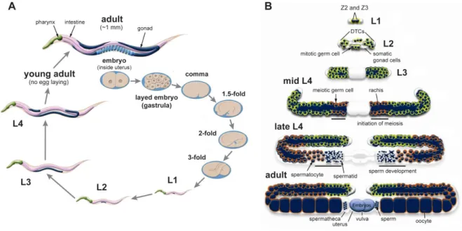
31 Hermaphrodite germline development begins at the L1 stage through the division of pluripotent germ precursor cells that originate during embryogenesis, Z2 and Z3 (Fig. 1.1B). From the L1 to L3 stages, germline growth occurs exclusively through mitotic divisions of germline cells. The distal tip cells (DTCs), which are of somatic origin, function at the growing ends of gonadal arms to continuously signal mitotic cell proliferation. Germ cells line the inner surface of the tubular structure of the germline and are only partially enveloped by a plasma membrane, sharing a common cytoplasm contained in the rachis (or the hollow center of the germline), effectively constituting a syncytium (Figure 1.1B). Meiosis does not begin until the L4 stage at the proximal end of the germline, near the section that unites the two gonadal arms, where two spermathecae, a uterus, and a vulva develop. As the germline grows, a higher proportion of the cells become meiotic. During the late L4 stage and the beginning of adulthood, meiosis is dedicated to hermaphrodite sperm differentiation. This starts with the formation of cellularized primary spermatocytes that detach from the germline wall. Completion of meiosis I divides the 4N primary spermatocyte into two, 2N, secondary spermatocytes. After meiosis II is completed, four haploid spermatids are generated. The sequence of events that go from a primary spermatocyte to a spermatid is termed spermatogenesis. These cells are stored in the spermatheca of the adult until the passage of oocytes leads to their activation in a process termed spermiogenesis, in which they become highly motile, fertilization-competent spermatozoa. During adulthood, meiotic cells start differentiating exclusively into oocytes which arrest at the diakinesis stage of meiosis I. Upon passage through the spermatheca, fertilization occurs, oocyte meiosis is resumed, and embryonic development is initiated. Embryos are stored in the uterus until they reach the gastrula stage (~30 cells) at which time they are expelled through the vulva.
32
Figure 1.1. Life cycle of C. elegans and germline development. (A) Life cycle of a
self-fertilizing hermaphrodite. Major stages of embryogenesis are shown. Every larval stage is separated by molting to replace the cuticle. The last molting event occurs in the L4 to adult transition. Wild-type C.elegans grows over a range of temperatures (typically 15-25˚C in the laboratory), influencing brood size and the duration of each developmental stage. (B) General architecture and development of the hermaphrodite germline. In the L1 stage, the two pluripotent germ precursor cells Z2 and Z3 begin to divide mitotically. These mitotic divisions continue into adulthood, and are stimulated by signaling from the DTCs, one per each gonad arm. The DTCs, the cells that envelope the germline, and the cells composing the spermathecae, uterus and vulva are all part of the so called somatic germline. The core of each gonadal arm is termed the rachis and constitutes a common cytoplasm to the partially cellularized nuclei that line the inner surface of the germline. Meiosis initiates in the proximal part of the germline during the L4 stage, during which they undergo differentiation into spermatids that are stored in the spermatheca of the developing L4 larva and adult. The sperm to oocyte switch occurs during adulthood. Oocytes are fertilized upon passage through the spermatheca, and embryos accumulate in the uterus for some time before being laid. Figures were adapted from WormAtlas.org (Altun et al., 2002-2015).
The MicroRNA Pathway
MicroRNAs represent the most abundant and well-studied class of sRNAs. C.
elegans expresses ~253 distinct miRNAs (WormBase release WS248; WormBase.org,
Harris et al., 2014). Mature miRNAs are ~23-nt long, with 5'-monophosphate and 3'-hydroxyl termini. They act by binding to partially complementary sites in the 3' untranslated regions (UTR) of mRNAs, to direct their post-transcriptional silencing (Jonas and Izaurralde, 2015). mRNAs frequently have more than one miRNA binding site, such that one target may be subjected to simultaneous regulation by several different miRNAs. The rules that dictate the pairing of miRNAs with their targets, primarily based on studies of the most abundant and conserved miRNAs, are fairly consensual. However, as new exceptions to these rules continue to be discovered, the prediction that the majority of
33 protein-coding RNAs are susceptible to some degree of miRNA-mediated regulation is gaining ground (Bartel, 2009).
Each miRNA is genomically encoded and transcribed by RNA polymerase II (Pol II) into a long, capped and polyadenylated RNA termed primary miRNA (pri-miRNA; Fig. 1.2; Bracht et al., 2004). The miRNA sequence is embedded in the dsRNA portion of a stem-loop structure within the pri-miRNA, which is released by the joint activity of the endoribonuclease Drosha and the dsRNA binding protein Pasha/DGCR8 (DRSH-1 and PASH-1 in C. elegans, respectively; Denli et al., 2004). This stem-loop of ~70 nt, with a 3' 2-nt overhanging terminus, termed pre-miRNA, is recognized by an ortholog of the human nuclear export protein exportin 5 (possibly XPO-1/IMB-4 in C. elegans) that transports it to the cytoplasm (Parry et al., 2004).
The next processing steps require Dicer (DCR-1 in C. elegans) and two redundant Argonaute proteins, ALG-1 and ALG-2 (Argonaute-Like Gene 1 and 2); Grishok et al., 2001; Ketting et al., 2001; Hutvagner et al., 2001). Dicer is a large (~220 kDa in C.
elegans), highly conserved endoribonuclease belonging to the RNase III family of dsRNA
cleaving enzymes, to which Drosha also belongs. Dicer is composed of a helicase domain, a PAZ (Piwi Argonaute and Zwille) domain that binds the substrate 3' end, two RNase III domains and a dsRNA-binding domain (Bernstein et al., 2001). As in humans, C. elegans only expresses one Dicer enzyme which is central for the synthesis of not only miRNAs but also for small-interfering RNAs (siRNAs; discussed in the next section) and certain endogenous sRNAs (endo-sRNAs). Argonautes are evolutionarily conserved sRNA-binding proteins consisting of a highly variable N-terminal domain, a PAZ domain for 3'-end nucleotide binding, a MID (middle) domain, which makes contact with the 5'-phosphates of sRNAs, and a C-terminal PIWI (P-element induced wimpy testis) domain with an RNase H-like fold to promote RNA cleavage, an activity termed ‘slicing’ (reviewed in Hutvagner and Simard, 2008).
DCR-1 binds the pre-mRNA using its PAZ domain to anchor the 2-nt 3' overhang generated by DRSH-1, and cleaves the dsRNA stem 22-23 nt away from the 3' end (Zhang et al., 2002; Macrae et al., 2007), resulting in a 22-23-nt long duplex RNA bearing 5'-monophosphates and 2-nt 3'-overhanging termini. The duplex is loaded into ALG-1 or -2 and one of the strands is cleaved (the passenger strand) by the slicer activity of the Argonaute, leaving the guide strand free to interact with its target (Bouasker and Simard, 2012). In C. elegans the majority of mature miRNAs carry a 5' uridine, which is thought to promote the specific interaction of miRNAs with ALG-1/2 (Ruby et al., 2006).
34
MiRNA-loaded ALG-1/2 Argonautes interact with a variety of proteins to bind the partially complementary 3' UTR sites on target mRNAs. Collectively, these sets of proteins are termed miRNA-induced silencing complexes (or miRISCs). A variety of miRISCs exist, based on the composition of factors associating with the core Argonaute-miRNA complex. One type of miRISC includes the micrococcal nuclease-related TSN-1 (Tudor
Staphylococcal Nuclease ortholog) in association with the dsRNA-binding factor VIG-1
(ortholog of the Drosophila Vasa Intronic Gene) (Caudy et al., 2003). Another type is defined by the ortholog of human GW182, AIN-1 (ALG-1 Interacting Protein 1), which facilitates both miRNA loading and the interaction of miRNAs with their mRNA targets in association not only with ALG-1/2, but also with DCR-1 (Ding et al., 2005). AIN-2 acts redundantly with AIN-1 by also binding DCR-1 and ALG-1/2, but can additionally recruit TSN-1 and the mRNA-binding translational repressing protein GLD-1 (Defective in
Germline Development 1). Moreover, AIN-2 has been shown to interact with components
of the translation initiation machinery (Parry et al., 2007; Zhang et al., 2007). Finally, NHL-2 (carrying a TRIM-NHL domain, which defines a large class of metazoan proteins; Wulczyn et al., 2010) in association with the conserved DEAD-box (Asp-Glu-Ala-Asp motif) RNA helicase CGH-1 (Conserved Germline Helicase 1), is required to increase the strength of binding of miRISCs to their target mRNAs (Hammell et al., 2009). The DEAD-box motif is present in numerous enzymes acting in sRNA pathways (including Dicer). Proteins carrying this motif form a subgroup within a large family of proteins present in all organisms defined more generally by the DExD/H motif, and are involved in a wide range of processes pertaining mostly to RNA metabolism (reviewed in Fuller-Pace, 2006 and Linder and Jankowsky, 2011). CGH-1 orthologs have been demonstrated to be required for miRISC activity, and are involved in several other aspects of mRNA post-transcriptional regulation and turnover (Chu and Rana, 2006; Eulalio et al., 2007; Rajyaguru and Parker, 2009).
All of the aforementioned proteins are required for effective silencing of miRNA targets, whether it occurs by translational repression, mRNA degradation, or both (Wightman et al., 1993; Bagga et al., 2005; reviewed in Jonas and Izaurralde, 2015). Notably, both AIN-1 and NHL-2/CGH-1 complexes localize to cytoplasmic domains called processing bodies (P-bodies), which contain a variety of ribonucleoprotein (RNP) complexes dedicated to processes such as nonsense-mediated decay, mRNA decay upon decapping and translational repression (Parker and Sheth, 2007). Through these molecular activities, miRNAs control various aspects of somatic development and physiology,
35 namely in aging, nervous system patterning and function, and general regulation of cell fate and developmental timing (Ambros, 2003; Abbott, 2011; Inukai and Slack, 2013). Interestingly, while individually deleting most miRNAs does not result in perceivable deleterious phenotypes (Miska et al., 2005), the collective deletion of some of the 23 miRNA families found in C. elegans can have very dramatic developmental consequences (Alvarez-Saavedra and Horvitz, 2010). The best example is the mir-35-41 family, which is essential to enable proper embryonic development over a wide range of temperatures, and to ensure full reproductive capacity in adult animals (McJunkin and Ambros, 2014). Studies of miRNA function in C. elegans continue to support the increasingly consensual idea that miRNAs have evolved to confer robustness upon sensitive gene expression programs in the face of challenging environmental conditions (Burke et al., 2015; Ren and Ambros., 2015).
36
Figure 1.2. The microRNA pathway. Processing of miRNAs takes place in the nucleus and
cytoplasm. Loading into ALG-1 or ALG-2 is thought to be concomitant with Dicer cleavage, as the two proteins stably interact in vivo. Loaded ALG-1/2 complexes then bind additional factors that recruit them to sites of RNA regulation, such as cytoplasmic P-bodies, where thay associate with target mRNAs, predominately via imperfect base pairing within 3' UTRs. This can lead to translational impairment and/or to direct degradation, upon recruitment of destabilizing factors such as RNA deadenylases.
The RDE-1 Pathway: Canonical RNA Interference and Beyond
Experimentally, RNAi can be triggered in C. elegans by exposing cells to double-stranded RNA (dsRNA) directly by injection or through feeding of dsRNA-expressing bacteria, in a process termed exogenous RNAi or exo-RNAi (Fig. 1.3 and Table 1.1). The process is strictly dependent on the Argonaute RDE-1, which is loaded with siRNAs resulting from the cleavage of dsRNA by Dicer. RNAi is both specific, leading to the silencing of sequences identical to the original dsRNA, and systemic, in that it is able to spread to most cells via the SID-1 (Systemic RNAi Defective 1) dsRNA specific transmembrane channel (Winston et al., 2002; Shih and Hunter, 2011). In association with the dsRNA-binding protein RDE-4 (RNAi-deficient 4; Tabara et al., 2002), DCR-1 cleaves the dsRNA to generate 23-nt duplex RNAs with 2-nt 3'-hydroxyl overhangs and 5'-monophosphate termini (Grishok et al., 2001; Ketting et al., 2001; Knight and Bass, 2001). The small interfering RNA (siRNA) duplexes are loaded into the Argonaute protein RDE-1, which also stably interacts with DCR-1 (Tabara et al., 1999). Similarly to ALG-1/2, after binding the siRNA duplex, RDE-1 cleaves one of the strands (the passenger strand) using its RNase H slicer activity, to retain a single strand that will serve as the guide strand to recognize and bind RNA target molecules (Steiner et al., 2009). Target RNA is cleaved near the RDE-1 binding site by the endonuclease RDE-8, followed by the addition of a 3' poly-uridine tract synthesized by the polynucleotidyl transferase RDE-3 (Chen et al., 2005; Tsai et al., 2015). This event is thought to signal the recruitment of the RNA-dependent RNA polymerases (RdRP) RRF-1 (RdRP family 1), expressed in the soma and germline (Sijen et al., 2001), and EGO-1 (Enhancer of Glp-One 1), expressed exclusively in the germline (Smardon et al., 2001).
RdRPs use the RNA template to catalyze the primer-independent synthesis of antisense 22-23 nt siRNAs bearing a 5'-triphosphorylated guanosine, termed 22G-RNAs (Sijen et al., 2001; Aoki et al., 2007; Pak and Fire, 2007; Sijen et al., 2007). These siRNAs spread across the template RNA, 5' of the RDE-1 binding site, usually within a range of
37 100-180 nt (Alder et al., 2003). Both RRF-1 and EGO-1 must physically associate with two co-factors to produce 22G-RNAs: EKL-1 (Enhancer of KSR-1 Lethality 1), a Tudor domain protein (Kim et al., 2005; Robert et al., 2005; Rocheleau et al., 2008; Claycomb et
al., 2009), and the helicase DRH-3 (Dicer-Related Helicase 3; Duchaine et al., 2006; Aoki et al., 2007; Nakamura et al., 2007; Claycomb et al., 2009; Gu et al., 2009).
Loss-of-function mutants for either of these factors are completely sterile and unable to produce 22G-RNAs, including those that are triggered endogenously, as discussed ahead (Claycomb et al., 2009; Gu et al., 2009). 22G-RNAs are loaded into Argonautes belonging to a group of 12 semi-redundant worm-specific Argonautes (or WAGOs) to target complementary RNAs for silencing (Yigit et al., 2006; Gu et al., 2009; reviewed in Buck and Blaxter, 2013). C. elegans specifically requires four WAGOs for exo-RNAi silencing, as their combined deletion leads to complete RNAi resistance in somatic and germline cells (Yigit et al., 2006). The four Argonautes are 4, 7(PPW-1), WAGO-6(SAGO-2), and WAGO-8(SAGO-1). PPW-1 (PAZ/PIWI Domain-Containing 1) is required predominately for germline RNAi, while the remaining Argonautes are employed in somatic RNAi (Tijsterman et al., 2002; Yigit et al., 2006).
The RNAi process is thus divided into two main phases: the primary phase, in which low abundance siRNAs are directly derived from Dicer-mediated cleavage of the dsRNA trigger; and the secondary (or amplification) phase, in which primary siRNA-loaded RDE-1 initiates the abundant RdRP synthesis of antisense 22G-RNAs (Fig. 1.3 and Table 1.1). The exo-RNAi response has been shown to be self-contained, in that (1) secondary 22G-RNAs are unable to initiate new rounds of RdRP-mediated amplification (Pak et al., 2012), and (2) RDE-1 cannot engage secondary 22G-RNAs or silence targets in the absence of 22G-RNA synthesis (Sijen et al., 2001; Yigit et al., 2006). Since the most critical Argonautes for exo-RNAi are assumed to be predominately cytoplasmic, the bulk of silencing is thought to occur post-transcriptionally. However, exogenously triggered 22G-RNAs can also be loaded into WAGO proteins that translocate into the nucleus to elicit transcriptional silencing, a process termed nuclear RNAi (discussed ahead).
RNAi can be triggered naturally by ingestion and uptake of dsRNA from the environment through the intestinal SID-2 transmembrane channel (Winston et al., 2007) or through infection by RNA viruses (Felix et al., 2011; introduced in Chapter V), to cite two known examples. Cells also make use of this pathway from endogenous sources of dsRNA arising, for instance, from transposons or highly-repetitive transgene arrays (Ketting et al., 1999; Sijen and Plasterk, 2003; Vastenhouw et al., 2003; Grishok et al., 2005; Robert et
38
al., 2005). Additionally, the formation of regions of dsRNA within C. elegans transcripts
has recently been shown to be widespread (Whipple et al., 2015). Several of these sequences overlap with protein coding genes that are known targets of endogenous 22G-RNAs. In combination with other factors, Dicer is proposed to use these dsRNA regions as substrates to produce primary siRNAs that likely enter the RDE-1 pathway to elicit the silencing of endogenous RNAs.
Figure 1.3. The exogenous RNAi pathway. Exo-RNAi is triggered by the uptake of dsRNA from
39 direct injection of dsRNA. There are two types of siRNAs: the primary siRNAs, which are direct products of Dicer cleavage and are only as abundant as the dsRNA source; and the secondary siRNAs, the result of RdRP synthesis triggered by RDE-1-bound primary siRNAs in somatic cells (by RRF-1), and in the germline (by both RRF-1 and EGO-1). The diagram is a simplification of our current knowledge of the process, as many of the factors required for 22G-RNA accumulation have been omitted. The RDE-8/RDE-3 step is presumed, but not proven, to recruit RdRPs to the target RNA. Downstream silencing can occur through post-transcriptional mechanisms in germline P granules or somatic P-body-like granules, and through transcriptional mechanisms inside the nucleus, depending on which WAGOs secondary siRNAs associate with. ‘m7G’, 5' methylguanosine cap.
The 21U-RNA/piRNA Pathway
C. elegans expresses over 30,000 distinct, genomically encoded 21-nt RNAs
carrying 5'-monophosphorylated uridines and 2'-O-methyl modified 3' termini, referred to as 21U-RNAs (Fig. 1.4A and Table 1.1; Ruby et al., 2006; Batista et al., 2008; Gu et al., 2012). Owing to their predominant germline expression, 5' and 3' molecular features, and their Dicer-independent biogenesis, 21U-RNAs are considered the C. elegans equivalents of animal piRNAs (PIWI-interacting RNAs). This class of endogenous sRNAs has been demonstrated to promote proper germline development and gamete differentiation while protecting the integrity of germline genomes from the mutagenic effects of active transposing elements, from Drosophila to mice (reviewed in Luteijn and Ketting, 2013). In contrast to all other sRNAs that interact with either Argonaute-clade (RDE-1, ALG-1/2, ALG-3/4; conserved from plants to animals) or with C. elegans-specific WAGO-clade Argonautes, 21U-RNAs interact with a conserved PIWI-clade Argonaute named PRG-1 (Piwi Related Gene 1; Batista et al., 2008), similarly to piRNAs in other species.
21U-RNAs are transcribed by Pol II, and are subdivided into two groups depending on the elements that govern their expression (Fig. 1.4A; Ruby et al., 2006; Cecere et al., 2012; Gu et al., 2012). Type-I 21U-RNAs are encoded primarily in intergenic clusters spread throughout chromosome IV or from sequences overlapping with other Pol II genes. Each Type-I locus is preceded by a two-part motif 25-60 nt upstream of the locus, which is recognized by specific Forkhead-family transcription factors (Ruby et al., 2006; Batista et
al., 2008; Das et al., 2008; Cecere et al., 2012; Billi et al., 2013). Type-II 21U-RNAs arise
from a subset of abortive Pol II transcription products at transcription start sites (TSS) of active promoters. These short (10-40 nt), capped products termed capped small RNAs (csRNAs), are often transcribed bi-directionally around the TSS (Gu et al., 2012). As part of their upstream motif, both Type-I and Type-II 21U-RNAs have a short consensus YRNT sequence (where Y is a pyrimidine, R is a purine, N is any nucleotide and T