UNIVERSIDADE DA BEIRA INTERIOR
Ciências da Saúde
Design of an one-step platform purification of
STEAP1 using Hydrophobic Interaction
Chromatography
Diogo José Pinheiro Monteiro
Dissertação para obtenção do grau de Mestre em
Ciências Biomédicas
(2º Ciclo de estudos)
Orientador: Prof. Doutor Luís António Paulino Passarinha
Co-orientador: Prof. Doutor Cláudio Jorge Maia Batista
Acknowledgements
Firstly, I would like to make special thanks to my supervisors, Professor Luís Passarinha and Professor Cláudio Maia for the opportunity that was given to me to demonstrate my worth and work developing this work. Thank you also for all the time you have given for my guidance and for all the advice and reviews that have helped to greatly improve this dissertation. Also, to all my colleagues and friends at CICS, thank you for the friendship, support, mutual help, patience and encouragement. Special thanks to Fátima Santos, Diana Duarte and Jorge Ferreira for the main help, friendship, and teachings during the last year.
To all my dear friends from all over the world a huge thank for all the support and care. Friends with whom I shared the best and the worst moments of my life, helping me to relax and believe in a better future giving me an extra motivation. Also, a huge thank to all my cyclist friends to be an important daily distraction outdoor the lab, being the competition and sport a great school for life, teaching above all the highest ethical values.
My forever thankfulness goes to my family, especially my mom and dad, for whom I have the greatest respect and love in the world, for the fact that they have been present throughout my all life. They give me with much effort, the possible and the impossible, to get where I came and show with pride the person that I am today. This dissertation is entirely dedicated to you all.
Resumo
O cancro de próstata é um dos carcinomas mais letais e prevalentes entre homens idosos em todo o mundo. Atualmente, o diagnóstico do cancro da próstata baseado nos níveis de Prostate Specific
Antigen (PSA) é inespecífico e com eficiência limitada, principalmente em estágios avançados de
cancro. Assim, existe a necessidade de identificar e caracterizar biomarcadores proteicos específicos e confiáveis para o cancro da próstata. A Six-transmembrane Epithelial Antigen of the
Prostate 1 (STEAP1) é uma proteína transmembranar cujos altos níveis de expressão foram
correlacionados com o cancro da próstata. A STEAP1 pode participar na comunicação intracelular e intercelular em células cancerígenas através da modulação da proliferação celular e invasão tumoral através da sua potencial atividade como canal iónico ou transportador. Assim, a caracterização da estrutura da STEAP1 pode permitir a conceção de inibidores específicos que diminuem e modulam a sua função oncogénica. Os estudos estruturais e funcionais requerem quantidades elevadas de proteína purificada, que podem ser obtidas através de uma produção recombinante da proteína STEAP1 humana integrada com uma estratégia cromatográfica adequada. Neste trabalho, foi avaliado o desempenho da Octil- e Butyl-Sepharose de acordo com as condições de ligação e eluição necessárias para o isolamento da STEAP1 a partir de lisados celulares, obtidos em culturas induzidas com metanol num mini biorreator de Pichia pastoris X33. A concentração do tampão fosfato de sódio e fosfato monossódico com cloreto de sódio no tampão de equilíbrio foi otimizada para promover uma adsorção completa da STEAP1 nos suportes hidrofóbicos. Sucintamente, observou-se uma retenção mais elevada da STEAP1 com concentrações acima de 500 mM de tampão fosfato de sódio e fosfato monossódico com cloreto de sódio, pH 8,0. Se a adsorção for alcançada com altas concentrações de tampão fosfato de sódio ou fosfato monossódico com cloreto de sódio, a eluição deve ser realizada com concentrações crescentes de Triton X-100 em 50 mM de tampão fosfato. Os resultados obtidos indicam que a exposição dos domínios de ligação de membrana da STEAP1 à Octyl- e Butyl-Sepharose requerem à priori altas concentrações de sal devido às fortes interações estabelecidas entre eles. No entanto, após a sua adsorção completa, a eluição da STEAP1 requer agentes caotrópicos, como detergentes. Embora a aplicação da Cromatografia de Interação Hidrofóbica (HIC) na purificação de proteínas integrais de membrana seja incomum, os resultados obtidos no desenvolvimento da dissertação indicam que a utilização de matrizes hidrofóbicas tradicionais pode abrir uma alternativa promissora para o isolamento da STEAP1 a partir de lisados celulares.
Palavras-chave
Resumo Alargado
O cancro de próstata é um dos carcinomas mais letais e prevalentes entre homens idosos em todo o mundo, apresentando especial incidência em homens com idade superior a 50 anos. Atualmente, os meios de diagnóstico e terapia existentes do cancro da próstata, principalmente em estágios avançados de cancro, são invasivos, inespecíficos e com eficiência limitada, sendo predominantemente baseados nos níveis de Prostate Specific Antigen (PSA). Assim, existe a necessidade de identificar e caracterizar biomarcadores proteicos específicos e confiáveis para o cancro da próstata. The Six-transmembrane Epithelial Antigen of the Prostate 1 (STEAP1) é uma proteína constituída por seis domínios transmembranares interligados por loops extracelulares, geralmente localizada na membrana plasmática, cujos altos níveis de expressão foram correlacionados com o cancro da próstata. A STEAP1 pode participar na comunicação intracelular e intercelular em células cancerígenas através da modulação da proliferação celular e invasão tumoral através da sua potencial atividade como canal iónico ou transportador. Assim, a caracterização da estrutura da STEAP1 pode permitir a conceção de inibidores específicos que diminuem e modulam a sua função oncogénica, permitindo a sua utilização como alvo terapêutico. Os estudos estruturais e funcionais requerem quantidades elevadas de proteína purificada, que podem ser obtidas através de uma produção recombinante da proteína STEAP1 humana integrada com uma estratégia cromatográfica adequada. Assim, o principal objetivo desta tese de mestrado foi desenvolver uma estratégia cromatográfica sustentável de um passo para a purificação da STEAP1, recuperada a partir de lisados de Pichia pastoris, usando a Cromatografia de Interação Hidrofóbica. Para atingir este objetivo final, vários procedimentos foram desenvolvidos e otimizados, tais como: a) Otimização do processo de recuperação de lisados de Pichia pastoris através da determinação do detergente mais eficaz para a solubilização da STEAP1; b) Desenvolvimento de um procedimento por Cromatografia de Interação Hidrofóbica através da avaliação do desempenho da Octyl- e Butyl-Sepharose de acordo com as condições requeridas para a ligação e eluição da STEAP1 nestas matrizes. A proteína STEAP1 foi obtida através de produção recombinante realizada em mini-biorreator de culturas de Pichia pastoris X33 Mut+.
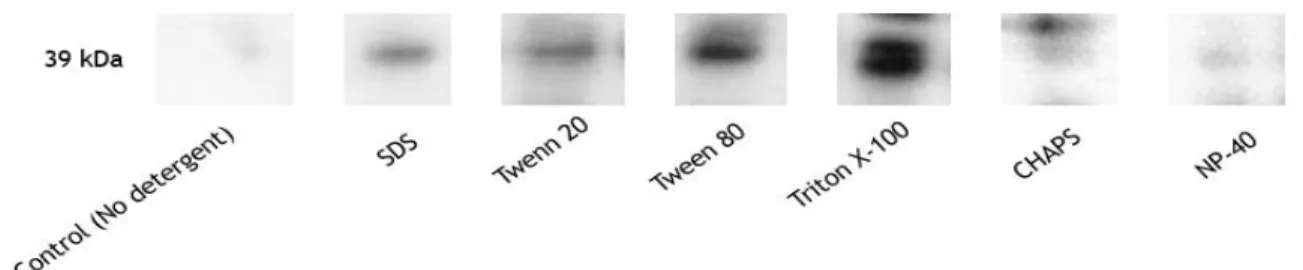
Fundamentalmente o processo fermentativo compreende três fases principais: fase batch de glicerol, fed-batch e indução com metanol. Assim, como já otimizado pelo nosso grupo de trabalho, a melhor estratégia para a obtenção da proteína com menor agregação é a fermentação com 20 horas de um batch de glicerol, seguida de 3 horas de fed-batch e posterior indução com metanol durante 10 horas. O passo seguinte consistiu em isolar o péptido de interesse no seu estado nativo adotando o método de lise por esferas, sendo que na etapa de solubilização, de entre cinco detergentes (SDS, Twenn-20, Tween-80, NP-40, Triton X-100 e CHAPS) foi estabelecido que o Triton X-100 alcançou o resultado mais eficiente, preservando a estrutura nativa da STEAP1 com os padrões de expressão mais elevados. Na etapa de purificação, a concentração do tampão fosfato de sódio e fosfato monossódico com cloreto de sódio no tampão de equilíbrio foi otimizada para promover uma adsorção completa da STEAP1 nos suportes hidrofóbicos. Observou-se uma retenção mais elevada da STEAP1 com concentrações acima de 500 mM de tampão fosfato de
viii
sódio e fosfato monossódico com cloreto de sódio, pH 8,0. Se a adsorção for alcançada com altas concentrações de tampão fosfato de sódio ou fosfato monossódico com cloreto de sódio, a eluição deve ser realizada com concentrações crescentes de Triton X-100 em 50 mM de tampão fosfato. Os resultados obtidos indicam que a exposição dos domínios de ligação de membrana da STEAP1 à Octyl- e Butyl-Sepharose requerem à priori altas concentrações de sal devido às fortes interações estabelecidas entre eles. No entanto, após a sua adsorção completa, a eluição da STEAP1 requer agentes caotrópicos, como detergentes. Embora a aplicação da Cromatografia de Interação Hidrofóbica (HIC) na purificação de proteínas integrais de membrana seja incomum, os resultados obtidos no desenvolvimento da dissertação indicam que a utilização de matrizes hidrofóbicas tradicionais pode abrir uma alternativa promissora para o isolamento da STEAP1 a partir de lisados celulares.
Abstract
Prostate cancer is one of the most lethal and prevalent carcinoma among elder men worldwide. Currently, prostate cancer diagnosis based on prostate-specific antigen levels is unspecific and with limited efficient, mainly in advanced stages of cancer. Thus, there is a need to identify and characterize specific and reliable protein biomarkers for prostate cancer. Six transmembrane epithelial antigen of the prostate 1 (STEAP1) is a transmembrane protein whose high expression levels were correlated with PCa. STEAP1 may take part in intracellular and intercellular communication in cancer cells by modulating cell proliferation and tumor invasiveness through its potential activity as ion channel or transporter. So, the characterization of STEAP1 structure might allow the design of specific inhibitors that decrease and modulate its oncogenic function. The structural and functional studies require high purified amounts of protein, which can be obtained through a recombinant production of human STEAP1 protein integrated with a properly chromatographic strategy. In this work, the performance of Octyl- and Butyl-Sepharose were evaluated according to binding and elution conditions required for STEAP1 isolation from cell lysates, obtained in mini-bioreactor Pichia pastoris X33 methanol-induced cultures. The concentration of sodium phosphate buffer and monosodium phosphate plus sodium chloride in the equilibration buffer was optimized in order to promote a complete STEAP1 adsorption on the hydrophobics supports. Succinctly, a higher retention of STEAP1 was observed with concentrations above 500 mM of sodium phosphate buffer and monosodium phosphate plus sodium chloride, pH 8.0. If the adsorption is achieved at high concentrations of sodium phosphate buffer, the elution must be performed with increasing concentrations of Triton X-100 in 50 mM phosphate buffer. The obtained results indicate that the exposition of membrane binding domains of STEAP1 to Octyl- and Butyl- Sepharose requires high salt concentrations due the strong interactions established between them. However, after its complete adsorption, STEAP1 elution requires chaotropic agents such as detergents. Although application of HIC in the purification of integral membrane proteins are uncommon, the obtained results in the development of this dissertation indicate that the use of traditional hydrophobic matrices may open a promising alternative for the isolation of STEAP1 from cell lysates.
Keywords
Index
Chapter I – Introduction 1
1. Human Prostate 1
1.1 Anatomy and Physiology 1
1.2. Prostate Cancer 2
1.2.1 Epidemiology 2
1.2.2 Risk Factors 3
1.2.3 Prostate Carcinogenesis 4
1.2.4 Diagnosis and Treatment 6
1.2.5 Potential Biomarkers of Prostate Cancer 7 2. Six-transmembrane Epithelial Antigen of Prostate Family 9
2.1 STEAP1 10
2.1.1 Structure, Function and Expression 10 2.1.2 STEAP1 as a Therapeutic Target? 11
3. Membrane Proteins 12
3.1 Protein Solubilization 12
3.2 Purification of Membrane Proteins 13
3.2.1 Hydrophobic Interaction Chromatography 15
Chapter II – Aims 19
Chapter III – Materials and Methods 21
1. Materials 21
2. Methods 21
2.2 Strain, plasmids, and media 21
2.3 STEAP1 Biosynthesis 22
2.4 Cell lysis and STEAP1 solubilization 22
2.5 Total protein quantification 23
2.6 STEAP1 purification by Hydrophobic Interaction Chromatography 23
2.7 SDS-PAGE and Western Blotting 24
Chapter IV – Results and Discussion 27
1. STEAP1 solubilization 27
2. Purification screening trials onto Octyl-Sepharose 28
3. STEAP1 isolation on Octyl-Sepharose 32
4. Purification trials on Butyl-Sepharose 36
Chapter V – Conclusion and Future Perspectives
39
References 41
List of Figures
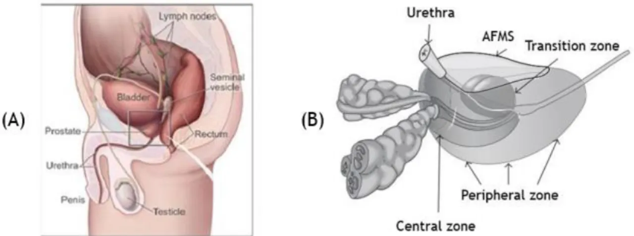
Figure 1. Anatomy of an adult human prostate showing urethra and bladder in relation to the four major glandular regions of the prostate. Central Zone (CZ) that surrounds the ejaculatory duct, peripheral zone (PZ) that consists in about 70% of prostate, and transitional zone (TZ) that surrounds proximal prostatic urethra and the anterior fibromuscular stroma (AFMS), which allows the connection between anterior and apical surfaces.
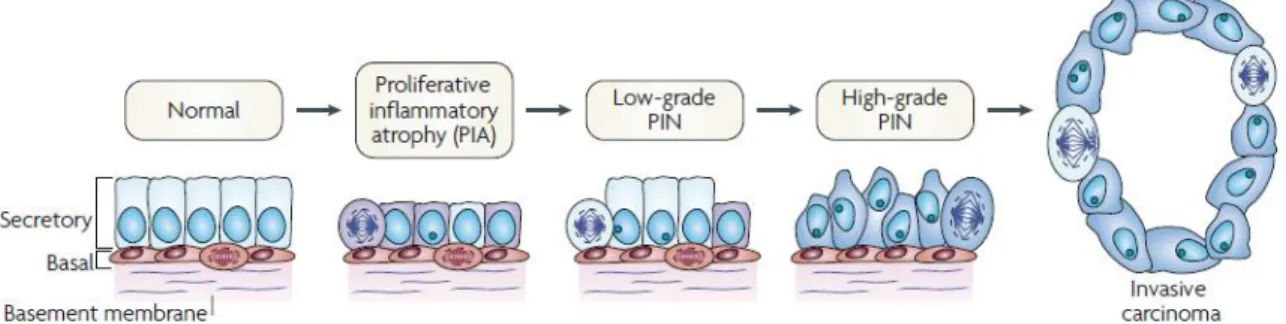
Figure 2. Cellular and molecular model of early prostate neoplasia progression. This stage is characterized by the infiltration of lymphocytes, macrophages and neutrophils. Phagocytes release reactive oxygen and nitrogen species causing DNA damage, cell injury and cell death, which initiate the beginning of epithelial cell regeneration. The downregulation of p27, phosphatase and tensin homologue (PTEN) and NKX3.1 in luminal cells stimulates cell-cycle progression. The continued proliferation of genetically unstable luminal cells and the further accumulation of genomic changes lead to progression towards invasive carcinomas.
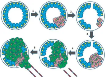
Figure 3. Progression from androgen-dependent to androgen-independent prostate cancers. 1. Various carcinogenic processes occur whereby some prostate cells proliferate out of control. 2. Prostate cancer cells are initially androgen dependent, and with androgen ablation therapy are successfully destroyed. 3. Some cells can survive to this treatment and continue proliferating. 4. Cells are now androgen independent and gain subsequent changes resulting in increased angiogenesis. 5. AIPC begins to metastasize to distant sites.
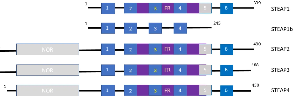
Figure 4. Schematic illustration of the domain organization of STEAP family. Superscript numbers indicate respectively the first and last amino acid. NOR: NADPH-oxidoreductase domain. The transmembrane domains are indicated as blue boxes. FR: ferric oxidoreductase domain. Heme-binding histidine residues within transmembrane domains 3 and 5 are indicated with orange lines.
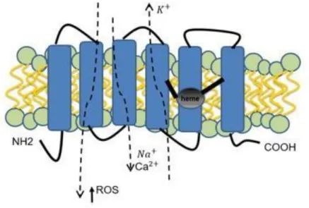
Figure 5. Schematic STEAP1 protein structure, cellular localization and physiologic functions. Presenting a six- transmembrane structure, intracellular COOH- and N-terminal, and intramembrane heme group. STEAP1 actively increases intra- and intercellular communication through the modulation of Na+, Ca2+ and
K+ concentration, as well as the concentration of small molecules. It stimulates cancer cell proliferation
and tumor invasiveness.
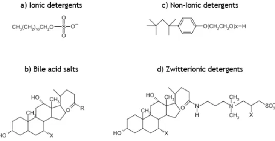
Figure 6. Structures of different types of detergents used in the solubilization of membrane proteins.
Figure 7. Mechanism of adsorption of biomolecules on hydrophobic interaction chromatography. Biomolecules of interest, with strong hydrophobic characteristics, interact with the ligand, while the ones with low hydrophobicity do not bind and are removed in the flow-through fractions.
Figure 8. Hofmeister series and effects of some anions and cations on proteins.
xiv
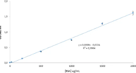
Figure 10. BSA calibration curve for total protein quantification (µg/mL) ranged between 25 –2000 μg/mL.
Figure 11. Western blot analysis of the STEAP1 solubilization using 1% (v/v) of some common detergents applied in the literature.
Figure 12. Schematic structures of Butyl- and Octyl-Sepharose ligands.
Figure 13. Initial purification screening trials on Octyl-Sepharose. (A) sodium chloride; (B) sodium phosphate buffer. Adsorption was performed at salt concentrations of 500 mM (pH 8.0). Desorption was performed with 50 mM sodium phosphate buffer (pH 8.0). Different color lines represent the absorbance at 280 nm, brown line the conductivity and the continuous green line represents sodium phosphate buffer concentration.
Figure 14. Dot blot analysis of samples collected on chromatographic profiles of figure 13. Lane L – Solubilized lysis pellet; Lane I – Peaks I obtained at 500 mM of sodium chloride and sodium phosphate buffer and Lane II – Peak II obtained with 50 mM sodium phosphate buffer on Octyl-Sepharose.
Figure 15. SDS-PAGE (A) and Western blot (B) analysis of samples collected on chromatographic profiles of figure 14. L – Solubilized lysis pellet; Lane I – Peaks I obtained at 500 mM and Lane II – Peak II obtained with 50mM sodium phosphate buffer on Octyl-Sepharose.
Figure 16. Chromatographic profile of STEAP1 isolation trials on Octyl-Sepharose. Adsorption was performed at 750 mM sodium chloride, pH 8.0 followed by 50 mM sodium phosphate buffer step. Blue line represents the absorbance at 280 nm, green line the sodium phosphate buffer concentration in mobile phase, and the brown line the conductivity.
Figure 17. SDS-PAGE (A) and Western Blot (B) analysis of samples collected on STEAP1 isolation chromatographic assay [figure 16]. Lane MW – molecular weight standards; Lane L – solubilized lysis pellet; Lane I – Peaks I obtained at 750 mM sodium chloride; Lane II – Peak II obtained with 50 mM sodium phosphate buffer.
Figure 18. Chromatographic profile of STEAP1 isolation on Octyl-Sepharose. Adsorption was performed at 1000 mM sodium phosphate buffer, pH 8.0 followed by 50 mM sodium phosphate buffer step. Desorption was performed at 1% Triton X-100, pH 8.0. Blue line represents the absorbance at 280 nm, green line the sodium phosphate buffer concentration in mobile phase, red line the Triton X-100 composition in mobile phase and the brown line the conductivity.
Figure 19. SDS-PAGE (A) and Western Blot (B) analysis of samples collected on STEAP1 isolation chromatographic assay [figure 18]. Lane MW – molecular weight standards; Lane L – solubilized lysis pellet; Lane I – Peaks I obtained at 1000 mM sodium chloride; Lane II – Peak II obtained with 50 mM sodium phosphate buffer. Lanes III and IV – Peaks III and IV obtained with a linear gradient of 1% Triton X-100.
at 750 mM monosodium phosphate with sodium chloride, pH 8.0 followed by 50 mM sodium phosphate buffer step. Desorption was performed in linear gradient (A) and step gradient (B) of 1% Triton X-100, pH 8.0. Blue line represents the absorbance at 280 nm, green line the monosodium phosphate with sodium chloride concentration in mobile phase, red line the Triton X-100 percentage in mobile phase and the brown line the conductivity.
Figure 21. SDS-PAGE (A) and Western Blot (B) analysis of samples collected on STEAP1 isolation chromatographic assay [figure 20 (A)]. Lane MW – molecular weight standards; Lane L – solubilized lysis pellet; Lane I – Peaks I obtained at 750 mM monosodium phosphate with sodium chloride; Lane II – Peak II obtained with 50Mm sodium phosphate buffer; Lanes III and IV – Peaks III and IV obtained at linear gradient of 1% Triton X-100.
Figure 22. SDS-PAGE (A) and Western Blot (B) analysis of samples collected on STEAP1 isolation chromatographic assay [figure 20 (B)]. Lane MW – molecular weight standards; Lane L – solubilized lysis pellet; Lane I – Peaks I obtained at 750 mM monosodium phosphate with sodium chloride; Lane II – Peak II obtained with 50Mm sodium phosphate buffer; Lanes III and IV – Peaks III and IV obtained at step gradient of 1% Triton X-100.
Figure 23. Chromatographic profiles of STEAP1 isolation trials on Octyl-Sepharose. Adsorption was performed at 750 mM monosodium phosphate with sodium chloride, pH 8.0 followed by 50 mM sodium phosphate buffer step. Desorption was performed in linear gradient (A) and step gradient (B) of 1% Triton X-100, pH 8.0. Blue line represents the absorbance at 280 nm, green line the monosodium phosphate with sodium chloride concentration in mobile phase, red line the Triton X-100 percentage in mobile phase and the brown line the conductivity.
Figure 24. SDS-PAGE (A) and Western Blot (B) analysis of samples collected on STEAP1 isolation chromatographic assay [figure 23 (A)]. Lane MW – molecular weight standards; Lane L – solubilized lysis pellet; Lane I – Peaks I obtained at 750 mM monosodium phosphate with sodium chloride; Lane II – Peak II obtained with 50 mM sodium phosphate buffer; Lanes III and IV – Peaks III and IV obtained at linear gradient of 1% Triton X-100.
Figure 25. SDS-PAGE (A) and Western Blot (B) analysis of samples collected on STEAP1 isolation chromatographic assay [figure 23 (B)]. Lane MW – molecular weight standards; Lane L – solubilized lysis pellet; Lane I – Peaks I obtained at 750 mM monosodium phosphate with sodium chloride; Lane II – Peak II obtained with 50 mM sodium phosphate buffer; Lanes III and IV – Peaks III and IV obtained at step gradient of 1% Triton X-100.
Figure 26. Chromatographic profile of STEAP1 isolation trial on Butyl-Sepharose (HIC). Adsorption was performed at 800 mM monosodium phosphate with sodium chloride, pH 8.0 followed by 50 mM sodium phosphate buffer step. Desorption was performed with a linear gradient of 1% Triton X-100, pH 8.0. Blue line represents the absorbance at 280 nm, green line the monosodium phosphate with sodium chloride concentration in mobile phase, red line the Triton X-100 percentage in mobile phase and the brown line the conductivity.
xvi
chromatographic assay [figure 26]. Lane MW – molecular weight standards; Lane L – solubilized lysis pellet; Lane I – Peaks I obtained at 800 mM monosodium phosphate with sodium chloride; Lane II – Peak II obtained with 50 mM sodium phosphate buffer; Lanes III and IV – Peaks III and IV obtained at linear gradient of 1% Triton X-100.
List of Tables
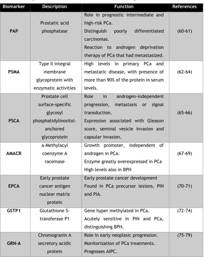
Table 1. Description of main biomarkers for PCa and their respective functions.
Table 2. Summary of salts concentrations used in isolation screening trials and STEAP1 elution behavior onto an Octyl-Sepharose support.
Table 3 – Summary of salt, Triton X-100 used in detergent gradient, and STEAP1 elution behavior onto an Octyl-Sepharose support.
Table 4 – Summary of salt dual system and Triton X-100, used in detergent gradient, concentrations and STEAP1 elution behavior onto an Octyl-Sepharose support.
Table 5 – Summary of salt dual system and Triton X-100, used in detergent gradient, concentrations and STEAP1 elution behavior onto a Butyl-Sepharose support.
List of abbreviations
PSA Prostate Specific Antigen
STEAP1 Six-transmembrane Epithelial Antigen of Prostate 1 HIC Hydrophobic Interaction Chromatography
CZ Central Zone
TZ Transition Zone
PZ Peripheral Zone
BPH Benign Prostatic Hyperplasia PCa Prostate Cancer
AFMS Anterior Fibromuscular Stroma UGS Urogenital Sinus
UGE Urogenital Sinus Epithelium UGM Urogenital Sinus Mesenchyme DHT Androgen 5α-dihydrotestosterone
AR Androgen Receptor
ROS Reactive Oxygen Species ED Endocrine Disruptors
PIN Prostatic Intraepithelial Neoplasia PIA Proliferative Inflammatory Atrophy PTEN Phosphatase and Tensin Homologue AIPC Androgen-independent Prostate Cancer EZH2 Enhancer of Zeste Homolog Gene 2 Upa Urokinase Plasminogen Activator TMPRSS2 Transmembrane Protease Serine 2 MCTs Monocarboxylate Transporters PCA3 Prostate Cancer Antigen 3 GOLPH2 Golgi Phosphoprotein 2
PTM Post-translational Modifications TFR1 Transferrin Receptor 1
TAAS Tumor-associated Antigens CTLs Cytotoxic T Lymphocytes PDB Protein Data Bank SDS Sodium Dodecyl Sulfate
IMAC Immobilized Metal-ion Affinity Chromatography SEC Size-exclusion Chromatography
IEC Ion-exchange Chromatography RPC Reverse-phase Chromatography BSA Bovine Serum Albumin
xx
CV Column Volume
SDS-PAGE Sodium Dodecyl Sulphate-Polyacrylamide Gel Electrophoresis PMSF Phenylmethylsulphonyl fluoride
hSCOMT Human Soluble Catechol-O-methyltransferase
Chapter 1
Introduction
1. Human Prostate
1.1 Anatomy and Physiology
The human prostate is a tubuloalveolar exocrine gland of the male reproductive system. The function of the prostate is to secrete a thin and, slightly alkaline fluid that forms a portion of the seminal fluid, an organic fluid that suspend the ejaculated sperm and maintain their mobility (1). The human adult prostate is a walnut-sized organ measuring 4x3x2 cm in wide, length and height, respectively, and weight of around 20 g. It is located posteriorly to the lower portion of the symphysis pubis, at the base of the urinary bladder, at the apex of the urogenital diaphragm and is separated anteriorly from rectum by the Denonvilliers’ fascia [Figure 1(A)]. At histological level, prostate is a branched duct gland with a pseudostratified epithelium composed of three differentiated epithelial cell types: luminal, basal and neuroendocrine. The inner layer of the prostate capsule is composed of smooth muscle with an outer layer covering of collagen (2-4).
McNeal divided the prostate into three major areas that are histologically distinct and anatomically separate, the Central Zone (CZ), Transition Zone (TZ) and Peripheral Zone (PZ) [figure 1(B)] (3). The CZ is a vertical wedge of glandular tissue, like a cone-shaped structure, with approximately 25% of the glandular tissue which surrounds the ejaculatory ducts and constitutes most of the apex of the prostate between the TZ and PZ. TZ is a smaller region with only 5% of the glandular tissue and consists of two equal small lobules portions of glandular tissue lateral to the urethra in the midgland (3) (5). This is the portion of the prostate involved in the development of Benign Prostatic Hyperplasia (BPH) and less commonly adenocarcinoma (1). The PZ is the largest area, comprising 70% of prostate, occupying from the base to the apex along the posterior surface and surrounds the distal urethra. This zone is the main site of prostate cancer (PCa), chronic prostatitis, postinflammatory atrophy and although not of BPH. Some authors also considered a fourth zone in the prostate, the Anterior Fibromuscular Stroma (AFMS) that forms the convexity of the anterior external surface (6). This apical area is rich in striated muscle, which blends into the muscle of pelvic diaphragm. The voluntary sphincter functions are performed by the distal portion of AFMS, while proximal portion plays a central role in involuntary sphincter functions (2) (5-7).
2
Figure 1. Anatomy of an adult human prostate showing urethra and bladder in relation to the four major glandular regions of the prostate. Central zone (CZ) that surrounds the ejaculatory duct, peripheral
zone (PZ) that consists in about 70 % of prostate, and transitional zone (TZ) that surrounds proximal prostatic urethra and the anterior fibromuscular stroma (AFMS), which allows the connection between anterior and apical surfaces (adapted from (8)).
Growth and development of the prostate begin at 10 weeks of gestation in humans, with the formation of prostatic buds from the fetal urogenital sinus (UGS), but only are completed at sexual maturity. The initial event is the outgrowth of solid epithelial buds from the Urogenital Sinus Epithelium (UGE) into the surrounding Urogenital Sinus Mesenchyme (UGM). The influence of testicular androgens is required to the proliferation of prostatic buds to originate solid cords of epithelial cells, which grow into the UGM in a particular spatial arrangement to establish the lobar divisions of the prostate (1). This androgen 5α-dihydrotestosterone (DHT), is synthesized from fetal testosterone by the action of 5α-reductase and is localized in the urogenital sinus and external genitalia of humans (9). Postnatally, under the influence of androgens, the epithelial cells undergo differentiation, produce protein secretions, and express characteristic markers such as cytokeratins 8 and 18, as well as high levels of Androgen Receptor (AR) and PSA. By observation of basal cells, it is possible to differentiate adenocarcinomas from benign conditions, since they are multipotent and can generate all epithelial lineages of prostate. Finally, neuroendocrine cells are rare cells of unclear function that express endocrine markers such as chromogranin A, synaptophysin and PSA, but not AR (10-11).
1.2 Prostate Cancer
1.2.1 Epidemiology
PCa is one of the most lethal and prevalent carcinoma affecting men worldwide and the second leading cause of cancer-related death of men in 2018 (29 430 estimated deaths) (12), with the highest number of new estimated cases (164 690 cases).
The highest prostate cancer incidence rates are found in developed regions, including Western and Northern Europe, Northern America and Australia/New Zeeland, whereas Asian and African countries have lower rates of incidence. The practice of PSA testing is the main reason to this discrepancy in incidence rates, which are able to detect even asymptomatic tumors, and in developing countries biopsy has become available for prostate cancer screening (13).
In Portugal is the main cancer diagnosed in men and the third cause of death. Although the incidence in Portugal is increasing, the mortality associated with prostate cancer seems to be decreasing constantly over the time (14-15).
1.2.2 Risk Factors
Several research studies have given insight into the causes and risk factors for prostate cancer (16-17). Although the specific causes remain unknown, several risk factors have been identified, which may contribute to the initiation and progression of this pathology.
The risk factors can be classified as endogenous or exogenous. In the first group there are factors such as age, family history, race, hormones and oxidative stress. The second group is constituted by diet, environmental agents and occupation (18). The major risk factor for PCa is age, once about 85% of cases are diagnosed after 65 years old (19). Family history can be associated with high risk of PCa, since have long known that cancer susceptibility can be inherited (20). In addition, race is also referred as a risk factor, since African-Americans have twice the risk of non-Hispanic white’s due differences in allelic frequencies of microsatellites or polymorphic variations at the AR locus (21-22). Reactive oxygen species (ROS) and the coupled oxidative stress have been associated with tumor formation, as ROS can act as secondary messengers and control several signaling cascades (22-23). High concentrations of sex steroids, in particular androgens such as testosterone and its metabolites, (e.g. dihydrotestosterone) have been implicated in the pathogenesis of prostate cancer (24-25). Fat consumption, especially polyunsaturated fat, shows a strong and positive correlation with prostate cancer incidence and mortality, perhaps resulting from fat-induced alterations in hormonal profiles, in proteins or DNA-reactive intermediates or in increasing of oxidative stress (26-28).The Endocrine Disruptors (ED) are a class of environmental agents which are highly correlated with PCa. An ED can be defined as an environmental agent that positively or negatively alters hormone activity and ultimately leads to effects on reproduction, development, and carcinogenesis, particularly of reproductive organs. Certain pesticide residues on foods, chemicals used in plastics production and phytoestrogens in dietary plant products behave as ED. Exposure to ED can occur through ingestion of food or water or through inhalation (29-31)
4
Finally, numerous other factors have shown some correlation with PCa, including smoking, energy intake, sexual activity, marital status, vasectomy, social factors (lifestyle, socioeconomic factors, and education), physical activity, and anthropometry (18).
1.2.3 Prostate Carcinogenesis
PCa develops through the accumulation of somatic genetic and epigenetic changes, resulting in the inactivation of tumor suppressor genes and caretaker genes and/or the activation of oncogenes and angiogenesis (32). Prostate cancer occurs when the rate of cell division overcomes cell death, leading to uncontrolled tumor growth. After the initial transformation event, additional mutations of a multitude of genes, including the genes for p53 and retinoblastoma, can result in tumor progression and metastasis (33).
More than 95% of PCa are adenocarcinomas that emerge from prostatic epithelial cells. Of these cases, 70% occur in the PZ, 15-20% in the CZ and 10-15% in the TZ. Most of cancer cells are multifocal and influenced simultaneously by numerous regions of the prostate gland (34). The pathophysiology of PCa englobes benign lesions, namely BPH or malignant, such as Prostatic Intraepithelial Neoplasia (PIN) or adenocarcinoma. Proliferative Inflammatory Atrophy (PIA) is characterized by atrophic lesions in which there is an increase in the fraction of epithelial cells that proliferate in focal atrophy lesions, when compared with normal epithelium (Figure 2) Normally, PIA is identified adjacent to high-grade PIN (35).
Figure 2. Cellular and molecular model of early prostate neoplasia progression. This stage is
characterized by the infiltration of lymphocytes, macrophages and neutrophils. Phagocytes release reactive oxygen and nitrogen species causing DNA damage, cell injury and cell death, which initiate the beginning of epithelial cell regeneration. The downregulation of p27, phosphatase and tensin homologue (PTEN) and NKX3.1 in luminal cells stimulates cell-cycle progression. The proliferation of genetically unstable luminal cells and the further accumulation of genomic changes lead to progression towards invasive carcinomas (adapted from (35)).
As represented in Figure 2, PIN is the most likely pre-invasive stage of adenocarcinoma (36). PIN is characterized by cellular proliferation within pre-existing ducts and by cytologic changes mimicking cancer. PIN coexists with adenocarcinoma in more than 85% of cases but retains an intact or fragmented basal cell layer, unlike carcinoma, which lacks a basal cell layer, as seen in Figure 2 (37-38).
Concerning molecular pathways, PIN and PCA have low levels of cytoplasmic protein p27, which is a modulator of cell-cycle progression by inhibiting the activity of cyclin–dependent kinase complexes in the nucleus (39). The deletion of tumor suppressor genes such as Phosphatase and Tensin Homologue (PTEN) and NKX3.1 are also linked with PCa. The PTEN is responsible for the dephosphorylation and inactivation of PIP3, a second messenger required for the activation of the protein kinase AKT, wherein this activation is relevant for the inhibition of apoptosis (40). NKX3.1 is a prostate-restricted homeodomain protein that often contains single copy deletions in prostate cancer. In addition to suppressing the prostate cells growth, decreased NKX3.1 protein levels result in increased oxidative DNA damage (41). Highly ROS, like superoxide, hydrogen peroxide and nitric oxide are released from inflammatory cells and can damage DNA and interfere with cells division with unpaired or misrepaired damages, which results in cell death (42). Inflammatory cells also secrete cytokines that promote epithelial cell proliferation and stimulate angiogenesis (43). In terms of disease progression, inflammatory cells can migrate quickly through the extracellular matrix as a consequence of proteolytic enzymes release and their inherent mobile nature. Therefore, they might facilitate epithelial cell invasion into the stromal and vasculature compartments and, ultimately, support the tumor cells metastasis (44).
In addition to its role in physiological architecture and homeostasis of the prostate, androgens play an important role in PCa growth and survival, since they are main regulators of cell proliferation and control cell survival/death ratio (45). In advanced-stage prostate cancer, hormone therapy is no longer effective because cancerous cells have gained the ability to grow in the absence of androgens. At this stage most of the patients develop Androgen-independent Prostate Cancer (AIPC) (46).The most prominent player in AIPC progression is AR, a protein that binds androgens and acts as a transcription factor to regulate the transcription of a wide array of genes involved in various processes, including proliferation and growth (9).
The Figure 3 shows the progression of PCa, from the initial stage (androgen-dependent) until it becomes a more aggressive and lethal form (androgen-independent), through androgen ablation therapy. Firstly, multiple carcinogenic processes occur, whereby some cells are altered and begin to proliferate out of control. If the cancer is detected early, androgen ablation can be used for therapy via chemical castration or by surgical removal of the testicles, the main producers of androgens. This therapy is very effective in the destruction of androgen dependent cells. However, over time, this continuous androgen ablation results in the choice of cell subpopulations that can survive in the absence of androgens, leading to AIPC (9) (47).
6
Figure 3. Progression from androgen-dependent to androgen-independent prostate cancers. 1. Various
carcinogenic processes occur whereby some prostate cells proliferate out of control. 2. Prostate cancer cells are initially androgen dependent, and with androgen ablation therapy are successfully destroyed. 3. Some cells can survive to this treatment and continue proliferating. 4. Cells are now androgen independent and gain subsequent changes resulting in increased angiogenesis. 5. AIPC begins to metastasize to distant sites (adapted from (47)).
1.2.4 Diagnosis and Treatment
Early diagnostic of PCa is essential to further treatments, since generally patients only present symptoms in more advanced stages or metastatic stages of the disease (48). The main diagnostic tools include digital rectal examination, measurement of PSA serum concentrations and transrectal ultrasound-guided biopsies. PCa is detected by digital rectal examination in about 18% of all patients. PSA is a serine protease secreted by epithelial cells of the prostate and is the most well-known human prostatic secretory protein used as an indicator of PCa. However, as PSA levels above 3 ng/ml only indicate approximately 30% of risk of having PCa, it is necessary to combine it with other diagnosis methods as biopsies, allowing the elimination of false positives and false negatives of PSA tests (49).
The PCa treatment directly depends on the age of patient and state of the disease (50). If the tumor is small, local and has not spread beyond the gland, it is recommended a watchful waiting, defined by an active surveillance with PSA serum measurements and prostate biopsies (51). In pre-metastatic stages, the most common treatments include androgen-deprivation therapy, prostatectomy and brachytherapy. Hormonal therapy, radiotherapy and chemotherapy are applied in more aggressive and more advanced stages of PCa, and metastatic cancer (50). PSA is considered the most important biomarker for detecting, staging, and monitoring PCa in its early stage (29) (52-53). The main advantage of PSA testing is its superior sensitivity. The main disadvantage is that is not very specific since common pathological conditions such as BPH and prostatitis can also cause moderately to perceptibly abnormal test results. These false-positive results may lead to further diagnostic evaluation, increasing costs and use of more invasive procedures (54-57).
Many of these traditional forms of treatment are aggressive, invasive and diminish the patient’s quality of life. Therefore, due to the limitations of the existing ones there is a need to discover and identify novel markers and therapeutic targets to improve the diagnostic and treatment minimizing the hazardous effects on patient health of existing methods.
1.2.5 Potential Biomarkers of Prostate Cancer
Nowadays, the identification of novel biomolecular markers and targets in PCa is critical for the development of improved diagnosis and therapeutic methods. Several proteins found overexpressed in PCa can act as potential biomarkers, but also can be considered immunotherapeutic targets, establishing new forms of treatment by targeting specifically cancer cells. These proteins show ability to modulate oncogenic functions through the cell surface (58-59).
In table 1 are summarized the description of other main biomarker for PCa and their respective functions.
8
Table 1. Description of main biomarkers for PCa and their respective functions.
Biomarker Description Function References
PAP
Prostatic acid phosphatase
Role in prognostic intermediate and high-risk PCa.
Distinguish poorly differentiated carcinomas.
Reaction to androgen deprivation therapy of PCa that had metastasized.
(60-61) PSMA Type II integral membrane glycoprotein with enzymatic activities
High levels in primary PCa and metastatic disease, with presence of more than 90% of the protein in serum levels. (62-64) PSCA Prostate cell surface-specific glycosyl phosphatidylinositol-anchored glycoprotein Role in androgen-independent progression, metastasis or signal transduction.
Expression associated with Gleason score, seminal vesicle invasion and capsular invasion. (65-66) AMACR a-Methylacyl coenzyme A racemase
Growth promoter, independent of androgen in PCa.
Enzyme greatly overexpressed in PCa High levels also in BPH
(67-69) EPCA Early prostate cancer antigen nuclear matrix protein
Early prostate cancer development Found in PCa precursor lesions, PIN and PIA.
(70-71)
GSTP1 Glutathione S-transferase P1
Gene hyper methylated in PCa. Acutely sensitive in PIN and PCa, distinguishing BPH. (72-74) GRN-A Chromogranin A secretory acidic protein
Role in early neoplasic progression. Monitorization of PCa treatments. Prognoses AIPC.
(75-79)
In addition to the biomarkers described in Table 1, there are also a lot of biomolecules like Enhancer of zeste homolog gene 2 (EZH2), Urokinase plasminogen activator (uPA), Transmembrane protease serine 2 (TMPRSS2), Monocarboxylate transporters (MCTs), B7-H3, Caveolin-I, prostate cancer antigen 3 (PCA3), Golgi phosphoprotein 2 (GOLPH2) and DAB2 interacting protein (DAB2IP) that are able to be a PCa biomarkers. However, there is a need to wait for more studies to further evaluate and determine its effectiveness as a clinical PCa marker.
Finally, the Six-transmembrane Epithelial Antigen of Prostate 1 (STEAP1) is considered the most suitable candidate to be a potential therapeutic target, since show high serum levels associated with PCa cases (80).
2. Six-transmembrane Epithelial Antigen of
Prostate Family
The STEAP protein family contains at least five homologous members. The STEAP family comprises STEAP1, STEAP2, STEAP3, and STEAP4. By the analysis of domain organization of STEAP family members (Figure 4), all proteins have in common a six-transmembrane domain, a COOH-terminal and an N-COOH-terminal. STEAP proteins uptake iron and copper because of two conserved histidine residues, where is predicted to bind at least an intramembrane heme group (81-82). The first role of STEAP protein was their contribution to metal homeostasis, through the reduction of iron and copper. Besides of contributing to metal homeostasis, STEAP family participates in maintenance of oxidative stress, cell-cell communication, proliferation and apoptosis. Nevertheless, the tissue-specific expression of STEAP family suggests they are assigned to distinct cellular functions and expression patterns (83). Indeed, STEAP3 seems to act as a potent metalloreductase essential for physiological iron absorption and STEAP4 appears to be rather involved in inflammatory stress, fatty acid and glucose metabolism (84). Finally, STEAP1, and in a lesser degree STEAP2, are highly overexpressed in different cancer types, but minimally expressed in normal tissues. Besides STEAP1, there is another related gene, STEAP1B, which may encode two different transcripts (STEAP1B1 and STEAP1B2) by transcriptional and post-translational mechanisms. Post-post-translational modifications (PTM) are intrinsically involved on regulating protein function and are crucial for a variety of cellular processes, such as transcription, replication, cell cycle, apoptosis and cell signaling (85). STEAP proteins possess important overlapping functions for growth and survival of cancer cells. Moreover, their subcellular localization diverges, since it is present either in plasma-membranous or in endosomal membranes. Due to their membrane localization and their high expression in many different cancers, including PCa, breast and bladder carcinoma and Ewing’s sarcoma, STEAP proteins have been recognized and utilized as promising targets for cell- and antibody-based immunotherapy (84).
10
Figure 4. Schematic illustration of the domain organization of STEAP family. Superscript numbers
indicate respectively the first and last amino acid. NOR: NADPH-oxidoreductase domain. The transmembrane domains are indicated as blue boxes. FR: ferric oxidoreductase domain. Heme-binding histidine residues within transmembrane domains 3 and 5 are indicated with orange lines (adapted from (84)).
2.1 STEAP1
2.1.1 Structure, Function and Expression
The STEAP1 protein was identified in 1999 by Hubert and coworkers as a novel marker and therapeutic target for PCa (89). The STEAP1 gene is located on chromosome 7q21.13, near of STEAP1b, STEAP2, STEAP3 and STEAP4, and comprises 10.4 kb, encompassing 5 exons and 4 introns. STEAP1 encodes a mRNA of 1.3 kb that is translated to a protein composed of 339 amino acids with a predicted molecular weight of 36 kDa. The protein contains 6-transmembrane domains with the COOH- and N-terminals located in the cytosol, and 3 extracellular and 2 intracellular loops (84) (86).
STEAP1 is mainly expressed in prostate epithelium, but high levels are also found in pericardium, peritoneum, fetal and adult liver, and human umbilical vein endothelial cells (83). Because of its localization on the cell membrane and its predicted 6-transmembrane domains, STEAP1 may presumably act as an ion channel or transporter protein in tight junctions and/or gap junctions, and thereby, it may be involved in cell adhesion and intercellular communication. As STEAP1 is overexpressed in cancer, it has been suggested that STEAP1 may facilitate cancer cell proliferation and invasion, perhaps through modulation of concentration of ions such as Na+, K+
and Ca2+ and small molecules (Figure 5). In addition, the modulation of K+ and Ca2+ levels seems
to be very important for the progression of prostate tumors toward androgen-independent stages, by conferring an apoptotic-resistant cellular phenotype (80) (82) (87). On the other hand, STEAP1 seems to facilitate cell growth by raising the intracellular level of ROS, showing that STEAP1 acts both on inter- and intracellular pathways (88). In addition, the fact that STEAP1 is found at endosomal membranes near to Transferrin Receptor 1 (TFR1) may possibly indicate its role in iron metabolism (83).
Figure 5. Schematic STEAP1 protein structure, cellular localization and physiologic functions.
Presenting a six- transmembrane structure, intracellular COOH- and N-terminal, and intramembrane heme group. STEAP1 actively increases intra- and intercellular communication through the modulation of Na+, Ca2+ and K+ concentration, as well as the concentration of small molecules. It stimulates cancer cell proliferation and tumor invasiveness.
2.1.2 STEAP1 as a therapeutic target?
As previously mentioned, STEAP1 is highly expressed in PCa but also in other different types of cancer (86) (89). Thus, given its increased expression in cancer tissues, STEAP1 could be a promising target for immunotherapy. In immunotherapy, the immune system is manipulated to boost the natural defenses against tumor-associated antigens (TAAs), proteins that are overexpressed in cancer cells (90). In a successful immunotherapy, the vaccine must be capable of generating a tumor-specific T-cell responses to weakly immunogenic “self-antigens” (91).
In prostate cancers, STEAP1-specific Cytotoxic T Lymphocytes (CTLs) were found to inhibit the growth of transplantable prostate tumor cells in murine models (92-93). STEAP1 peptides have been recently demonstrated to induce antigen-specific CTLs able to recognize and destroy STEAP1-expressing tumor cells in vitro (93-94). Appropriate immunotherapy techniques require an increased expression or cross-presentation of self-peptides to naïve T-cells. Therefore, the ultimate purpose of tumor immunotherapy is the production of an effective CD8+ and CD4+ T-cell
immune responses, leading to tumor regression. This vaccine should be administrated to patients with cancer without using invasive techniques (95). The application of an effective therapeutic vaccination based on STEAP1 is still at an early stage of development.
As a result, STEAP1 is see as a promising candidate biomarker to be imposed as a viable alternative for the therapeutics and diagnosis of PCa. So, the characterization of STEAP1 structure might allow the design of specific inhibitors that decrease and modulate its oncogenic function. However, high amounts of purified protein are required for structural, functional and interactions studies with potential drugs. For this it is necessary a sustainable biotechnological procedure that can deliver large amounts of proteins through a recombinant production of human
12
STEAP1 protein combined with a suitable chromatographic strategy.
3. Membrane Proteins
Membrane proteins may be classified according to their association with the membrane. The integral proteins that are embedded or cross the membrane are strongly associated through hydrophobic interactions. Integral proteins also contain more hydrophilic segments that contact with the cytosolic and exoplasmic sides of the membrane. The segments passing through the membrane may be composed of one or more α-helices or several β-sheets. These proteins can only be extracted from the membrane with the use of organic solvents, denaturing agents or detergents, which interfere with hydrophobic interactions and disrupt the structure of the lipid bilayer (96). Integral membrane proteins can be solubilized with detergents (amphipathic molecules), which have a hydrophobic part that replaces membrane lipids and binds to protein hydrophobic portion, leaving the hydrophilic part of the detergent exposed to the aqueous medium (97).
Membrane proteins play a major role in many biological processes such as signaling, metabolism, solute and macro-molecular transport and bioenergetics. Thus, they are frequently pharmaceutical targets in many diseases. However, a deeper understanding of structure–function relationship of membrane proteins requires high-resolution structural information. To date, the Protein Data Bank (PDB) contains >26,000 structures, of which approximately 50% are annotated as distinct integral membrane proteins. Considering that 20%–25% of all proteins in a typical cell are integral membrane proteins, the number of known membrane protein structures represents a small fraction of all existing ones (98-99). The recombinant expression and subsequent purification of integral membrane proteins are already considered a major challenge but, combined with crystallization, they represent the biggest issue towards routine structure determination of membrane proteins (100). Consequently, the number of membrane proteins with known structure has remained negligible when compared to soluble proteins (101).
3.1 Solubilization of Membrane Proteins
Membrane proteins are naturally embedded in a mosaic lipid bilayer, which is a complex, heterogeneous and dynamic environment. This limits the use of many standard biophysical techniques to determine structure and function such as NMR, X-ray crystallography, circular dichroism, ligand-binding studies and, classical kinetic characterization. Such biophysical methods are impossible to conduct in the native environment since they require the protein extraction from its native membrane and solubilization in a detergent or lipid environment in
vitro (102-103). Considering the complexities of the lipid bilayer, it is highly desirable to transfer
membrane proteins to a more manageable environment for experimental studies. Such systems will consist of solubilizing components that must satisfy the hydrophobic nature of the
transmembrane segments, while loop regions stay into contact with an aqueous phase. The application of detergent micelles, mixed lipid/detergent micelles and liposomes are some of the systems applied in the reconstitution and crystallization of membrane proteins (102) (104-105).
Detergents are amphipathic molecules consisting of a polar head group and a hydrophobic chain. In aqueous solutions, they exhibit unique properties and spontaneously form spherical micellar structures. Membrane proteins are frequently soluble in micelles formed by amphiphilic detergents. Membrane proteins are solubilized by detergents creating a mimic of the natural lipid bilayer environment normally inhabited by the protein. They are usually crucial in protein isolation and purification (106).
Typically, membrane protein solubilization implies the use of detergents (96). Detergents are classified into four major categories according to their structure: ionic detergents, nonionic detergents, bile acid salts, and zwitterionic detergents. Figure 6 gives an example of each class of detergent.
Figure 6. Structures of different types of detergents used in the solubilization of membrane proteins.
Ionic detergents contain a head group with a net charge that can be either cationic or anionic. They also contain a hydrophobic hydrocarbon chain or steroidal backbone. Ionic detergents, such as sodium dodecyl sulfate (SDS), are extremely effective in membrane proteins solubilization but are almost always denaturing (107). Bile acid salts are also ionic detergents but differ from SDS in their backbone, which consists of rigid steroidal groups. As a result, these bile acid salts have a polar and nonpolar face, instead of a well-defined head group, and they form small kidney-shaped aggregates. Bile acids are relatively mild detergents (108). Nonionic detergents contain uncharged hydrophilic head groups of either polyoxyethylene or glycosidic groups. They are generally considered to be mild and relatively non-denaturing. This allows many membrane proteins to be solubilized in nonionic detergents without affecting the protein’s structural features (109). Finally, zwitterion detergents combine the properties of ionic and nonionic detergents and are in general more deactivating than nonionic detergents (109) (110).
14
3.2 Purification of Membrane Proteins
The protein purity is an essential pre-requisite for posterior structural and functional studies, like crystallization, or for therapeutic applications. Besides of requiring significant amounts and a high degree of purity, the protein must be in their active, stable and native conformation (111-113). In order to obtain these amounts, it is necessary the development of a suitable overexpression system, followed by a purification procedure. As a basic rule for any crystallization attempt, the protein should be chemically and conformationally homogeneous (99). The larger amount of protein produced, the higher chance of fulfilling these conditions. A larger quantity usually means a favorable protein/impurity ratio, and furthermore, it allows the isolation of only the purest fractions during purification (114).
Chromatography is the preferential separation technique, perhaps due to its high resolving power and the existence of several chromatographic strategies with different selectivity. There are various methods for the purification of biomolecules, however different types of chromatography have become dominant due to their resolution power. In contrast to other separation approaches that are limited to certain types of substances, chromatography can be applied to an extensive spectrum of compounds (115). The acceptance of chromatographic techniques can be attributed to their versatility. Each stationary phase has the ability to separate analytes by exploiting the affinity that each analyte has for the different ligands immobilized on the chromatographic support. On the other hand, the composition of the mobile phase, temperature and pH are main variables which can also be explored to purify the desired biomolecule (116). During the last years, several chromatographic techniques have been used for MPs purification, as single step or in combination with other techniques (117). The Immobilized metal-affinity chromatography (IMAC) is an affinity technique of chromatographic separation based in affinity between the immobilized metal ions on a solid matrix and the biomolecule in solution. This affinity results of reversible linkages formed between a metal ion chelated and certain groups in amino acids naturally presented or in residues of tags incorporated biotechnologically in target protein (118-119). These methods require often the addition of an affinity tag to protein during production step, which facilitates target protein binding to chromatographic matrix. Consequently, affinity tag removal is necessary after the purification step, which usually implies a significant reduction in process yield and irreversible activity losses. In addition, size exclusion chromatography or gel filtration is also applied to membrane proteins purification, in which fractionation is totally based on molecular size. The size exclusion chromatography (SEC) is considered appropriate for a final purification step, such as polishing and final desalting in a downstream bioprocess. This technique has the main gain that can be employed for purification of any kind of protein and permitted that target protein retains its bioactivity since no molecular interactions are established. Nevertheless, resolution is very low, and is not able to distinguish proteins with small differences in their molecular weight (120-121). Ion-exchange chromatography (IEC) is a suitable separation technique often used in early stages of purification. The biomolecules are separated based on electrostatic interactions between protein and the charged ligands, cation or anion exchangers (122). Thus,
there are two types of IEC: a cation exchange chromatography where anion exchangers interact to positive charged molecules and anion exchange chromatography where cation exchangers bind anionic molecules (123). Generally, compounds of the load sample are retained at low salt concentrations and the elution can be achieved by increasing the ionic strength or by changing pH. The many advantages of this method are the support low cost, high flow rates that allows large-scale trials and the well described protein-matrix interactions and binding/elution conditions. However, detergents must be used with some careful during protein purification by IEC, since ionic detergents might interfere with the ionic chromatographic performance step (122) (124).
HIC is nowadays established as a powerful bio separation technique, both on a laboratory scale and on an industrial scale, for the purification of biomolecules (115) (125-126).
3.2.1 Hydrophobic Interaction Chromatography
The Hydrophobic Interaction Chromatography is one of the classical preparative chromatography method and it has been successfully used for the isolation of therapeutic biomolecules, including proteins (125) (127-131) and DNA plasmids (132-134). HIC promotes the separation of biomolecules based on the interaction between hydrophobic ligands immobilized on the support and non-polar regions of biomolecules that are exposed on surface at higher concentrations of salt, especially antichaotropic salt (115). In HIC, as shown in Figure 7, biomolecules retention is promoted with high concentrations of salt in the mobile phase, being the elution generally promoted by simply decreasing the ionic strength of buffer and/or by adding organic solvents or some detergents. The mobile phase properties (salt type, ionic strength, pH), the stationary phase characteristics (matrix chemical nature, type of hydrophobic ligand, chain length and degree of ligand substitution) and temperature are factors that generally affect the chromatographic behavior of biomolecules in HIC type (135-136).
Figure 7. Mechanism of adsorption of biomolecules on hydrophobic interaction chromatography.
Biomolecules of interest, with strong hydrophobic characteristics, interact with the ligand, while the ones with low hydrophobicity do not bind and are removed in the flow-through.
16
situation, an increase in entropy is observed (ΔS>0), resulting from a displacement of the ordered water molecules around the non-associated hydrophobic groups to free bulk water (137). Employing this chromatographic technique, the protein structural alterations are minimal, since the forces involved are relatively weak (Van der Walls forces), which allow the maintaining of their biological activity, (118). Hydrophobic interactions are the most important noncovalent forces responsible for protein structure stabilization, binding of enzymes to substrates and protein folding (138-139)
The effect of salt type on protein retention has been shown to follow the Hofmeister series (Figure 8) for the precipitation of proteins in aqueous solutions (140). Antichaotropic salts, at the begin of the series, has higher polarity and bind water strongly, which induces exclusion of water on the protein and ligand surface, promoting hydrophobic interactions and/or protein precipitation (salting-out effect). Additionally, the presence of this type of salts has a stabilizing effect on protein structure. In contrast, chaotropic salts, at the end of the series, have less polarity and bind water loosely, which induces inclusion of water on the protein and ligand surface, and thus tend to decrease the strength of hydrophobic interactions (salting-in effect) (141).
Figure 8. Hofmeister series and effects of some anions and cations on proteins (adapted from (140)).
Proper selection of salt type in the eluent results in significant changes not only in the retention of the total protein, but also in the selectivity of the separation (142-143). Different authors have shown that selectivity changes when different types of salt are applied in the mobile phase (144-145). For neutral and cosmotropic salts, such as ammonium sulfate and sodium chloride, protein retention increases with increasing salt concentration (141). Ligands with an intermediate hydrophobic character are more efficient than ligands that promote strong hydrophobic interactions, since they apply moderate forces and the elution of biomolecules can be achieved by a simple decrease of salt concentration, avoiding the use of organic solvents or detergents (115). The most commonly used ligands in HIC are straight chain alkanes (such as Butyl, Octyl) and some aromatic groups (such as phenyl) as demonstrated in figure 9.
Figure 9. Hydrophobicity scale of n-alkane ligands (115)
With increasing length of the n-alkyl chain there is an increase in hydrophobicity and strength of the interaction between the protein and the matrix, but the selectivity of the adsorption can decrease. In opposition, an increase in the degree of substitution of the support leads to an increase in the binding capacity of the stationary phase, due to the high chance of multi-point bonds formation leading to denaturation due to the use of harsh conditions in protein elution (143).
Indeed, HIC appears to be an excellent approach for membrane protein purification since exploits the hydrophobic properties on a more polar and less denaturing environment than RPC, in which there is an application of non-polar solvents for protein elution. Furthermore, biomolecules damage is smallest than on IMAC, SEC, IEC or RPC due to the weakly interactions such Van der Waals forces accountable for membrane proteins support onto cell membrane. These forces are also the main reason for hydrophobic interactions and in conjunction with application of mild conditions, to keep biological activity of proteins in HIC (115).
Chapter 2
Aims
Currently, STEAP1 is a promising candidate biomarker to be imposed as an alternative therapeutic and diagnosis target, due its overexpression and potential role in PCa. Thereupon, the resolution of its 3D structure and the further functional and biointeraction studies may shed some light on the actual role of STEAP1 in PCa. For this, the development of sustainable biotechnological procedure is required to obtain enough quantities of highly pure STEAP1.
The main aim of this master thesis was to develop a one-step chromatographic strategy for the purification of STEAP1, recovered from Pichia pastoris lysates, using hydrophobic interaction chromatography. In order to achieve this objective, two intermediate goals were define to:
➢ Improve the recovery yield of STEAP1 from Pichia pastoris lysates by establishing the most effective detergent for the solubilization of the target protein.
➢ Develop a hydrophobic interaction isolation procedure by evaluate the performance of Octyl- and Butyl-Sepharose according to the conditions required for the binding and elution of STEAP1 on these matrices.
Chapter 3
Materials and Methods
1. Materials
Ultrapure reagent-grade water was obtained from a Milli-Q system (Millipore/Waters). Calcium chloride dihydrate, dithiothreitol (DTT), sulfuric acid (H2SO4), SDS and Phenylmethylsulphonyl
fluoride (PMSF) were obtained from PancReac AppliChem (Darmstadt, Germany). ZeocinTM was
purchased from InvivoGen (Toulouse, France). Deoxyribonuclease I (DNase), glass beads (500 μm) and proline were obtained from Sigma-Aldrich Co. (St Louis, MO, USA). Yeast nitrogen base (YNB) was obtained Pronadisa (Malaysia). Yeast extract and glycerol were acquired from HiMedia Laboratories (Mumbai, India). Glacial acetic acid and potassium hydroxide were obtained from CHEM-LAB N.V (Zedelgem). CHAPS were obtained from Fisher scientific (Epson, United Kingdom). Agar, ammonium hydroxide, glucose, hydrochloric acid, tris-base, methanol, dimethyl sulfoxide, phosphoric acid, Tween-20, bovine serum albumin (BSA) were obtained from Thermo Fisher Scientific UK (Loughborough, UK). Biotin was obtained from Roche (Basileia, Swiss). The NZYColour protein marker II was purchased from NZYTech (Lisbon, Portugal). Antifoam A was obtained from Sigma-Aldrich (St. Louis, MO, USA), Bis-Acrylamide 30% was obtained from Grisp Research Solutions (Porto, Portugal). All other chemicals were of analytical grade commercial available and used without further purification.
2. Methods
2.1. Strain, plasmids, and media
The plasmid pICZαB-STEAP1_His6 (Invitrogen Corporation, Carlsbad, CA, USA) was previously produced by our research group and used for recombinant STEAP1 production into Pichia pastoris X33 Mut+ strain (from Invitrogen, EasySelectTM Pichia Expression Kit no. 25, 2010). The Pichia
pastoris transformants were selected on YPD plates (1% yeast extract, 2% peptone, 2% glucose
and 2% Agar) supplemented with 200 μg/mL ZeocinTM.
Pre-fermentation process was carried out in BMGH medium (2% YNB, 4x10⁻⁴ g/L biotin and 1% glycerol, 1 M potassium phosphate buffer pH 6.0) supplemented with 200 μg/mL ZeocinTM. The
bioreactor fermentation was performed in BSM medium (20.3 mL/L H3PO4, 0.5g/L CaCl2, 11.3 g/L
MgSO4.7H2O, 3.1 g/L KOH, 40 g/L Glycerol), supplemented with a trace elements solution, SMT
(27 g/L FeCl3·6H2O, 2 g/L ZnCl2, 2 g/L CoCl2·6H2O, 2 g/L Na2MoO4·2H2O, 1 g/L CaCl2·2H2O, 1.2 g/L