*Correspondence: Gabriel Hancu. Disciplina Chimie Farmaceutică, Facultatea
de Farmacie, Universitatea de Medicină şi Farmacie, 540139 Tîrgu Mureş, România. E-mail: [email protected]; [email protected]
A
vol. 50, n. 3, jul./sep., 2014 http://dx.doi.org/10.1590/S1984-82502014000300010
Application of capillary electrophoresis to the simultaneous
determination and stability study of four extensively used penicillin
derivatives
Brigitta Simon, Gabriel Hancu
*, Árpád Gyéresi
Department of Pharmaceutical Chemistry, Faculty of Pharmacy, University of Medicine and Pharmacy, Tîrgu Mureş, Romania
The applicability of capillary electrophoresis for the analysis of four extensively used penicillin derivatives (benzylpenicillin, ampicillin, amoxicillin, oxacilllin) has been studied. Because of structural similarities, the electrophoretic behavior of these derivatives is very similar; consequently an eficient separation using the conventional capillary zone electrophoresis is hard to be achieved. Their simultaneous separation was solved by using micellar electrokinetic capillary chromatography, the separation being based on the differential partition of the analytes between the micellar and aqueous phase. Using a buffer solution containing 25 mM sodium tetraborate and 100 mM sodium dodecyl sulfate as surfactant, at a pH of 9.3, applying a voltage of + 25 kV at a temperature of 25 °C, we achieved the simultaneous separation of the studied penicillin derivatives in less then 5 minutes. The separation conditions were optimized and the analytical performance of the method was evaluated in terms of precision, linearity, limit of detection, and quantiication. Also, a simple capillary zone electrophoresis method was applied to study the stability of the studied penicillin derivatives in water at different temperatures, using ciproloxacin hydrochloride as internal standard. It was observed that the extent of the hydrolysis of penicillins in water is highly dependent on the time and also temperature.
Uniterms: Penicillin/derivatives/stability study. Capillary electrophoresis/drugs analysis.
Estudou-se a aplicabilidade de electroforese capilar para a análise de quatro derivados de penicilina (benzilpenicilina, ampicilina, amoxicilina, oxacilina) amplamente utilizados. Em razão das semelhanças estruturais, o comportamento electroforético destes derivados é muito semelhante e, por conseguinte, a separação eicaz utilizando a electroforese capilar de zona convencional é difícil de ser efetuada. A separação simultânea foi realizada por cromatograia capilar electrocinética micelar, que se baseia na partição diferencial entre os analitos na fase micelar e aquosa. Utilizando-se solução tampão contendo 25 mM de tetraborato de sódio e 100 mM de dodecil sulfato de sódio, como agente tensioativo, com pH de 9,3, voltagem de +25 kV, à temperatura de 25 °C, obteve-se a separação simultânea das penicilinas estudadas em menos de 5 minutos. As condições de separação foram otimizadas e o desempenho do método analítico foi avaliado em termos de precisão, linearidade, limite de detecção e de quantiicação. Além disso, aplicou-se método de electroforese capilar de zona simples para estudar a estabilidade de penicilinas em água a diferentes temperaturas, utilizando cloridrato de ciproloxacino como padrão interno. Estabeleceu-se que o grau de hidrólise de penicilinas em água é altamente dependente do tempo e também da temperatura.
INTRODUCTION
Penicillin derivatives are still among the most widely used antibiotics worldwide, as they are generally well tolerated, apart from hypersensitivity reactions, and are usually bactericidal due to their inhibitory action on the synthesis of the bacterial cell wall, but increased resistance has limited their use (Sweetman, 2011).
A mixture of penicillins is rarely administered simultaneously, but complex mixtures are often found in environmental samples (water, soil). The extensive use of penicillins causes their presence in environment and food products of animal origin, which may be responsible for allergic reactions in humans and promote the occurrence of antibiotic resistant bacteria (Kummerer, 2004).
In this study we analyzed four penicillin derivatives with different structural characteristics: benzylpenicillin (PEN) – the irst natural penicillin introduced in therapy; ampicillin (AMP) and amoxicillin (AMO) – two semisynthetic aminopenicillins; oxacillin (OXA) – a semisynthetic izoxazolilpenicillin. The basic structure of these compounds consists of a β-lactamic ring fused with a thiazolidine ring. Penicillins differ from one another only by the substituent attached to the 6-aminopenicillanic acid residue. The nature of this side chain affects the antimicrobial spectrum, stability to stomach acid, and susceptibility to bacterial degradative enzymes (Block, Beale, 2004).
The chemical structures of the studied penicillins are presented in Table I.
The great therapeutic importance of penicillins is closely linked to their analytical aspects; consequently, elaboration of new analytical methods for their analysis is always a necessity and also a permanent challenge.
Capillary electrophoresis (CE) is an oficinal method in the European Pharmacopoeia 7 (2011), comprising a family of related techniques that employ narrow-bore silica capillaries; the separation being facilitated by the use of high voltage, which generates electroosmotic and electrophoretic low of the buffer solution and ionic analytes, respectively, within the capillary.
Because of its separation eficiency, low amount of sample and reagents consumption, speed of analysis and applications to a wider selection of analytes, CE is gaining momentum in the analysis of pharmaceutical substances, being regarded nowadays as an alternative and also a complementary method for the more frequently used high performance liquid chromatography (HPLC) methods (Ahuja, Jimidar, 2008; Landers, 2008).
In recent years, quite a few CE studies on the determination of penicillins by means of capillary zone
electrophoresis (CZE) (Pajchel, Michalska, Tyski, 2005; Yongxin et al., 1997a) and micellar electrokinetic capillary
chromatography (MEKC) (Bailon Perez, Cuadros Rodriguez, Crusses Blanco,2007; Nozal, Arce, Rios, 2004; Yongxinet al.,1997b) have been published. Usually CZE has been used to analyze a single compound and/or its metabolite(s), while MEKC was the method applied for the simultaneous determination of several penicillins (Hernandez, Borrull, Calull,2003; Nozal, Arce, Rios, 2004; Bailon Perez, Cuadros Rodriguez, Crusses Blanco, 2007; Tian et al., 2011).
Also MEKC can be especially useful for the determination of penicillins from samples having high protein content (clinical samples, biological fluids), reducing the disadvantageous matrix effects caused by organic materials, while CZE through its simplicity and versatility can be successfully used for the determination of penicillins from pharmaceutical formulations (Hernandez, Borrull, Calull,2003; Puig et al., 2005). CE methods for the specific penicillin residues determination from environmental (Bailon Perez et al., 2009) and food
(Kowalski, Konieczna, 2007; Bailon Perez et al.,2009) samples were also reported lately.
Due to common structural characteristics, the studied penicillins exhibit very similar electrophoretic mobilities, therefore an eficient separation by a conventional CZE (method based on the differences between the own electrophoretic mobilities of the analytes) cannot be TABLE I - The chemical structure of the studied penicillins
Penicillin derivative R
Benzylpenicillin C
H2
Oxacillin
N
O CH3
Ampicillin CH
NH2
Amoxicillin HO CH
achieved (Yongxin et al., 1997 a). From the beginning, it was clear that the separation of the two aminopenicillins would raise dificulties, as the only difference between AMO and AMP is the presence of the hydroxyl group attached to the aromatic ring in the side chain.
Their separation can be solved by using MEKC, where surfactants are added to the buffer solution in a concentration above their critical micellar concentration (CMC) in order to form micelles, the separation being based on the partition of the analytes between the micellar and aqueous phase (Nozal, Arce, Rios,2004; Landers 2008).
The aim of our work was the elaboration of a simple, rapid and eficient CE method for the separation of pencillins from a complex mixture, and also to study the stability in solution of these antibiotics, depending on time and temperature.
MATERIAL AND METHOD
The studied compounds (amoxicillin trihydrate, ampicillin trihydrate, benzylpenicillin sodium, oxacillin sodium monohydrate) were supplied by Antiobiotice Iaşi, Romania. All the substances were of pharmaceutical grade.
Reagents of analytical grade were obtained from various distributors: sodium tetraborate, sodium dodecyl sulfate (Merck, Germany), phosphoric acid (Fluka, Germany), sodium hydroxide solution 0.1 N (Agilent). Deionized water was prepared with a Milli-Q system (Millipore).
We conducted our experiments on Agilent 6100 CE system. The electropherograms were recorded and processed by Chemstation 7.01 (Agilent). The pH of the buffer solutions was determined with the Terminal 740 pH–meter (Inolab). The samples were introduced in the system at the anodic end of the capillary by hydrodynamic injection. Separations were performed using polyimide-coated fused silica-capillaries of 56 cm (effective length: 48 cm) x 50 μm I.D. (Agilent).
The sample solutions were prepared by dissolving solid salts in water just before the analyses. The electrophoretic runs were performed as quickly as
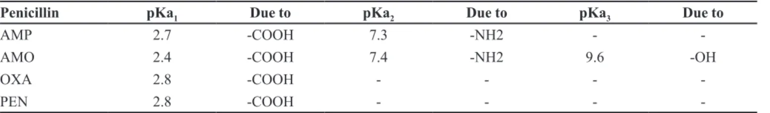
TABLE II - The pKa values of the studied penicillins
Penicillin pKa1 Due to pKa2 Due to pKa3 Due to
AMP 2.7 -COOH 7.3 -NH2 -
-AMO 2.4 -COOH 7.4 -NH2 9.6 -OH
OXA 2.8 -COOH - - -
-PEN 2.8 -COOH - - -
-possible, due to the instability of penicillins in solution. The capillaries were preconditioned with 0.1 M NaOH (2 min), distilled water (2 min) and buffer solution (2 min).
The detection was carried out by on-column photometric measurement at 210nm. Each component was identiied from the mixture based on their individual migration time and UV spectra. Previously, we recorded the UV spectra for the studied penicillins, which are relatively similar, but deinite small differences can be observed in the case of all components.
RESULTS AND DISCUSSION
Optimization of the separation conditions
Penicillins have acidic character (due to – COOH substituent) and are consequently ionizable in an alkaline environment. Aminopenicillins can be detected also in acid environment, due to the ionization of the – NH2 substituent,
but OXA and PEN could not be detected at acid pH values. The pKa values of the studied penicillins are presented in Table II (Yongxinet al.,1997 a; Hernandez, Borrull, Calull,2003).
In the preliminary analysis we used 25 mM phosphoric acid (pH –2.1), 25 mM disodium hydrogenophosphate – 25 mM sodium didydrogenophosphate (pH – 7) and 25 mM sodium tetraborate (pH – 9.3) background electrolytes (BGEs), respectively, and we also modiied the pH of the buffer by adding a 0.1 M sodium hydroxide solution. We applied some “standard” electrophoretic conditions for a CE analysis: temperature 20 ˚C, applied voltage + 20 kV, injection pressure/time 50 mbar/3 sec, sample concentration 10 μg/mL. After the initial runs, in order to obtain a good electrophoretic signal for all four penicillins, we chose a buffer containing sodium tetraborate (pH = 9.3), as an acid buffer can only be used for the simultaneous separation of AMP and AMO.
number of carboxyl and amino groups of each analyte, respectively, but also on the pH of the buffer electrolyte, as dissociation of these groups is pH dependent.
Efforts were focused on the optimization of the analytical conditions (effects of buffer concentration and pH, the presence of possible modiiers), in order to obtain better resolutions and shorter analysis times.
An increase in buffer concentration modiied only on the migration times of the analytes, but had only a slight effect on the resolution of the separation. The higher the buffer concentration, the later the migration time of each penicillin, because the electroosmotic low (EOF) decreases with an increase in ionic strength. The optimum buffer concentration was set at 25 mM.
In order to improve separation, we added an anionic surfactant, sodium dodecyl sulphate (SDS), to the buffer solution. MEKC is based on a micellar “pseudostationary” phase added to the buffer solution, which interacts with the analytes according to partitioning mechanisms, in a chromatography-like mode; the EOF acting as the chromatographic “mobile phase”. The anionic SDS micelles are electrostatically attracted towards the positive electrode, but the EOF transports the bulk solution towards the negative electrode due to the negative charge on the internal surface of the silica-fused capillaries. However, the EOF is stronger than the electrophoretic mobility of the micelle under alkaline condition; therefore, the anionic micelle will travel also towards the negative electrode with a retarded velocity (Landers, 2008).
Depending on the individual partitioning equilibria of the different analytes between the micellar and the aqueous phase, a different retarding effect on the electrophoretic mobility of the analytes will be observed. The greater the percentage of the analyte distributed into the micelles, the more slowly it migrates (Silva, 2013).
The migration time increased with the increase of SDS concentration, due to the solubilization of the analytes in the micellar phase. In the presence of SDS the resolution of the separation improved considerably. The optimum surfactant concentration was set at 100 mM SDS.
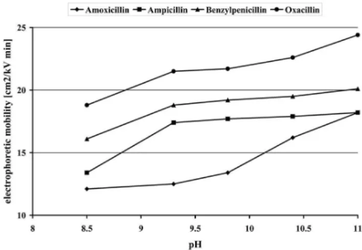
The pH of the buffer is the main factor affecting resolution. The pH was adjusted by adding 0.1 M NaOH respectively 0.1 M HCl solutions to the buffer solution. Migration times had the tendency to increase at high pH values, but the resolution became poor. The optimum pH value for the separation was set at 9.3. The inluence of pH values of the background electrolyte on the effective electrophoretic mobilities of the studied penicillins is presented in Figure 1.
In order to optimize the electrophoretic conditions, we studied the influence of the applied voltage and
temperature on the separation. The increase of the voltage and temperature, respectively, results in the decrease of the migration times, but with little effect on the resolution. The optimum voltage was set at +25 kV while the optimum temperature was set at 25 °C, in order to obtain good resolutions and short analysis times.
Using buffer solution containing 25 mM sodium tetraborate and 100 mM SDS, at a pH of 9.3, applying a voltage of +25 kV at a temperature of 25°C, we achieved the simultaneous separation of the studied penicillins in less then 5 minutes, the order of separation being: AMO, AMP, BENZ, OXA (Figure 2). The migration order can be explained as AMO is the more polar molecule in the mixture (because of the – OH substituent), it consequently has the lowest affinity towards micelles and migrates fastest; while OXA is the less polar molecule in the mixture (because of the large izoxazolyl substituent), it has the best afinity towards micelles and migrates last.
Analytical performance
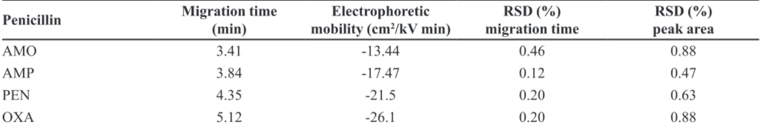
The optimized separation method was evaluated based on precision (migration times and peak areas), linear range, limit of detection (LD) and limit of quantiication (LQ) (Table III/Table IV).
Very similar migration times and peak areas were obtained for six repeated measurements of the 4 analytes, as the RSD values were smaller than 1%. As it is usual, the precision for migration times was better than of peak areas.
The LD and LQ were calculated as the sample concentration that produces a peak signal-to-noise ratio of 3:1 and 10:1, respectively
FIGURE 1 - The inluence of pH of the background electrolyte
on the separation of penicillins (separation conditions:
capillary 56 cm x 50 μm I.D.; buffer electrolyte: 25 mm sodium tetraborate + 100 mM SDS; temperature 20 ˚C; applied voltage +
TABLE IV - Linearity regression dataof penicillins determination
Penicillin Correlation coeficient LD (μg/ml) LQ (μg/ml) F
AMO 0.994 3.22 10.73 3.34
AMP 0.997 2.15 7.18 3.03
PEN 0.995 2.81 9.37 3.18
OXA 0.996 2.69 8.99 3.23
concentration range=1-100 μg/mL, LD – S/N=3, LQ – S/N=10 TABLE III - Analytical parameters of penicillins determination
Penicillin Migration time
(min) mobility (cmElectrophoretic 2/kV min)
RSD (%)
migration time
RSD (%) peak area
AMO 3.41 -13.44 0.46 0.88
AMP 3.84 -17.47 0.12 0.47
PEN 4.35 -21.5 0.20 0.63
OXA 5.12 -26.1 0.20 0.88
c=1 mg/mL, n=6
FIGURE 2 - Electropherogram of the separation of the 4 studied penicillins (separation conditions: capillary 56 cm x 50 μm I.D.; buffer electrolyte: 25 mm sodium tetraborate + 100 mM SDS; pH – 9.3; temperature 25 ˚C; applied voltage + 25 kV; UV detection
at 210 nm).
The individual linear regression equations were calculated according to six concentrations in a speciic range and three replicates per concentration. Linearity was evaluated taking into account the correlation coeficients; as correlation coeficients higher than 0.99 are considered to be evidence of good data itting to line regression.
Stability study of penicillins
The instability of pencillins, due to the presence of β-lactamic ring, is well known and represents a controversial problem. Penicillins dissolved in water undergo a rapid hydrolysis, being gradually converted to different degradation products (Block, Beale, 2004; Kummerer, 2004).
After dissolution of the penicillins in water, the sample solutions were reinjected several times over the duration of two weeks, using ciproloxacin hydrochloride as internal standard.
Ciprofloxacin is a fluoroquinolone derivative, a zwitterionic compound, which can ionize in both acid and alkaline environment. Ciproloxacin exhibits a smaller electrophoretic mobility than penicillin derivatives; consequently, it will migrate after the last studied penicillin. Its stability in water is relatively good (the rate of decrease over two weeks was less than 5%).
N N
F
N O
COOH
H
Ciprofloxacin
The stability of penicillins was evaluated applying a simple CZE method, using a 25 mM sodium tetraborate buffer, at a pH=9.3, a voltage of +25 kV, hydrodynamic sample injection, injection pressure/time 30 mbar/5 seconds, detection at 210 nm. We performed the separation using a short capillary of 38 cm (effective length: 30 cm) x 50 μm I.D, in order to obtain shorter analysis times.
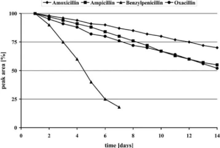
The degradation of the compounds depends highly on the temperature of the medium, with an increase of the degradation rate at higher temperatures. In our experiments, room temperature (25 °C) and refrigerator temperature (4 °C) were tested.
Figure 3 and Figure 4 present the stability diagrams of the four studied penicillins at 25 °C and 4 °C. Peaks areas obtained for the irst analysis were regarded as 100%.
From the curves of Figures 3 and 4 it is obvious that the three semisynthetic penicillins (AMO, AMP, OXA) exhibit a quite similar decomposition proile, as their degradation was around 20% after two weeks at 4°C respectively 30-40% at 25 °C. PEN is by far the most unstable of the four studied substances, as its degradation
was generally around 50% at 4 °C and complete after a week at 25 °C.
At 4 °C the degradation of compounds is considerably lower in comparison with the degradation of the same compounds at 25 °C. The degradation depends also on the pH, but further investigations are necessary.
This stability investigation was facilitated by the automatic measurement repetition/ time-programming mode of the CE system.
CONCLUSIONS
CE has proven to be an important and versatile FIGURE 3 - Solution stability diagram of the four penicillins
stored at refrigerator temperature (4 °C) (separation conditions: capillary 38 cm x 50 μm I.D.; buffer electrolyte: 25 mm sodium tetraborate; pH – 9.3, temperature 20 °C; applied voltage +
25 kV; UV detection at 210 nm).
FIGURE 4 - Solution stability diagram of the four penicillins at
room temperature (25 °C) (separation conditions: capillary 38 cm x 50 μm I.D.; buffer electrolyte: 25 mm sodium tetraborate; pH – 9.3, temperature 20 °C; applied voltage + 25 kV; UV
technique for the analysis of the investigated penicillin derivatives. The reason for studying a mixture of penicillins was to prove the applicability of CE for the analysis of penicillins in general, and for the analysis of the studied penicillins in particular. Using the optimized analytical conditions the method can be used for the analysis and identification of drugs in formulated products and also resolving separations from complex mixtures of drugs.
The stability studies showed that the extent of the hydrolysis of penicillins is highly dependent on time and temperature. The degradation rate is much smaller in the case of samples stored at lower temperatures; consequently, the samples should be stored at 4 °C and analyzed within 24 h of dissolution. This is especially important if the separation method is being applied for direct determination of penicillins from clinical and environmental samples.
REFERENCES
AHUJA, S.; JIMIDAR, I.M. Capillary electrophoresis methods for pharmaceutical analysis. London: Academic Press
Elsevier, 2008. p.33-40.
BAILON PEREZ, M.I.; GARCIA-CAMPANA, A.M.; DEL OLMO IRUELA, M.; CRUCES BLANCO, C.; GRAMIS GRACIA, L. Multiresidue determination of penicillins in environmental waters and chicken muscle samples
by means of capillary electrophoresis-tandem mass spectrometry. Electrophoresis, v.30, p.1708-1717, 2009.
BAILON PEREZ, M.I.; CUADROS RODRIGUEZ, L.; CRUSSES BLANCO, C. Analysis of different beta-lactams
antibiotics in pharmaceutical preparations using micellar
electrokinetic capillary chromatography. J. Pharm. Biomed. Anal., v.43, p.746-752, 2007.
BLOCK, J.H.; BEALE, J.M. Wilson and Gisvold’s textbook of organic medicinal and pharmaceutical chemistry. 11ed.
Philadelphia: Lippincott Williams&Wilkins, 2004. p.311-329.
HERNANDEZ, M.; BORRULL, F.; CALULL, M. Analysis of
antibiotic in biological samples by capillary electrophoresis.
Trac-Trend. Anal. Chem., v.22, p.416-427, 2003.
KOWALSKI, P.; KONIECZNA, L. Determination of penicillin
antibiotics in poultry muscle by capillary electrophoresis.
Bull. Vet. Inst. Pulawy, v.51, p.595-598, 2007.
KUMMERER, K. Pharmaceuticals in the environment, sources, fate, effects and risks. Berlin-Heidelberg: Springer-Verlag,
2004. p.58-67.
LANDERS, J.P. Handbook of capillary and microchip electrophoresis and associated microtechniques. New York:
CRC Press, 2008. p.109-135.
SWEETMAN, S. Martindale: the complete drug reference. 37.ed. London: The Pharmaceutica Press, 2011. 4160 p.
NOZAL, L.; ARCE, L.; RIOS, A. Development of a screening
method for analytical control of antibiotic residues by
micellar electrokinetic capillary electrophoresis. Anal. Chim. Acta., v.523, p.21-28, 2004.
PAJCHEL, G.; MICHALSKA, K.; TYSKI, S. Analysis of phenoxymethylpenicillin potassium by capillary
electrophoresis. J. Chromatogr. A., v.1087, p.197-202, 2005.
PUIG, P.; BORRULL, F.; CALULL, M.; AGUILAR, C. Sample stacking for the analysis of eight penicillin antibiotics by micellar electrokinetic capillary chromatography.
Electrophoresis, v.26, p.954-961, 2005.
SCHMIDT-KOPPLINS, P. Capillary electrophoresis: methods
and protocols. New Jersey: Humana Press, 2008. p.205-247.
SILVA, M. Micellar electrokinetic chromatography: a review of methodological and instrumental innovations focusing on
practical aspects. Electrophoresis, v.34, p.141-158, 2013.
TIAN, C.; TAN, H.; GAO, L.; SHEN, H., QI, K. Determination of penicillin intermediate and three penicillins in milk by
high performance capillary electrophoresis. Se Pu, v.29,
p.1128-1132, 2011.
YONGXIN, Z.; DALLE, J.; VAN SCHEPDAEL, A.; ROETS, E.; HOOGMARTENS, J. Analysis of benzylpenicillin by
capillary electrophoresis. J. Chromatogr. A., v.792,
p.83-88, 1997a.
YONGXIN, Z.; VAN SCHEPDAEL, A.; ROETS, E.; HOOGMARTENS, J. Micellar electrokinetic chromato-graphy for the separation of phenoxymethylpenicillin and
related substances. J. Chromatogr. A, v.781, p.417-422,
1997b.
Received for publication on 13th April 2013