Human dental pulp stem cell adhesion and
detachment in polycaprolactone electrospun
scaffolds under direct perfusion
A. Paim
1,2,3, D.I. Braghirolli
3, N.S.M. Cardozo
2, P. Pranke
3,4and I.C. Tessaro
1 1Laboratório de Separac¸ão por Membranas, Departamento de Engenharia Química, Universidade Federal do Rio Grande do Sul, Porto Alegre, RS, Brasil 2
Laboratório de Simulac¸ão, Departamento de Engenharia Química, Universidade Federal do Rio Grande do Sul, Porto Alegre, RS, Brasil 3
Laboratório de Hematologia e Células-Tronco, Faculdade de Farmácia, Universidade Federal do Rio Grande do Sul, Porto Alegre, RS, Brasil 4Instituto de Pesquisa com Células-Tronco, Porto Alegre, RS, Brasil
Abstract
Cell adhesion in three-dimensional scaffolds plays a key role in tissue development. However, stem cell behavior in electrospun scaffolds under perfusion is not fully understood. Thus, an investigation was made on the effect offlow rate and shear stress, adhesion time, and seeding density under direct perfusion in polycaprolactone electrospun scaffolds on human dental pulp stem cell detachment. Polycaprolactone scaffolds were electrospun using a solvent mixture of chloroform and methanol. The viable cell number was determined at each tested condition. Cell morphology was analyzed by confocal microscopy after various incubation times for static cell adhesion with a high seeding density. Scanning electron microscopy images were obtained before and after perfusion for the highestflow rate tested. The wall pore shear stress was calculated for all testedflow rates (0.005–3 mL/min). An inversely proportional relationship between adhesion time with cell detachment under perfusion was observed. Lowerflow rates and lower seeding densities reduced the drag of cells by shear stress. However, there was an operational limit for the lowestflow rate that can be used without compromising cell viability, indicating that aflow rate of 0.05 mL/min might be more suitable for the tested cell culture in electrospun scaffolds under direct perfusion.
Key words: Cell adhesion; Perfusion; Shear stress; Stem cell; Electrospun scaffolds
Introduction
In tissue engineering, scaffolds are used as substitutes for damaged tissue and act as a support for cell prolifera-tion, differentiaprolifera-tion, and migration. In order to promote the formation of natural extracellular-matrix, a scaffold must be designed with appropriate biocompatibility, biodegrad-ability, architecture, and mechanical properties (1).
An important class of scaffolds for tissue engineering is based on electrospun polymer-based structures com-prising solid microfibers or nanofibers, which can present high packing density and interconnected pore network (2). Nanofiber scaffolds favor higher mesenchymal stem cell viability than smooth surfaces (3). However, nanofiber scaffolds usually present small pores (4) that can hinder cell infiltration through three-dimensional structures (2). On the other hand, microfiber scaffolds can provide structures with bigger pores, allowing the cell migration and coloni-zation inside the matrix (5).
Perfusion culture systems enhance mass transfer in scaffold-containing bioreactors and provide increased nutri-ent transport and cell viability (6), migration (7), growth, and differentiation (8). In addition, perfusion bioreactors can reduce the accumulation of toxic metabolites and degra-dation byproducts and the polymer degradegra-dation rate (9). Nevertheless, high shear stress can provoke cell detach-ment followed by cell death (10). Consequently, the cell number in three-dimensional (3D) scaffolds under perfu-sion is influenced by the cell detachment provoked by shear stress (11) and the capability of the cells to remain adhered to the scaffold and to proliferate, differentiate, and migrate is strongly dependent on the flow rate and the pore size employed. This is important because in order to obtain a homogeneous and effective regenera-tion of damaged tissue, it is essential to produce a bio-material with an adequate cell number for implantation.
Correspondence: A. Paim:<[email protected]>
Received September 12, 2017 | Accepted January 11, 2018
Therefore, it is necessary to quantify the cell drag and the
final cell number in perfusion bioreactors to produce tissue substitutes thatfit the quality standard required in a medical environment. Despite this, many studies on perfusion systems based on 3D scaffolds focus on the flow rate and shear stress effect on nutrient transport and stem cell proliferation and differentiation (12–14), without evaluating
the cell detachment from the scaffold.
This work addressed the reduction of the shear stress effects inside the scaffold pores under perfusion to pro-duce cellularized electrospun structures for clinical appli-cation. An investigation was made offlow rate and shear stress under direct perfusion in polycaprolactone electro-spun scaffolds on human dental pulp stem cell detachment. The influence of the adhesion time on cell adhesion and detachment under static conditions was also evaluated. Different seeding densities were tested under perfusion to evaluate the detachment.
Material and Methods
Scaffold production
The scaffolds were produced in an electrospinning apparatus with temperature and humidity control (EC-CLI, IME Technologies, Netherlands). A 16% w/w solution of polycaprolactone (Sigma-Aldrich, USA; Mw 70-90 kDa) in a chloroform:methanol 9:1 vol% mixture was electrospun at 38% humidity, 19°C, 35 cm distance between the needle and the collector,flow rate of 0.1 mL/min, and voltage of 17 kV. The scaffolds were cut into 16 mm diameter disks and sterilized by ultraviolet radiation (UV) for 1 h.
Cell isolation and expansion
The pulp of human deciduous teeth was used to obtain dental pulp stem cells with the approval of the Research Committee and the Ethics Committee of the Universidade Federal do Rio Grande do Sul (project No. 33177214.1. 3001.5330), according to the methodology described by Werle et al. (15). Human deciduous teeth with physiologic root resorption were extracted and immersed in DMEM (Dulbecco’s modified Eagle’s culture medium)/Hepes (Sigma-Aldrich), supplemented with 10% fetal bovine serum (FBS; Gibco, USA), 100 U/mL penicillin and 100 mg/mL streptomycin (Gibco), for transportation. The dental pulp tissue was removed with the use of endodontic instruments and the cells were isolated from the pulp by a mechanic and enzymatic process. The isolated cells were incubated for 24 h at 37°C and 5% CO2. The primary cultures and further passages were subcultured when a confluence of 90% was reached, with medium exchange every 3 or 4 days. Five primary culture cells (between the third and eighth passages) were used in this work. The cells were characterized as mesenchymal stem cells in terms of immunophenotypic profile and differentiation potential, as presented in the Supplementary Material (Table S1 and Figure S1, respectively).
Cell viability
A colorimetric assay with water-soluble tetrazolium salts (WST-8, [2-(2-methoxy-4-nitrophenyl)-3-(4-nitrophenyl)-5-(2,4-disulfophenyl)-2H-tetrazolium]) was used to deter-mine the number of viable cells. As opposed to other common colorimetric assays, the WST-8 assay does not kill the cells, which allows continuous culturing of the cells, performing other assays with the living cells, and preserv-ing the samples after the measurements. For this test, the culture medium was removed and the scaffolds were incubated with 20mL of Cell Counting Kit-8 solution (CCK-8, Sigma-Aldrich) and 180mL of fresh culture medium at 5% CO2and 37°C for 1 h. The absorbance was read in 450 nm in a microplate reader (Multiskan FC, Thermo Scientific, USA). Standard curves relating absorbance readings (450 nm) with cell number were constructed for each cell culture to determine the viable cell number.
Cell morphology
After the viability assay, the scaffolds were washed with phosphate-buffered saline solution (PBS) and fixed with 4% (w/v) paraformaldehyde (PFA; Sigma-Aldrich) for 30 min. The PFA was then removed and the scaffolds were washed again with PBS. Cytoskeleton and nuclei cells were stained with 50mg/mL rhodamine-phalloidin (Molecular Probes, USA) for 40 min and 0.5 mg/mL 40 ,6-diamidino-2-phenylindole (DAPI) for 5 min, respectively, for further imaging using a confocal microscope (Olympus FV1000, Japan). For scanning electron microscopy (SEM; JEOL JSM 6060, Japan), the scaffolds were washed with PBS,
fixed with 3% glutaraldehyde and dehydrated in graded ethanol baths before being sputtered with gold and imaged.
Scaffold properties
The porosity of scaffolds was calculated from the volume offibers and the total volume of the scaffold. The volume of the fibers (Vfibers) of the scaffold was deter-mined by dividing the weight of the scaffold by the PCL density, and the total volume of the scaffold (Vtotal) was determined taking into account its geometry. The total porous fraction (y), thereby, is given by Equation 1.
y¼Vtotal Vfibers Vtotal
ðEq:1Þ
The average shear stress on the wall of the pores was calculated considering a cylindrical pore approximation (16), using Equation 2.
s¼ 8mQ dpyp D2
2 ðEq:2Þ
where m is the medium viscosity (Pa.s) (similar to that of water at 37°C), dpis the pore diameter (m) (measured through analysis of SEM images of the scaffolds with the software ImageJ), Q is the flow rate (m3/s), and D is the scaffold diameter (m).
Cell adhesion and drag
Initially, 1.5105 or 0.5105 cells were seeded in each scaffold and the volume was completed to 1 mL with culture medium in each well. Different adhesion times -3 h (17), 6 h (18), and 24 h (19,20) - were used for mesenchy-mal stem cell attachment in the 3D scaffolds. In order to evaluate the impact of the adhesion time on cell adhesion and morphology, cells were then incubated at 5% CO2and 37°C for 3, 6, and 24 h for cell adhesion. Three cell cul-tures were used in this experiment and the number of scaffolds analyzed for each adhesion time was 3 per culture. Firstly, the scaffolds seeded with 0.5105cells and incubated for 3, 6, and 24 h were perfused with culture medium for 18 h at 0.05 mL/min, in order to evaluate the adhesion strength of each adhesion time group. Two scaf-folds per culture were analyzed for each adhesion time under flow and three cell cultures were used. The bio-reactor system used for the culture medium perfusion is shown in Figure 1.
Considering that increased cell adhesion and spread-ing have been reported in the literature with longer adhe-sion times (19–21), the effect of the flow rate in cell
detachment was evaluated following an adhesion time of 24 h under static conditions, to guarantee that the cells were fully adhered and would not be detached withflow perfusion due to poor adhesion. In addition, the perfusion time used in further experiments was changed from 18 to 24 h to guarantee that maximum cell drag was achieved. Therefore, the scaffolds with 24 h cell adhesion were transferred to the bioreactor chambers (Figure 1) and incubated at 5% CO2and 37°C for 24 h of perfusion in a bioreactor system.
As cell drag was expected to increase with the flow rate, the scaffolds with high and low cell densities were
perfused at high and low flow rates, respectively, to guarantee that the viability measurements remained above the detection limit. Thus, the scaffolds seeded with 0.5 105cells were perfused with culture medium at theflow rates of 0.005, 0.01, 0.05, and 0.1 mL/min (Figure 2), while those seeded with 1.5105cells were perfused at 0.75, 1.5, and 3 mL/min (Figure 3). Two cell cultures were used in these experiments and the number of scaffolds analyzed for eachflow rate was 4 per culture.
The schemes of the experimental procedure for cell adhesion and perfusion experiments are presented in Figures 2 and 3, with an indication of the steps in which cell viability and morphological characteristics were determined.
It was also considered that with the reduction of the
flow rate, the time required to fill the perfusion system increased. Hence, if the arrival of culture medium to the scaffold occurs with an increased delay, the cells could starve due to the lack of nutrients. Thus, at lowflow rates (0.005–0.1 mL/min), the perfusion chambers were atfirst
partially filled with culture medium at a higher flow rate (0.5 mL/min). Theflow rate was then decreased to 0.005, 0.01, 0.05, or 0.1 mL/min before the fluid reached the scaffold in any of the chambers.
The average drag ratio was calculated by subtracting the average cell number (determined with the CCK-8 kit) after perfusion from the control average cell number (non-perfused scaffold).
Statistical analysis
Normal distribution was verified with the combina-tion of the Shapiro–Wilk test and visual inspection of Q-Q
plots. Statistical analyses were performed using one-way ANOVA followed by Tukey’s post hoc test, and were
carried out with R Statistical Software (version 3.3.2; R Foundation for Statistical Computing, Austria).
Results and Discussion
Cell morphology
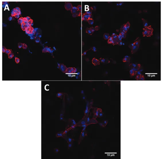
Figure 4 presents the confocal images of scaffolds seeded with 1.5105cells and incubated for 3, 6, and 24 h. Additionally, a similar set of images with smaller
magni-fication can be seen as Supplementary Material (Figure S2) to show that the effects observed in Figure 4 do not depend on the specifically focused region. It can be observed that the cell shape was still round after 3 h of adhesion (Figure 4A). At 6 h (Figure 4B), the area of actin fibers stained with phalloidin was higher and after 24 h of adhesion, a spread morphology can be observed (Figure 4C). These results indicate that cytoskeleton spreading was increased with longer adhesion times. As larger cell spreading has been associated with increased focal adhesion size (22) and strength (23), it can be expected that after 24-h adhesion, the cells will be more strongly attached to thefibers of the scaffold.
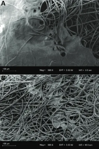
To evaluate the cell distribution on the scaffold surface, SEM analyses were performed before and after perfusion.
Figure 5 presents SEM images of the scaffolds seeded with 1.5105cells after 24-h cell adhesion followed by 24-h perfusion. As can be seen, after 24-h adhesion, the cells organize themselves in agglomerates, forming regions containing a layer of cells on the matrix surface, obstructing the pores. Several studies support that the pore size is responsible for cell infiltration and shear stress is applied to the cells due to the passage offlow in perfusion bioreactors (24,25). In this study, the porosity (93±1%) and pore diameter of the scaffolds (11.97±
4.36 mm) did not necessarily limit cell penetration into the scaffold, if the cell diameter range of 12.2–16.6mm
is considered, as suggested by Suchanek et al. (26). However, with the static seeding, the cells adhered on the
fibers covering the scaffold surface and occupying the pore spaces (as shown in Figure 5A). Due tofluidflow, the cells obstructing large pores were detached and dragged with the culture medium under high velocities (Figure 5B), and eventually detached because of the shear stress applied, even under lowflow rates.
Cell adhesion and drag
Figure 6 presents the results of cell number, deter-mined by WST-8, for the adhesion times of 3, 6, and 24 h
Figure 2.Scheme of the experimental procedure for cell culture with seeding density of 1.5 105cells/scaffold (WST-8, PFA, GTA, and DAPI are, respectively, the viability assay, paraformal-dehyde, glutaralparaformal-dehyde, and 40 ,6-diamidino-2-phenylindole).
Figure 3.Scheme of the experimental procedure for cell culture with seeding density of 0.5105 cells/scaffold (WST-8 is the viability assay).
for the scaffolds seeded with a low seeding density (0.5105cells/scaffold) with the cell cultures I, III, and IV (Figure 6A) and with a high seeding density (1.5105cells/ scaffold) with the cell cultures I and II (Figure 6B). For low seeding density, there was no significant effect of adhe-sion time on cell number determined by cell viability for cultures III and IV, while for culture I a significant (Po0.05)
decrease of mean cell number occurred after 24-h adhe-sion compared to the lower adheadhe-sion times (3 and 6 h). For high seeding density, there was no significant dif-ference between the values of the mean cell number obtained at the different adhesion times for culture I. For culture II, a decrease of this parameter was also observed between 6 and 24 h, after an initial increase between the adhesion times of 3 and 6 h. However, these results are probably exhibiting a behavior related to the intrinsic characteristics of this specific cell culture, which can be an outcome with primary cell cultures due to the variability
between donors (27–31). Harumi Miyagi et al. (31) observed
donor-to-donor variation of the expression of extracellular matrix proteins with different human dental pulp stem cells from deciduous teeth, which could justify the different adhesion behavior between the cultures presented in Figure 6. This is in agreement with the fact that mesen-chymal stem cells, as anchorage-dependent cells, can undergo cell death by the lack of appropriated attachment to a substrate (32).
A further aspect to be mentioned about Figure 6 is that at both low and high seeding density, significant dif-ferences between the cultures regarding the number of cells were observed. This can be a result of the use of cells derived from different individuals. Donor-to-donor variability can occur due to several factors such as donor age and gender, and it has been reported in several studies with primary cultures of human mesenchymal stem cells (27–31).
Figure 7 presents the cell drag percentage calculated from the viable cell numbers (determined by WST-8) obtained for the scaffolds seeded with 0.5105cells and perfused at a flow rate of 0.05 mL/min for 18 h. As can be seen, there was no effect of adhesion time in cell loss under perfusion at 0.05 mL/min for cultures I and IV because no significant difference was observed for the different adhesion time groups. In addition, mean cell drag, calculated as the average drag from the three cultures, presented no significant difference between the different adhesion time groups (mean cell drag of 17±11,
20±28, and 5±6% for scaffolds with 3, 6, and 24 h of
adhesion time, respectively). However, culture III pre-sented significantly different cell drag when seeded with 6-h adhesion compared to the other cultures with the same adhesion time (Po0.001), and to the same culture with
other adhesion times (Po0.001). Furthermore, culture I
presented no cell loss for 6 and 24 h (0% cell drag). These reduced cell losses can be related to a higher cell
spreading observed at 6 and 24 h of adhesion, observed in Figure 4. Similar results to those obtained for cultures I and IV were observed by van Kooten et al. (33) in bi-dimensional studies using parallel-plateflow chambers, where tangentialflow was used to induce shear stress and detach a cell population from a surface. The authors observed that cell adhesion strength, determined as the shear stress level that promotes 50% of cell detachment, was not sensitive to adhesion time. However, 3D attach-ment results in different cell morphology (bridged form) than cell adhesion in 2D structures (flat shape) (34). Furthermore, reduced cell adhesion strength and resis-tance to shear stress can be observed in 3D scaffolds under perfusion conditions because the cells can adhere in an orientation normal for theflow and lead to increased cell detachment under lowflow rates (35). However, cell attachment in bi-dimensional structures result inflat form morphology (34). In this study, with the increase of adhe-sion time, the cells, initially adhered to thefibers (Figure 4A),
Figure 5.SEM images of scaffolds seeded with 1.5105cells for 24 h with cell culture V before (A) and after (B) culture medium perfusion for 24 h with a flow rate of 1.5 mL/min. Magnification 500.
Figure 6.Cell number for different adhesion times for cell cultures I, III, and IV seeded with 0.5105 cells (A) and for cell cul-tures I and II seeded with 1.5105cells (B). Data are reported as means±SD. Different capital letters represent significantly dif-ferent means for the groups with the same adhesion time. Difdif-ferent lowercase letters preceded by the culture number represent sig-nificantly different means for the groups of the same culture with different adhesion times (one-way ANOVA withpost hocTukey test, Po0.05).
stretched through thefibers and the pore space to adhere to other cells andfibers (Figure 4C). This is in accordance with the bridged form morphology (cells attached to more than one fiber) obtained by Binulal et al. (34) for cell attachment in 3D electrospun scaffolds. Furthermore, since the cells adheredfilling the pore space (Figure 5), the main orientation to cell attachment in the studied system is expected to be perpendicular to theflow direc-tion, which differs from the parallel flow used by van Kooten et al. (33). According to McCoy and O’Brien (35), reduced cell adhesion strength and resistance to shear stress can be observed in 3D scaffolds under perfusion conditions, because cells can adhere in an orientation normal for theflow and lead to increased cell detachment under lowflow rates. This could explain the mechanism of cell drag in a direct perfusion system and the distribution of the cells after perfusion, observed in Figure 5B. Thus, it could be that cultures I and IV presented no enhance-ment in adhesion strength, with larger adhesion times due to this relationship between cell morphology andflow direction.
Figure 8 presents the values of cell drag percent-age calculated from viable cell numbers (determined with WST-8) in the scaffolds from cell cultures I, III, and IV, seeded with 0.5105cells and perfused with
flow rates varying from 0.005 to 0.1 mL/min (Figure 8A) and from cultures I, II, III, and V, seeded with 1.5105cells and perfused withflow rates from 0.75 to 3 mL/min (Figure 8B). In all cases, the cell drag percentages were determined after 24-h adhesion followed by 24-h perfusion. As can be seen, at low seeding density, higher flow rates led to a significant increase in cell loss for culture IV (Po0.001).
This was also observed for the scaffolds seeded
(0.5105cells) with cultures I and III (Po0.01) compared
to the results withflow rates of 0.05 and 0.1 mL/min. On the other hand, culture I, when seeded at a low density, presented higher cell loss for theflow rate of 0.005 mL/min compared to the flow rate of 0.05 mL/min (Po0.0001).
For very lowflow rates as 0.005 mL/min, the loss of cell viability (observed for culture I) is probably not provoked by shear stress but by the reduction of oxygen delivery inside the perfusion chamber, because of the decreased convection. Decreased oxygen concentrations have already been reported with reduced convection in perfusion bio-reactors (36,37). Furthermore, the cell drag differences observed between the cultures can be a result of the use of cells derived from different individuals, as previously mentioned. Interestingly, at higherflow rates and seeding density there was no significant difference in cell loss between the different groups with different flow rates
Figure 7. Cell drag in scaffolds seeded with 0.5105 cells with different adhesion times and perfused with aflow rate of 0.05 mL/min (shear stress of 2.1 mPa) for 18 h. Data are reported as means±SD. Different capital letters represent significantly
different means for the groups with the same adhesion time. Dif-ferent lowercase letters preceded by the culture number repre-sent significantly different means for the groups of the same culture with different adhesion times (one-way ANOVA withpost hocTukey test, Po0.05).
and/or cultures (Figure 8B). This can be due to the higher seeding density, which results in higher cell number at the beginning of the perfusion and in an initial reduction of the permeability of the scaffold due to superficial pore obstruction. With less free space for fluid flow, the pore diameter and porosity of the scaffold are reduced, increas-ing shear stress levels (as in accordance with Equation 2) and cell drag. Additionally, it was observed that as cells are detached by the passage of flow through the pores, the amount of cells and debris in suspension is increased (results not shown). It is possible that, with a high flow rate, the high quantity of cellular particles in suspension affected the viscosity of the culture medium, also increas-ing the shear stress levels (as in accordance with Equation 2). The combination of these factors with the variability in seeding efficiency between the cultures with high seeding density (observed in Figure 6B) could have homogenized the cell drag with differentflow rates.
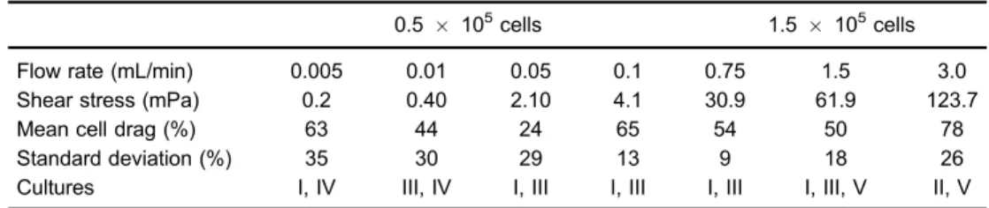
Table 1 shows the values of mean cell drag per-centage and shear stress (Equation 2) obtained with the scaffolds seeded with 0.5 and 1.5105 cells for 24-h adhesion and perfused withflow rates from 0.005 to 3 mL/min for 24 h. The lowest cell drag (24%) was obtained with a
flow rate of 0.05 mL/min, which results in a shear stress of 2.1 mPa on the pore walls. This result indicates that there is an optimalflow rate value for each system, providing thisflow rate does not provoke cell starvation or high cell detachment. However, no significant difference was observed for mean cell drag with different flow rates, which could be due to the high standard deviation of each group and donor-to-donor variability. Fibrous (but not electrospun) scaffolds with fiber diameters of 20 mm presented cell detachment values (62 and 69%) atflow rates of 0.5 and 1 mL/min (38) similar to those presented in Table 1 at
flow rates of 0.75 and 1.5 mL/min. On the other hand, high
shear stresses (values up to 56 and 57 mPa), which are close to those calculated in this work for a flow rate of 1.5 mL/min, have been shown to provoke cell washout and apoptosis (10,39). This could justify the high cell loss presented in Table 1 at high flow rates, as cell drag was calculated based on cell viability, being the possible reason for not observing the general trend reported in direct perfusion systems (i.e., increase in cell detachment with the increase offlow rate) (40) in the present work.
The detachment of human dental pulp stem cells from polycaprolactone electrospun scaffolds under direct per-fusion was studied for different flow rates, adhesion times and seeding densities. Higher adhesion time led to higher cell spreading in static conditions and reduced cell detachment under perfusion. The seeding density affected cell distribution on the scaffold surface and the sensitivity of the cells to theflow rate. High shear stress andflow rate values resulted in high cell detachments, but too lowflow rates were closer to operational constraints that could result in loss of cell viability. Thus, the lowest flow rate within a safe operating range might be more suitable for the culture of human dental pulp stem cells in electrospun scaffolds.
Supplementary material
Click here to view [pdf].
Acknowledgments
The authors wish to thank the Stem Cell Research Institute (Instituto de Pesquisa com Células-tronco), the Coordination for the Improvement of Higher Level Personnel (CAPES), the Study and Project Financer (FINEP) forfinancial support.
References
1. Khorshidi S, Solouk A, Mirzadeh H, Mazinani S, Lagaron JM, SharifiS, et al. A review of key challenges of electro-spun scaffolds for tissue-engineering applications.J Tissue Eng Regen Med2016; 10: 715–738, doi: 10.1002/term.1978. 2. Vaquette C, Cooper-White JJ. Increasing electrospun scaf-fold pore size with tailored collectors for improved cell penetration.
Acta Biomater2011; 7: 2544–2557, doi: 10.1016/j.actbio.2011. 02.036.
3. Ruckh TT, Kumar K, Kipper MJ, Popat KC. Osteogenic differ-entiation of bone marrow stromal cells on poly(ε-caprolactone) nanofiber scaffolds. Acta Biomater 2010; 6: 2949–2959, doi: 10.1016/j.actbio.2010.02.006.
Table 1.Mean cell drag and shear stress in scaffolds seeded with 0.5105and 1.5105cells with 24 h of cell adhesion and perfused for 24 h.
0.5 105cells 1.5
105cells
Flow rate (mL/min) 0.005 0.01 0.05 0.1 0.75 1.5 3.0
Shear stress (mPa) 0.2 0.40 2.10 4.1 30.9 61.9 123.7
Mean cell drag (%) 63 44 24 65 54 50 78
Standard deviation (%) 35 30 29 13 9 18 26
Cultures I, IV III, IV I, III I, III I, III I, III, V II, V
4. Gluck JM. Electrospun nanofibrous poly(ε-caprolactone) (PCL) scaffolds for liver tissue engineering. [Master’s thesis]. Raleigh: Graduate Faculty of North Carolina State University; 2007.
5. Pham QP, Sharma U, Mikos AG. Electrospun Poly(ε -caprol-actone) microfiber and multilayer nanofiber/microfiber scaf-folds: characterization of scaffolds and measurement of cellular infiltration.Biomacromolecules2006; 7: 2796–2805, doi: 10.1021/bm060680j.
6. Bancroft GN, Sikavitsas VI, Mikos AG. Design of a flow perfusion bioreactor system for bone tissue-engineering applications.Tissue Eng 2003; 9: 549–554, doi: 10.1089/ 107632703322066723.
7. Markhoff J, Wieding J, Weissmann V, Pasold J, Jonitz-Heincke A, Bader R. Influence of different three-dimensional open porous titanium scaffold designs on human osteo-blasts behavior in static and dynamic cell investigations.
Materials2015; 8: 5490–5507, doi: 10.3390/ma8085259. 8. Yeatts AB. Tubular perfusion system bioreactor for the dynamic culture of human mesenchymal stem cells. [PhD thesis]. College Park: Faculty of the Graduate School of the University of Maryland; 2012.
9. An J, Leeuwenburgh SCG, Wolke JGC, Jansen JA. Effects of stirring andfluid perfusion on thein vitrodegradation of calcium phosphate cement/PLGA composites.Tissue Eng Part C Methods 2015; 21: 1171–1177, doi: 10.1089/ten. tec.2015.0016.
10. Cartmell SH, Porter BD, García AJ, Guldberg RE. Effects of medium perfusion rate on cell-seeded three-dimensional bone constructsin vitro. tissue Eng 2003; 9: 1197–1203, doi: 10.1089/10763270360728107.
11. Sinlapabodin S, Amornsudthiwat P, Damrongsakkul S, Kanokpanont S. An axial distribution of seeding, prolifera-tion, and osteogenic differentiation of MC3T3-E1 cells and rat bone marrow-derived mesenchymal stem cells across a 3D Thai silk fibroin/gelatin/hydroxyapatite scaffold in a perfusion bioreactor.Mater Sci Eng C2016; 58: 960–970, doi: 10.1016/j.msec.2015.09.034.
12. Datta N, Pham QP, Sharma U, Sikavitsas VI, Jansen JA, Mikos AG. In vitro generated extracellular matrix andfluid shear stress synergistically enhance 3D osteoblastic dif-ferentiation. Proc Natl Acad Sci 2006; 103: 2488–2493, doi: 10.1073/pnas.0505661103.
13. Grayson WL, Bhumiratana S, Cannizzaro C, Chao P-HG, Lennon DP, Caplan AI, et al. Effects of initial seeding density andfluid perfusion rate on formation of tissue-engineered bone.Tissue Eng Part A2008; 14: 1809–1820, doi: 10.1089/ ten.tea.2007.0255.
14. Jagodzinski M, Breitbart A, Wehmeier M, Hesse E, Haasper C, Krettek C, et al. Influence of perfusion and cyclic com-pression on proliferation and differentiation of bone marrow stromal cells in 3-dimensional culture.J Biomech2008; 41: 1885–1891, doi: 10.1016/j.jbiomech.2008.04.001.
15. Werle SB, Lindemann D, Steffens D, Demarco FF, de Araujo FB, Pranke P, et al. Carious deciduous teeth are a potential source for dental pulp stem cells.Clin Oral Investig2016; 20: 75–81, doi: 10.1007/s00784-015-1477-5.
16. Santoro M, Lamhamedi-Cherradi S-E, Menegaz BA, Ludwig JA, Mikos AG. Flow perfusion effects on three-dimensional culture and drug sensitivity of Ewing sarcoma.Proc Natl Acad Sci2015; 112: 10304–10309, doi: 10.1073/pnas.1506684112.
17. Yassin MA, Leknes KN, Pedersen TO, Xing Z, Sun Y, Lie SA, et al. Cell seeding density is a critical determinant for copolymer scaffolds-induced bone regeneration.J Biomed Mater Res Part A2015; 103: 3649–3658, doi: 10.1002/jbm. a.35505.
18. Cheng Y-L, Chen Y-W, Wang K, Shie M-Y. Enhanced adhesion and differentiation of human mesenchymal stem cell inside apatite-mineralized/poly(dopamine)-coated poly (ε-caprolactone) scaffolds by stereolithography. J Mater
Chem B2016; 4: 6307–6615, doi: 10.1039/C6TB01377E. 19. Kafi MA, Phanny Y, Nakamuta Y, Todo M. Proliferation
behavior of mesenchymal stem cells in peptide functiona-lized chitosan scaffolds.The 15th International Conference on Biomedical Engineering: ICBME 2013, 2013 Dec 4–7, Singapore Cham: Springer International Publishing; 2014. p. 279–282.
20. Serra T. Development of 3d-printed biodegradable compo-site scaffolds for tissue engineering applications. [PhD thesis]. Barcelona: Universitat Politècnica de Cataluny; 2014. 21. Banik BL, Riley TR, Platt CJ, Brown JL. Human
mesench-ymal stem cell morphology and migration on microtextured titanium.Front Bioeng Biotechnol2016; 4: 41, doi: 10.3389/ fbioe.2016.00041.
22. Kim D-H, Wirtz D. Predicting how cells spread and migrate.
Cell Adh Migr2013; 7: 293–296, doi: 10.4161/cam.24804. 23. Christophis C, Grunze M, Rosenhahn A. Quantification of
the adhesion strength offibroblast cells on ethylene glycol terminated self-assembled monolayers by a microfluidic shear force assay.Phys Chem Chem Phys2010; 12: 4498, doi: 10.1039/b924304f.
24. Balguid A, Mol A, van Marion MH, Bank RA, Bouten CVC, Baaijens FPT. Tailoringfiber diameter in electrospun poly(ε-Caprolactone) scaffolds for optimal cellular infiltration in cardiovascular tissue engineering. Tissue Eng Part A
2009; 15: 437–444, doi: 10.1089/ten.tea.2007.0294. 25. Lynch ME, Fischbach C. Biomechanical forces in the
skeleton and their relevance to bone metastasis: Biology and engineering considerations.Adv Drug Deliv Rev2014; 79–80: 119–134, doi: 10.1016/j.addr.2014.08.009.
26. Suchanek J, Soukup T, Visek B, Ivancakova R, Kucerova L, Mokry J. Dental pulp stem cells and their characterization.
Biomed Pap Med Fac Univ Palacky Olomouc Czech Repub
2009; 153: 31–35, doi: 10.5507/bp.2009.005.
27. Capra E, Beretta R, Parazzi V, Viganò M, Lazzari L, Baldi A, et al. Changes in the proteomic profile of adipose tissue-derived mesenchymal stem cells during passages. Pro-teome Sci2012; 10: 46, doi: 10.1186/1477-5956-10-46. 28. Siddappa R, Licht R, van Blitterswijk C, de Boer J. Donor
variation and loss of multipotency during in vitro expansion of human mesenchymal stem cells for bone tissue engi-neering.J Orthop Res2007; 25: 1029–1041, doi: 10.1002/ jor.20402.
29. Heathman TRJ, Rafiq QA, Chan AKC, Coopman K, Nienow AW, Kara B, et al. Characterization of human mesenchymal stem cells from multiple donors and the implications for large scale bioprocess development.Biochem Eng J2016; 108: 14–23, doi: 10.1016/j.bej.2015.06.018.
31. Harumi Miyagi SP, Kerkis I, da Costa Maranduba CM, Gomes CM, Martins MD, Marques MM. Expression of extracellular matrix proteins in human dental pulp stem cells depends on the donor tooth conditions.J Endod2010; 36: 826–831, doi: 10.1016/j.joen.2010.02.020.
32. Engbers-Buijtenhuijs P, Kamphuis M, van der Sluijs Veer G, Haanen C, Poot AA, Feijen J, et al. A novel time resolved
fluorometric assay of anoikis using Europium-labelled Annexin V in cultured adherent cells. Apoptosis 2005; 10: 429–437, doi: 10.1007/s10495-005-0816-4.
33. van Kooten TG, Schakenraad JM, van der Mei HC, Dekker A, Kirkpatrick CJ, Busscher HJ. Fluid shear induced endo-thelial cell detachment from glass - influence of adhesion time and shear stress.Med Eng Phys1994; 16: 506–512, doi: 10.1016/1350-4533(94)90077-9.
34. Binulal NS, Deepthy M, Selvamurugan N, Shalumon KT, Suja S, Mony U, et al. Role of nanofibrous poly(Caprolactone) scaffolds in human mesenchymal stem cell attachment and spreading forin vitro bone tissue engineering-response to osteogenic regulators.Tissue Eng Part A2010; 16: 393–404, doi: 10.1089/ten.tea.2009.0242.
35. McCoy RJ, O’Brien FJ. Influence of shear stress in perfusion bioreactor cultures for the development of
three-dimensional bone tissue constructs: a review.Tissue Eng Part B Rev2010; 16: 587–601, doi: 10.1089/ten.teb. 2010.0370.
36. Coletti F, Macchietto S, Elvassore N. Mathematical modeling of three-dimensional cell cultures in perfusion bioreactors. Ind Eng Chem Res2006; 45: 8158–8169, doi: 10.1021/ie051144v. 37. Pathi P, Ma T, Locke BR. Role of nutrient supply on cell growth in bioreactor design for tissue engineering of hematopoietic cells.Biotechnol Bioeng2005; 89: 743–758, doi: 10.1002/bit.20367.
38. Alvarez-Barreto JF, Linehan SM, Shambaugh RL, Sikavitsas VI. Flow perfusion improves seeding of tissue engineering scaffolds with different architectures.Ann Biomed Eng2007; 35: 429–442, doi: 10.1007/s10439-006-9244-z.
39. Raimondi MT, Moretti M, CioffiM, Giordano C, Boschetti F, Laganà K, et al. The effect of hydrodynamic shear on 3D engineered chondrocyte systems subject to direct perfusion.
Biorheology2006; 43: 215–222.
40. Raimondi MT, Bertoldi S, Caddeo S, Farè S, Arrigoni C, Moretti M. The effect of polyurethane scaffold surface treat-ments on the adhesion of chondrocytes subjected to inter-stitial perfusion culture.Tissue Eng Regen Med2016; 13: 364–374, doi: 10.1007/s13770-016-9047-8.