Morphological changes in rat rectus abdominis
muscle induced by diabetes and pregnancy
G. Vesentini
1, G. Marini
1,2, F. Piculo
1, D.C. Damasceno
1, S.M.M. Matheus
3, S.L. Felisbino
4,
I.M.P. Calderon
1, A. Hijaz
5, A.M.P. Barbosa
6and M.V.C. Rudge
11
Departamento de Ginecologia e Obstetrícia, Faculdade de Medicina de Botucatu, Universidade Estadual Paulista, SP, Brasil 2Departamento de Ciências da Saúde, Universidade do Sagrado Corac
¸ão, SP, Brasil
3Departamento de Anatomia, Instituto de Biociências de Botucatu, Universidade Estadual Paulista, SP, Brasil 4Departamento de Morfologia, Instituto de Biociências de Botucatu, Universidade Estadual Paulista, SP, Brasil 5Department of Urology, Urology Institute, University Hospitals Case Medical Center, Cleveland, OH, USA 6Departamento de Fisioterapia e Terapia Ocupacional, Universidade Estadual Paulista, SP, Brasil
Abstract
The urethral muscle of diabetic pregnant rats is affected by long-term mild diabetes and short-term severe diabetes, which plays a crucial role in the pathogenesis of pelvic floor disorders. We hypothesized that muscles outside the pelvis are subject to similar changes. The current study aimed at analyzing the effects of long-term mild and short-term severe diabetes on the structure and ultrastructure offiber muscles and collagen in rats’rectus abdominis (RA) muscle. Therefore, the RA muscle of virgin, pregnant, long-term mild diabetic, short-term severe diabetic, long-term mild diabetic pregnant and short-term severe diabetic pregnant 3-month-old Wistar rats were collected. The structure was analyzed by picrosirius red staining, immunohistochemistry for fast and slow muscle fibers and transmission electron microscopy. We investigated two levels of STZ- induced diabetes: long-term mild diabetes (blood glucose level: 120-200 mg/dL) and short-term severe diabetes (blood glucose level4300 mg/dL). Long-term mild diabetic pregnant and short-term severe diabetic pregnant rats had decreased fast
fibers and increased slowfibers, disrupted areas of sarcomere, intermyofibrillar mitochondria and myelinfigures in the RA muscle. Both groups enabled us to analyze the specific influence of pregnancy, separately from diabetes. The current study demonstrated that diabetes and pregnancy induced intramuscular transformation and reorganization of RA muscle with a switch offiber type adjusting their architecture according to intensity and duration of hyperglycemic insult within pregnancy.
Key words: Diabetes; Pregnancy; Collagen; Skeletal muscle; Rats
Introduction
Screening strategy, diagnosis, and treatment of gesta-tional diabetes by American Diabetes Association are unable to prevent the high prevalence of urinary incon-tinence (UI) in women with gestational diabetes mellitus (GDM) (1,2). Suchfindings motivated our hypothesis that hyperglycemia in pregnancy negatively impacts the pelvic floor muscle function, which plays an important role in the pathogenesis of pelvicfloor disorders (1,2). Diabetes is a major factor in total economic costs, i.e. US$322 billion in 2012 (3). In addition, the direct management of UI leads to a cost of US$19.5 billion, which will continue to advance (4). It is possible to infer that GDM and UI would increase women health costs and represent a substantial economic burden for not only individual patients, but also health care systems. Diabetes-related chronic hyperglycemia has been associated with damage in different tissues, including lower
urinary tract and striated skeletal muscle (5). The term "diabetic myopathy" refers to function, metabolic and structural changes that are induced by diabetes mellitus (DM) in skeletal muscle (6,7).
A high prevalence of pelvicfloor muscle (PFM) dysfunc-tion and decreased vaginal squeeze pressure two years after cesarean delivery in women with GDM (1) inspired us to conduct translational studies using diabetic pregnant rats (8–10), since ethical issues for collection of human tissue might be impeding; therefore, animal models become valuable for studying UI pathophysiology (11). Previous studies have demonstrated that intramuscular changes occur in urethral striated muscles of streptozotocin (STZ)-induced pregnant rats using two different models: short-term severe diabetes (blood glucose level 4300 mg/dL) (8,10) and long-term mild diabetes (blood glucose level
Correspondence: G. Vesentini:<gi.vesentini@hotmail.com>
between from 120 to 300 mg/dL) (9,10). The urethral striated muscle was thin, atrophic, disorganized and asso-ciated with decreased expression of fast fibers, as well as increased expression of slow fibers. These findings suggested that PFM dysfunction detected in diabetic pregnant women might reflect changes in urethral stri-ated muscle (1,8–10). Furthermore, research groups have reported a co-contraction mechanism between the abdom-inal wall muscles and PFM in women (12,13) and the physiological participation of the abdominal wall during voiding in rats (14), showing an important interaction between abdominal wall muscles and PFM both in rats and humans. Thus, morphology and architecture of the rat’s abdominal muscles represent a valid model of the human abdominal wall musculature (15).
Some studies have already shown changes in the skeletal muscle as a result of DM (16,17). In order to assess such changes, these studies have used several types of diabetes induction, hyperglycemic levels and diabetes exposure times. The understanding of the impact of hyperglycemia intensity and duration within pregnancy on skeletal muscle morphology is still not clear, limiting the offer of means for prevention.
Due to the many considerable ethical constraints in assessing human pelvicfloor tissue to perform bench-to-bed approach, we used a rat model to observe whether changes are specific to urethral striated skeletal muscle or other muscles.
We hypothesized that the levels and/or duration of hyperglycemic insult associated with pregnancy would lead to similar changes in rectus abdominis (RA) muscle and extracellular matrix content, specifically collagen. The current study aimed to analyze the effects of intensity and duration of hyperglycemic insult within pregnancy on the structure and ultrastructure offiber muscles and collagen of RA muscle in rats.
Material and Methods
Animals
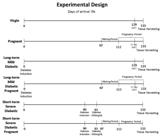
Institutional Animal Care and Use Committee in the Faculdade de Medicina de Botucatu (Universidade Esta-dual Paulista) approved the current study (protocol #1003-2013). Male and female Wistar rats (12–13 weeks, 250– 300 g) were obtained from Multidisciplinary Center for Biological Investigation (Campinas, SP, Brazil). Animals were kept at the Gynecology and Obstetrics Laboratory of Experimental Research in polypropylene cages under controlled conditions at room temperature 22±2°C, relative humidity of 55±5%, 12/12 light/dark cycle and fedad libitum
(Figure 1).
Seventy-eight rats were randomly distributed into 6 groups with 13 animals each: virgin, pregnant, long-term mild diabetic, short-term severe diabetic, long-term mild diabetic pregnant, and short-term severe diabetic preg-nant. All animals were euthanized at the end of the
experiment. Exposure time, blood glucose levels and pregnancy determined the aforementioned groups.
Long-term mild diabetic rats. On the first day of life, female newborns received STZ (Sigma Chemical Co., USA), diluted in citrate buffer (0.1 M; pH 4.5) at a dose of 100 mg/kg, using a subcutaneous route according to Iessi et al. (18). The STZ-treated rats that presented blood glucose levels higher than 400 mg/dL at the 5th day of life were included in the long-term mild diabetic group. Blood glucose concentrations were measured using a One-Touch Ultra glucometer (LifeScan, Johnson and Johnsons, USA),
and values are reported as mg/dL (18).
Short-term severe diabetic rats. Diabetes was induced in adult female rats (90 days of age) using STZ injection (Sigma Chemical Co.). The STZ was administered intra-peritoneally at doses of 50 mg/kg to induce a permanent and severe diabetic state. At 72 h after STZ injection, blood samples were obtained to confirm severe diabetic levels, and rats that presented blood glucose levels superior to 300 mg/dL were included in the short-term severe diabetic groups (19).
Virgin and pregnant rats. Both non-diabetic groups received citrate buffer at a similar volume and period according to the long-term mild and short-term severe diabetic groups. These rats presented glycemia lower than 120 mg/dL and were included in either virgin or pregnant groups.
Mating period
Adult female rats were kept overnight with non-diabetic adult male rats. To verify if mating had occurred, female rats were checked early the following morning for spermatozoa in vaginal smear. If positive, it was considered day zero of pregnancy.
Tissue harvesting
On the 21st day of pregnancy, pregnant and non-pregnant rats were euthanized by sodium thiopental injection (Thiopentaxs, Brazil 80 mg/kg dose). Fragments
of the lower third of the RA muscle were immediately obtained. For structural examination, samples were rolled in talcum powder, frozen in liquid nitrogen and stored at
–80°C. For ultrastructure analyses, samples were immersed in Karnovsky’sfixative.
Structural examination
The frozen muscle specimens were cut into 10-mm thick cross-sections using a cryostat (Leica CM 1800, Germany). The cross-sections werefixed on microscope glass slides in cold acetone for 10 min, stained with hematoxylin and eosin (H&E) or picrosirius red, and processed for immunohistochemical analysis (n=10 sam-ples/group). The slides were examined using a light microscopy and subsequently photographed (DMR, Leicas
coupled with digital camera CCD-IRIS/RGB, Sonys,
Germany).
H&E-stained slides were used to observe the general morphology. Slides stained with picrosirius red were analyzed with the color-segmentation method to deter-mine the red- (collagen) and yellow (musclefiber)-stained tissue in the same section. For analysis, 10 sections/animal were selected for morphometric analysis of collagen area and muscle area (20 magnification) using Image Pro Plus 7.0 image analysis software (Media Cybernetics, at Case Western Reserve University, USA). This software can distinguish and accurately measurefields stained with different colors by counting and converting the pixels in the image into a value for the area (mm2).
Immunohistochemistry. Immunohistochemical analysis was used to stain fast and slow-type skeletal musclefibers. Frozen sections were hydrated for 10 min; endogenous peroxidase was blocked by using H2O2(3%) in a methanol solution (97%). After washing, sections were incubated with 3% fetal bovine serum at 37°C. The sections were incubated overnight at 4°C with antibodies against WB-myosin heavy chain, fast (WB-MHCf, Novocastra, 1:120) or WB-MHC slow (WB-MHCs, Novocastra, 1:180). Then, samples were washed three times with 0.01 M PBS. Dako LSABs System-HRP Detection System (Denmark) was
utilized for further incubation at room temperature, and slides were washed three times with 0.01 M PBS. For staining, the samples were incubated with 3.3-diamino-benzidine tetrahydrochloride (Sigma-Aldrich) for 5 min, 0.01 M PBS for 10 min and hematoxylin for 10 min. The localization of fast and slowfibers was done using a light microscopy (DMR, Leicascoupled with a CCD-IRIS/RGB
digital camera, Sonys), and the
fiber type area was quantified using the image processing and analysis Java software ImageJ (National Institutes of Health, USA). This software measures and counts each fiber area after manually defining and converting the pixels in the image into a value for the area (mm2). The area was determined after measuringB200 musclefibers from each animal. In addition, the fiber type number in 4 sections/animal was counted using the ImageJ software.
Statistical analysis
Results are reported as means±SD. Comparisons between muscles and conditions were performed by repeated measures 2-way analysis of variance to avoid over influence from a single group (virgin, pregnant, long-term mild diabetes, short-long-term severe diabetes, long-long-term mild diabetes pregnant and short-term severe diabetes pregnant). Comparisons between individual groups were made using multiple comparisons with Student’st-test, as appropriate. Poisson distribution was performed when data presented no homogeneous distribution, such as fiber type number. Pearson’s correlation test was used to assess the correlation between variables (offspring number, litter weight, weight gain during pregnancy, collagen area, muscle area, slowfiber number and area, fast fiber number and area, and blood glucose levels) in pregnant groups. The significance level was set at Po0.05. All statistical analyses were performed using SAS software version 9.2. (Statistical Analysis System Institute Inc., USA).
Results
Results indicated that long-term mild diabetic pregnant and short-term severe diabetic pregnant had decreased fastfibers and increased slowfibers in RA muscle. The four
control groups, i.e., virgin, pregnant, long-term mild diabetic and short-term severe diabetic, allowed us to analyze the influence of pregnancy, separated from diabetes.
Fibers presented no morphological differences using color segmentation analysis; however, further character-ization using picrosirius red, fast and slow immunostaining and ultrastructural analysis was effective to identify such differences.
General morphologic characteristics
Virgin group. The RA of healthy adult rat females was comprised of different sizefibers with polygonal and peripheral myonuclei. By immunolocalization, a high propor-tion of fastfibers were detected, which were also generally larger than slow fibers (Figure 2a). Each fiber was surrounded by collagen, and a thick layer of connective tissue involving a small group offibers (Figure 3a). In the ultrastructural analysis, the RA showed well-organized myofibrils forming intact sarcomeres and organized triads, which are a system formed by sarcoplasmic reticulum and t tubules, with morphological traits associated with different musclefiber types and a normal distribution of intermyofi -brillar mitochondria (Figure 4a).
Pregnant group. The RA of this group were morpho-logically different from those obtained in the virgin group, with an increased number of slow fibers and increased
Figure 2.Photomicrographs of rat rectus abdominis after picrosirius red staining of striated muscle (yellow) and collagen (red). Virgin (a), long-term mild diabetic (b), short-term severe diabetic (c), pregnant (d), long-term mild diabetic pregnant (e), and short-term severe diabetic pregnant (f). Magnification 20.
collagen area (Figures 2d, 3d, 5a and 5d). Disorganized Z lines, thinned sarcomeres, and a usual form and quantity distribution of intermyofibrillar mitochondria were observed (Figure 4d).
Long-term mild diabetic group. This group showed a decrease in muscle and fastfiber area compared to the virgin group (Figures 2b, 3b, 5a and 5c). There was no difference in the collagen area (Figures 3b and 5d). In the ultrastructure analysis, the long-term mild diabetic group presented well-organized myofibrils and myelin figures associated with degenerated organelles (Figure 4b).
Short-term severe diabetic group. This group pre-sented a significantly decreased number of fastfibers and a reduced slow fiber area compared to virgin and long-term mild diabetic groups. In addition, the number of slow fibers was higher than virgin and long-term mild diabetic groups. However, the fastfiber area decreased compared to virgin and long-term mild diabetic groups (Figures 2c, 5a and 5b). The muscle area was decreased compared to virgin and short-term severe diabetic pregnant groups. The collagen area was increased compared to virgin group (Figures 3c and 5d). The ultrastructural analysis revealed numerous subsarcolemmal, intermyofibrillar mitochondria and striated muscle cells thinning in the muscle fibers (Figure 4c).
Long-term mild diabetic pregnant group. This group had a decreased number of fastfibers and area compared
to long-term mild diabetic and pregnant groups. Moreover, the slowfibers number and area were higher than long-term mild diabetic group, whereas a higher number of slow fibers was observed compared to the pregnant group (Figures 2e, 5a and 5b). There was no difference regard-ing the collagen and muscle area in the two-way analysis between the groups (Figures 3e, 5c and 5d). Ultrastruc-tural analysis showed swollen sarcoplasmic reticulum, dilated t tubes and areas with sarcomere disruption (Figure 4e).
Short-term severe diabetic pregnant group. The quan-titative analysis showed an increased number of fastfibers and a decrease in fastfiber area compared to short-term severe diabetic pregnant and long-term mild diabetic pregnant groups. The slow fiber number and area were higher than those in short-term severe diabetic group. In addition, a higher number of slow fibers compared to pregnant and long-term mild diabetic pregnant groups were observed (Figures 2f, 5a and 5b). The muscle area was greater compared to the short-term severe diabetic group and smaller compared to the pregnant group. In the collagen area, no difference was found between groups (Figures 3f and 5d). Ultrastructural analysis showed an increase in intermyofibrillar mitochondria and myelinfigures (Figure 4f).
number (r=–0.585, P=0.0007; Figure 6A). Blood glucose level was negatively correlated with fast fiber area (r=
–0.792, P=o0.0001; Figure 6B), however it was positively correlated with slow fiber number (r=0.498, P=o0.005; Figure 6C).
Discussion
Many studies have contributed to the understanding of the additive or synergistic effects of muscle function in diabetic women or rats. Also, many hypotheses have been proposed based on scientific evidence obtained from translational studies to explain the relationship between DM and muscle dysfunction (1,2,8–10). However, the current study is thefirst investigation of structural and ultrastructural alterations of RA muscles of STZ-induced diabetic pregnant rats.
Current data demonstrated that pregnancy and DM induce adaptations in rat RA muscle by systematically adjusting architectural design in eachfiber type. An increase infiber type area and number was detected in the integrated morphological analysis of rat RA in long-term mild or short-term severe diabetic pregnant rats. Our results showed an increased number of slowfibers in both diabetic pregnant models. Although a significant decreased in fast fiber
number occurred in long-term mild diabetic, fastfiber number increased in short-term severe diabetic. Therefore, we were somewhat surprised tofind a negative change offiber types in the long-term mild diabetic pregnant group compared to short-term. Possibly, a long-term mild hyperglycemic insult can have more severe detrimental impact in architectural parameters than short-term severe hyperglycemic insult. Such finding suggested a potential influence of the dura-tion of hyperglycemic insult. Moreover, a switch from fast to slowfibers supposedly represents an adaptive response to hyperglycemic status on muscle more related to hypergly-cemic duration (10).
The pathophysiological cycle of diabetic myopathy was established after Pearson correlation analysis (Figure 6). High blood glucose levels directly caused a decrease in fast fiber area during pregnancy both in diabetic and non-diabetic rats; such persistent decrease in fastfiber area led to an increase in slow fiber number that was associated with high blood glucose concentrations during diabetic pregnancy. Skeletal muscle nutrient-related atrophy, such as that observed in diabetes, reflects different intracellular pathways that are associated with protein degradation abnormalities in fastfibers (20). Increased slowfibers may result from higher glucose handling capacity from this type offiber (21). Similar changes in the normal architecture of Figure 4.Electron micrographs of rat rectus abdominis musclefibers from virgin (a), long-term mild diabetic (b), short-term severe diabetic (c), pregnant (d), long-term mild diabetic pregnant (e), and short-term severe diabetic pregnant (f) animals. The micrographs show mitochondria (m), myelin figures (M), well-organized myofibrils forming intact sarcomeres (S) and a system formed by sarcoplasmic reticulum and t tubule-triads (t), areas with disorganized Z lines and thinning sarcomeres (arrows), swollen sarcoplasmic reticulum and dilated T tubes (arrowheads) and areas with disrupted sarcomeres (*). Inset scale bar: 500 nm.
muscles subjected to a hyperglycemic environment were observed in the current study, which are consistent with previous literature (7–10,16,17).
Diabetes is associated with increased collagen, which plays a crucial role in muscle regeneration (22). In the current study, there was no change in the collagen area of the RA muscle in both diabetic pregnant groups, contrary to changes observed in collagen content of other tissues using the same diabetic model. Previously, we showed an increased collagen area in urethral striated muscle of the long-term mild diabetic pregnant group (8–10). The reason for the discrepancy between different tissue com-positions remains elusive. The striated urethral muscle closely contacts urothelium and smooth muscle. Con-versely, the skeletal muscle extracellular matrix (ECM) surrounds the muscle fibers. Krause et al. (23) demon-strated that increased expression of collagen is important for maintaining muscle integrity. In contrast, excessive colla-gen levels are pathological, leading tofibrosis and affecting normal regenerative process, with impaired infiltration of macrophages and muscle satellite cells into damaged
tissue areas. These changes might be associated with a negative impact on muscle function, such as fibrotic muscles, leading to muscle atrophy (24). The current results showed that combined diabetes and pregnancy, two important factors for increased collagen, were not enough to develop skeletal musclefibrosis in RA.
pregnant group corresponds to the impact that pregnancy has on the skeletal muscle architecture, as the ECM is a passive structure with capacity of sustaining load. ECM collagen stabilizes the elongated sarcomeres and protects the musclefibers from excessive stretching of the abdom-inal wall during pregnancy and parturition, supplying an elastic element that limits fiber tension (26,29). Ultra-structure images indicated stretched areas with disorga-nized Z lines and thinned sarcomeres (Figure 4d), as a result of pregnancy.
The interpretation of the results obtained using the STZ-induced diabetic model should be cautiously made because this diabetic induction occurred before preg-nancy in contrast with GDM that is developed during pregnancy. Future animal studies should use alternative models, considering the current knowledge of the effects of diabetes on skeletal muscle. Even though there are inherent limitations in the use of a quadrupedal animal model, the abdominal muscles of rats and humans have been previously described as similar concerning archi-tectural and morphological properties (15). Due to the differences in posture of rats and humans and to the pre-pregnancy diabetes induction, such model is valuable to establish structural adaptations in rat RA muscle. The current study represents a significant step towards future studies examining GDM effects in RA muscle during a cesarean section.
In conclusion, ourfindings demonstrated an important adaptation to excessive mechanical tension, showing intramuscular transformation and reorganization infiber types of diabetic pregnant rat RA. The adjustment of muscle architecture according to the metabolic or mechanical environment could contribute to muscle dysfunction. These results confirm RA musclefiber adaptation in pregnant rats with short-term severe diabetes, as well as pregnant rats with long-term mild diabetes showing that muscles outside the pelvis are subjected to similar structural changes related to diabetic myopathy (8–10). Understanding the pathophy-siological mechanisms that underlie diabetic myopathy, as a systemic disease, is relevant to the development of appropriate and successful long-term therapeutic strate-gies to improve quality of life.
Acknowledgments
The authors would like to thank the staff from the Gynecology and Obstetrics Laboratory of Experimental Research, particularly T. Moretto, G. Rodrigues, who is a technician in the Anatomy and Morphology Laboratory, (UNESP, campus of Botucatu), and Dr. J.E. Corrente for statistical analyses. The study wasfinancially supported by fellowship grants from Fundac¸ão de Amparo à Pesquisa do Estado de São Paulo (FAPESP 2012/25053-7 and 2014/ 14144-7).
Figure 6.Pearson’s correlation for fastfiber area and slowfiber number (A), fastfiber area and blood glucose level (B), and slowfiber number and blood glucose level (C).
References
1. Barbosa AM, Dias A, Marini G, Calderon IM, Witkin S, Rudge MV. Urinary incontinence and vaginal squeeze pressure two years post-cesarean delivery in primiparous women with previous gestational diabetes mellitus.Clinics 2011; 66: 1341–1346.
2. Chuang CM, Lin IF, Horng HC, Hsiao YH, Shyu IL, Chou P. The impact of gestational diabetes mellitus on postpartum urinary incontinence: a longitudinal cohort study on singleton pregnancies. BJOG 2012; 119: 1334–1343, doi: 10.1111/ j.1471-0528.2012.03468.x.
3. Dall TM, Yang W, Halder P, Pang B, Massoudi M, et al. The economic burden of elevated blood glucose levels in 2012: diagnosed and undiagnosed diabetes, gestational diabetes mellitus, and prediabetes. Diabetes care2014; 37: 3172– 3179, doi: 10.2337/dc14-1036.
4. Hu TW, Wagner TH, Bentkover JD, Leblanc K, Zhou SZ, Hunt T. Costs of urinary incontinence and overactive bladder in the United States: a comparative study.Urology2004; 63: 461–465, doi: 10.1016/j.urology.2003.10.037.
5. American Diabetes A. 2. Classification and Diagnosis of Diabetes.Diabetes care2017; 40: S11–S24, doi: 10.2337/ dc17-S005.
6. Krause MP, Riddell MC, Gordon CS, Imam SA, Cafarelli E, Hawke TJ. Diabetic myopathy differs between Ins2Akita+ /-and streptozotocin-induced Type 1 diabetic models.J Appl
Physiol (1985)2009; 106: 1650–1659.
7. D’Souza DM, Al-Sajee D, Hawke TJ. Diabetic myopathy: impact of diabetes mellitus on skeletal muscle progenitor cells.Front Physiol2013; 4: 379, doi: 10.3389/fphys.2013. 00379.
8. Marini G, Piculo F, Vesentini G, Damasceno D, Delella F, Calderon IMP, et al. The influence of hyperglycemia on the remodeling of urethral connective tissue in pregnant rats.
Eur J Obstet Gynecol Reprod Biol 2017; 221: 81–88,
doi: 10.1016/j.ejogrb.2017.12.032.
9. Piculo F, Marini G, Barbosa AM, Damasceno DC, Matheus SM, Felisbino SL, et al. Urethral striated muscle and extracellular matrix morphological characteristics among mildly diabetic pregnant rats: translational approach. Int
Urogynecol J2014; 25: 403–415, doi:
10.1007/s00192-013-2218-4.
10. Marini G, Piculo F, Vesentini G, Barbosa AM, Damasceno DC, Matheus SM, et al. Effects of short-term severe and long-term mild STZ-induced diabetes in urethral tissue of female rats. Neurourol Urodyn 2017; 36: 574–579, doi: 10.1002/nau.22974.
11. Hijaz A, Daneshgari F, Sievert KD, Damaser MS. Animal models of female stress urinary incontinence.J Urol2008; 179: 2103–2110, doi: 10.1016/j.juro.2008.01.096.
12. Madill SJ, McLean L. Relationship between abdominal and pelvic floor muscle activation and intravaginal pressure during pelvicfloor muscle contractions in healthy continent women.Neurourol Urodyn2006; 25: 722–730, doi: 10.1002/ nau.20285.
13. Ptaszkowski K, Paprocka-Borowicz M, Slupska L, Bartnicki J, Dymarek R, Rosinczuk J, et al. Assessment of bioelec-trical activity of synergistic muscles during pelvic floor
muscles activation in postmenopausal women with and without stress urinary incontinence: a preliminary observa-tional study. Clin Interv Aging2015; 10: 1521–1528, doi: 10.2147/CIA.S89852.
14. Smith PP, Smith CP, Boone TB, Somogyi GT. Is abdominal wall contraction important for normal voiding in the female
rat?BMC Urology2007; 7: 5, doi: 10.1186/1471-2490-7-5.
15. Brown SH, Banuelos K, Ward SR, Lieber RL. Architectural and morphological assessment of rat abdominal wall mus-cles: comparison for use as a human model.J Anat2010; 217: 196–202, doi: 10.1111/j.1469-7580.2010.01271.x. 16. Kelleher AR, Fairchild TJ, Keslacy S. STZ-induced skeletal
muscle atrophy is associated with increased p65 content and downregulation of insulin pathway without NF-kappaB canonical cascade activation.Acta Diabetol2010; 47: 315– 323, doi: 10.1007/s00592-010-0209-1.
17. Nonaka K, Une S, Tatsuta N, Ito K, Akiyama J. Changes in antioxidant enzymes and lipid peroxidation in extensor digitorum longus muscles of streptozotocin-diabetic rats may contribute to muscle atrophy.Acta Physiol Hung2014; 101: 421–428, doi: 10.1556/APhysiol.101.2014.007. 18. Iessi IL, Bueno A, Sinzato YK, Taylor KN, Rudge MV,
Damasceno DC. Evaluation of neonatally-induced mild diabetes in rats: Maternal and fetal repercussions.Diabetol
Metab Syndr2010; 2: 37, doi: 10.1186/1758-5996-2-37.
19. Yalcin O, Sen S, Usta U, Huseyinova G, Puyan FO, Kutlu K, et al. The effects of enalapril and irbesatan in experimental diabetic nephropathy.Biotechnology & Biotechnological Equip-ment2007; 21: 366–371.
20. Wang Y, Pessin JE. Mechanisms forfiber-type specificity of skeletal muscle atrophy.Curr Opin Clin Nutr Metab Care 2013; 16: 243–250, doi: 10.1097/MCO.0b013e328360272d. 21. Albers PH, Pedersen AJ, Birk JB, Kristensen DE, Vind BF, Baba O, et al. Human muscle fiber type-specific insulin signaling: impact of obesity and type 2 diabetes.Diabetes 2015; 64: 485–497, doi: 10.2337/db14-0590.
22. Berria R, Wang L, Richardson DK, Finlayson J, Belfort R, Pratipanawatr T, et al. Increased collagen content in insulin-resistant skeletal muscle.Am J Physiol Endocrinol Metab 2006; 290: E560–E565, doi: 10.1152/ajpendo.00202.2005. 23. Krause MP, Al-Sajee D, D’Souza DM, Rebalka IA, Moradi J,
Riddell MC, et al. Impaired macrophage and satellite cell infiltration occurs in a muscle-specific fashion following injury in diabetic skeletal muscle.PloS one2013; 8: e70971, doi: 10.1371/journal.pone.0070971.
24. Lieber RL, Ward SR. Cellular mechanisms of tissuefibrosis. 4. Structural and functional consequences of skeletal muscle fibrosis.Am J Physiol Cell Physiol2013; 305: C241–C252, doi: 10.1152/ajpcell.00173.2013.
25. Lalatta Costerbosa G, Barazzoni AM, Lucchi ML, Bortolami R. Histochemical types and sizes of fibers in the rectus abdominis muscle of guinea pig: adaptive response to pregnancy. Anat Rec 1987; 217: 23–29, doi: 10.1002/ar. 1092170105.
27. Crowther GJ, Jubrias SA, Gronka RK, Conley KE. A "functional biopsy" of muscle properties in sprinters and distance runners.
Med Sci Sports Exerc 2002; 34: 1719–1724, doi: 10.1097/
00005768-200211000-00005.
28. Wiik A, Gustafsson T, Esbjornsson M, Johansson O, Ekman M, Sundberg CJ, et al. Expression of oestrogen receptor alpha and beta is higher in skeletal muscle of highly
endurance-trained than of moderately active men. Acta
Physiol Scand2005; 184: 105–112, doi:
10.1111/j.1365-201X.2005.01433.x.
29. Alperin M, Kaddis T, Pichika R, Esparza MC, Lieber RL. Pregnancy-induced adaptations in intramuscular extracellu-lar matrix of rat pelvicfloor muscles.Am J Obstet Gynecol 2016; 215: 210 e1–e7, doi: 10.1016/j.ajog.2016.02.018.