Dement Neuropsychol 2015 December;9(4):369-379
369
Rocha et al. MRI in dementia with MND
Views
&
Reviews
Dementia in motor neuron disease
Reviewing the role of MRI in diagnosis
Antonio José da Rocha1,2, Renato Hoffmann Nunes1,2, Antonio Carlos Martins Maia Jr.1,2
ABSTRACT. The superimposed clinical features of motor neuron disease (MND) and frontotemporal dementia (FTD) comprise a distinct, yet not fully understood, neurological overlap syndrome whose clinicopathological basis has recently been reviewed. Here, we present a review of the clinical, pathological and genetic basis of MND-FTD and the role of MRI in its diagnosis. In doing so, we discuss current techniques that depict the involvement of the selective corticospinal tract (CST) and temporal lobe in MND-FTD.
Key words: frontotemporal dementia, magnetic resonance, motor neuron disease, amyotrophic lateral sclerosis, frontotemporal lobe degeneration.
DEMÊNCIA EM DOENÇA DO NEURÔNIO MOTOR: REVISÃO DO PAPEL DA RM NO DIAGNÓSTICO
RESUMO. As características clínicas sobreposta da doença do neurônio motor (DNM) e demência frontotemporal (DFT) compreendem um distinto ainda não totalmente compreendido, base neurológica síndrome de sobreposição clínico-patológico foi recentemente revisto. Aqui, apresentamos uma revisão das bases clínicas, patológicas e genética de DNM-DFT e o papel da ressonância magnética no diagnóstico STI. Ao fazê-lo, discutimos as técnicas atuais que retratam o envolvimento do trato corticoespinhal seletiva (TCS) e lobo temporal em DNM-DFT.
Palavras-chave: demência frontotemporal, ressonância magnética, doença do neurônio motor, eslerose lateral amiotrófica, degeneração do lobo frontotemporal.
AMYOTROPHIC LATERAL SCLEROSIS
A
myotrophic lateral sclerosis (ALS) is afatal, late onset neurological disorder characterized by motor neuron degeneration in the primary motor cortex, brainstem and spinal cord. ALS is also known as Lou Geh-rig’s disease.1,2 he term “amyotrophic lateral
sclerosis” was coined by the French
neurolo-gist Jean-Martin Charcot.3 Early studies of
ALS, beginning in the 1880s, recognized that dementia often accompanied ALS, although this association has been largely neglected until recent years.4
ALS is the most common adult-onset MND and is one of the most common neu-rodegenerative diseases. Although familial forms of ALS have been identiied, approxi-mately 90% of cases of ALS are sporadic. Men
are slightly more frequently afected than
women (1.4:1).5 It is assumed that ALS has a
relatively even distribution worldwide. It has a mean prevalence of 5.40/100,000 in Europe
and 3.40/100,000 in North America.6 In South
America, there is little information available on ALS, but it has a reported prevalence of 5.0/100,000 in Porto Alegre, Brazil.7 In most
cases, disease onset occurs during late-adult-hood (61.8 ± 3.8 years).6
he main neuropathological features of ALS include degeneration of the corticospi-nal tract (CST), extensive loss of lower motor neurons (LMN) from the anterior horns of the spinal cord and brainstem, as well as degen-eration and loss of Betz cells in the primary
motor cortex and reactive gliosis.1
Grow-ing evidence suggests that ALS is a non-cell
This study was conducted at the Santa Casa de Misericórdia de São Paulo.
1,2PhD, Division of Neuroradiology - Santa Casa de Misericórdia de São Paulo, Brazil. 1,2MD, Division of Neuroradiology - Fleury Medicina e Saúde (São Paulo - Brazil).
Antonio José da Rocha. Santa Casa de Misericordia de São Paulo / Serviço de Diagnostico por Imagem – Rua Dr. Cesário Motta Junior, 112 – 01221-020 São Paulo SP – Brazil. E-mail: [email protected]
Disclosure: The authors report no conflits of interest.
Received September 03, 2015. Accepted in final form November 10, 2015.
autonomous disease and that dysfunctional glia have an important role in the death of motor neurons. Originally, astrocytes were proposed as a central contributor to the
disease,8 but recent data have identiied equally
impor-tant contributions from microglia and oligodendrocytes.9
A common feature of many neurodegenerative diseases, including ALS, is the formation of protein aggregates/inclusions in degenerating motor neu-rons. Although these pathological structures were irst observed several decades ago, their presence still remains an issue of considerable debate. he exact composition of these protein structures remains largely unknown, but observed cytoplasmic inclusions containing a trans-active response DNA-binding protein with a molecular weight of 43 kD (TDP-43) or fused in sarcoma (FUS), as well as their association with other ALS associated pro-teins, have now become hallmark pathological features of the disease.1 he neuronal distribution and prion-like
propagation of phosphorylated TDP-43 inclusions have enabled pathologists to currently distinguish four patho-logical stages for ALS.10
In 1993, mutations in the superoxide dismutase 1
gene (SOD1) became the irst known genetic cause of
familial ALS. hese mutations account for approximately 10% of all familial ALS cases.11 Since then, mutations in
several genes have been identiied as causative in ALS.
Mutations in four genes (C9orf72, SOD1, TDP-43, and
FUS) account for approximately 65% of familial ALS
cases. Other rare genes that are causal to familial ALS
include microtubule-associated protein tau (MAPT),
pro-granulin (PGRN), valosin containing protein (VCP),
ubiq-uilin2 (UBQLN2), and charged multivesicular protein 2B
(CHMP2B) (Table 1).1
ALS is generally a pure motor disorder without any signiicant evidence of sensory symptoms, extraocular movement disturbances, bladder and bowel dysfunction, or cognitive impairment. he clinical diagnosis of ALS is supported by a combination of upper and LMN signs following the exclusion of “ALS mimic syndromes.” ALS symptoms typically start focally, in a particular segment of the body, usually asymmetrically, before spreading to other regions over time. Bulbar onset occurs in approxi-mately 25% of patients while respiratory onset is very rare.1
Upper motor neuron (UMN) signs include slow speech, brisk relexes (brisk gag and jaw jerk, brisk limb
relexes), and Hofman’s or Babinski’s signs.2,12,13 LMN
signs include atrophy, fasciculations, and weakness. Clas-sical ALS is diagnosed based upon the El Escorial
crite-ria12 when evidence of LMN degeneration by clinical,
electrophysiological or neuropathological examination
is demonstrated, along with evidence of UMN degenera-tion by clinical examinadegenera-tion in the absence of neuroim-aging, electrophysiological or pathological evidence of a better explanation.12,13 Awaji criteria have recently been
introduced to better deine lower motor neuron degen-eration, which has improved the sensitivity of early diag-nostic methods for ALS. Under the Awaji criteria, needle electromyography is considered an extension of the clini-cal examination, but the general principles of previous criteria are maintained.13
FRONTOTEMPORAL DEMENTIA
FTD is a progressive neurodegenerative condition characterized by selective involvement of the frontal and temporal lobes and is associated with changes in behavior and personality, frontal executive deicits, and
language dysfunction.14
he irst description of FTD came from Arnold Pick in 1892, who reported a patient with progressive
aphasia and anterior temporal lobar atrophy.15Alois
Alzheimer in 1911 described the pathological indings
of FTD patients,16 speciically identifying the absence
of senile plaques and the neuroibrillary tangles the had described in a disease in 1907 that bears his name. Instead, Alzheimer reported the presence of argyrophilic neural inclusions and swollen cells in FTD, later called Pick bodies and Pick cells, respectively.16
Once considered rare, FTD is now recognized as the second-most common early-onset dementia, afecting
individuals under 65 years of age.17 Furthermore, there
is clinical and neuropathological evidence that this
con-dition also occurs in individuals of an advanced age.18
he mean age of onset of FTD is typically in the ifth to seventh decades of life.17
Pathologically, there is progressive degeneration of frontal and/or anterior temporal lobe neurons, which is characterized by frontotemporal lobar degeneration
(FTLD).14 It can be divided into two major subtypes:
FTLD with tau+ inclusions (FTLD-tau) and FTLD with ubiquitin+ and TDP-43+ but tau–inclusions (FTLD-TDP). Roughly, 90% of FTD syndromes show either TDP-43 proteinopathy (50%) or tauopathy. Consensus opinion currently recognizes ive major pathological subtypes of FTLD (FTLD-tau, FTLD-TDP, FTLD-FUS, FTLD-UPS, and
FTLD-no inclusions).19 he MAPT, PGRN and, recently,
C9orf72 genes represent the three main genetic markers associated with FTD. In addition, genetic variability in
TDP-43, CHMP2B, VCP, FUS and transmembrane protein
10 6B (TMEM106B) genes contribute to <5% of cases.
MAPT, PGRN and C9orf72 are the major (95%) genetic
Dement Neuropsyc
hol 2015 December;9(4):369-379
371
Rocha
et
al.
MRI in dementia with MND
Table 1. A summary of genetic, clinical and brain histopathology data together with the possible target of mutations in ALS and/or FTD.20,82
Genes Frequency in familial cases Type of mutations Brain pathologya Likely pathological effect Clinical presentation Imaging presentation
SOD (21q22.11)
~ 20% Mainly missense SOD1/p62 Toxic aggregation - Classical ALS - Signs of UMN degeneration
FUS (16p11.2)
~5% Mainly missense and in-frame small deletions/insertions
FUS/p62 DNA/RNA metabolism - ALS (both juvenile- and adult-onset ALS; predominantly lower motor neuron involve-ment; rarely reported cognitive impairment)
- Signs of UMN degeneration
TARDBP (TDP43) (1p36.22)
~3% Mainly missense and one truncating TDP43/p62 DNA/RNA metabolism - ALS (25% bulbar-onset; cognitive impair-ment is rarely seen)
- Signs of UMN degeneration
C9orf72 (9p21.2)
~30% G4 C2 - repeat expansion TDP43/p62, p62/
repeat-dipeptides, UBQLN2
Unknown (toxic RNA, toxic aggregation, low C9orf72 expression)
- ALS (bulbar ALS > 40%); - FTD (bvFTD >80%)
- Signs of UMN degeneration,
- Global atrophy, may involve parieto-occipital region, thalamus and cerebellum
- Less frontotemporal atrophy; VCP
(9p13.3)
Rare Missense TDP43/p62 Autophagy - FTD (FTD symptoms in 30% of cases; aphasia/language deficits common); - ALS (isolated motor neuron involvement is rare, less than 2% of familial ALS cases); - Myopathy with Paget disease of bone and frontotemporal dementia
- Frontotemporal atrophy
SQSTM1 (p62) (5q35)
~3% Missense and nonsense TDP43/p62 Autophagy - FTD (behavioural disorder); - ALS (limb or bulbar ALS);
- Paget disease of bone (>1/3 of patients)
- Frontotemporal atrophy (may be asym-metric)
- May have signs of UMN degeneration OPTN Rare Missense and nonsense
(haploinsufficiency)
TDP43/p62 Autophagy - ALS; - FTD; - Glaucoma; - Paget disease of bone
–
UBQLN2 (Xp11.21)
Rare Missense TDP43/p62, UBQLN2, FUS, OPTN
Autophagy - ALS, FTD (1–2% of apparent sporadic ALS and FTD; behavioural disorders precede motor symptoms);
- Spastic paraplegia; - Multiple sclerosis
- Frontotemporal atrophy
- May have signs of UMN degeneration
GRN (17q21.32)
~10% Nonsense (haploinsufficiency) TDP43/p62 Autophagy / lysosomal pathway
- FTD (Usually bvFTD (>50%); psychosis and parkinsonism are common);
- Neuronal ceroid lipofuscinosis-11
- Asymmetrical frontotemporoparietal atrophy
CHMP2B (3p11.2)
Rare C-terminal truncation of the CHMP2B p62 Autophagy / lysosomal pathway
- FTD (early behavioural features; progressive dynamic aphasia; parkinsonism, dystonia, myoclonus, pyramidal signs later on)
- Generalized cortical atrophy at diagnosis, most marked in frontal, parietal and occipital lobes
MAPT (17q21.32)
~10% Missense and splicing of exon 10 Abnormal tau fila-ments (tangles)
Toxic aggregation (defect in neuronal cytoskeleton)
- FTD (usually bvFTD; may be associated with other tauopathies, such as progressive supra-nuclear palsy and corticobasal degeneration)
- Relatively symmetrical orbitofrontal, medial temporal atrophy
FTD is clinically characterized by diferent combina-tions of frontal lobe or frontotemporal abnormalities, including behavior changes (bvFTD) as well as gradual impairment of language skills. In this setting, primary progressive aphasia (PPA) is further subclassiied into three subtypes. he most common type is a nonluent variant, while rare logopenic and semantic dementia variants also exist.22
DEMENTIA IN NOTOR NEURON DISEASE
MND is generally considered separately, and is more often free of cognitive impairment, but a growing body of evidence supports an association between MND and frontal lobe or frontotemporal dysfunction. Cognitive impairment in MND patients is correlated with patho-logic and imaging abnormalities in the cerebral cortex beyond the motor regions. MND is now considered a complex multisystem neurodegenerative disease due to the discovery that areas other than the motor cortices
of the brain undergo degeneration.23
More than 100 years after its irst description, links between ALS and dementia were described as
associa-tions of ALS and dementia in Guam in speciic families.24
he modern age of FTD and MND research began in the 1990s, when the irst patients were recognized, and this
marked a paradigm shift for the ield.25,26 hese reports
helped to clarify that MND was associated with a speciic type of dementia that is in turn associated with frontal lobe dysfunction. Conversely, the realization that MND-FTD had distinctive neuropathology began in the 1980s with the irst reports of ubiquitin+ immunoreactive
(UI) inclusions in the cytoplasm of motor neurons.27,28
In addition, evidence of UI inclusions in the extramotor cortex was shown in both pure ALS patients and ALS
patients with dementia.28 hese UI inclusions became the
pathological hallmark of the combined FTD and MND syndrome.
Furthermore, in 2006 TDP-43 was identiied as the major inclusion protein in this condition and is asso-ciated with UI inclusions in the vast majority of ALS patients as well as in the most common pathological subtype of FTD, now referred to as FTLD with TDP-43 pathology.19,29 Recognition of this mutation in TDP-43 as
being causal to ALS and FTD quickly led to screening for other RNA binding proteins.
Mutations in the FUS gene are now shown to account
for an additional 5% of familial ALS cases and some cases
of FTD.30 Recently, the most convincing direct
molecu-lar link between ALS and FTD has been the identiica-tion of a large, intronic hexanucleotide expansion in the
previously uncharacterized C9orf72 gene of unknown
function in families with ALS, FTD, and overlapping
syndrome.31-35 his mutation accounts for approximately
40% of familial ALS, 10% of sporadic ALS, 5% of spo-radic FTD, and up to 80% of familial ALS-FTD cases, thus making it the most common cause of ALS and FTD. Many clinical MND phenotypes, including classical ALS, progressive muscular atrophy and primary lateral
sclerosis, are linked to the C9orf72 gene mutation, but
generally it is characterized by bulbar-onset, cognitive impairment at a relatively early age, and accelerated dis-ease progression.21,33-35 More recent insights revealing
that the products of these identiied genes are involved in RNA metabolism and protein homeostasis pro-vides a further mechanistic link in the pathogenesis of this spectrum.36
he frequency of FTD in MND patients varies in the literature, with symptoms of FTD observed in 5–50% of
ALS patients.37,38 Similarly, approximately 15% of FTD
patients develop clinical symptoms of motor neuron
dysfunction.37 he exact phenotype and natural history
of impaired cognition in ALS remains unclear due to the heterogeneity in patient ascertainment and meth-ods used to assess cognition. Current estimates suggest that more than half of patients with ALS have cognitive impairment. In addition to familial associations between ALS and FTD, sporadic cases of FTD in association with
ALS also seem to be common,39 although the prevalence
and etiology for this co-association remain unknown. In some instances, FTD precedes ALS by many years;
in others, ALS precedes FTD.40 It has been noted that
a percentage of ALS patients with no previous diagno-sis of FTD have early behavioural changes that precede
the onset of symptoms of ALS.41 Several suggested risk
factors for dementia in ALS include older age, male sex, lower educational level, family history of dementia, low forced vital capacity, pseudobulbar palsy, and bulbar site of onset.38,42
One possibility to explain the phenotypic split between the similar genetics of FTD and MND is the efect of pathological mutations on the speciic function of the gene product. he genetic and pathological data, as well as mutation efect, are briely summarized in Table 1. he strongest clinical, brain histopathology and func-tional overlap is observed for VCP, OPTN, SQSTM1 and
UBQLN2 genes, suggesting that these genes represent the core of the disease continuum.21 Intriguingly,
muta-tions in three of these (VCP, OPTN and SQSTM1) cause
Paget disease, in addition to ALS and FTD. hese muta-tions are believed to cause disease by inhibiting protein degradation through autophagy and the
Dement Neuropsychol 2015 December;9(4):369-379
373
Rocha et al. MRI in dementia with MND Table 2. Defining cognitive and behavioural subtypes in ALS.83
ALS-FTD ALS-bvFTD ALS patient meeting either the Neary criteria or Hodge’s criteria for FTD
ALS-PNFA ALS patient meeting Neary criteria for PNFA
ALS-SD ALS patient meeting Neary criteria for SD
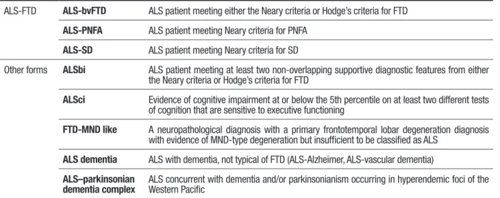
Other forms ALSbi ALS patient meeting at least two non-overlapping supportive diagnostic features from either the Neary criteria or Hodge’s criteria for FTD
ALSci Evidence of cognitive impairment at or below the 5th percentile on at least two different tests of cognition that are sensitive to executive functioning
FTD-MND like A neuropathological diagnosis with a primary frontotemporal lobar degeneration diagnosis with evidence of MND-type degeneration but insufficient to be classified as ALS
ALS dementia ALS with dementia, not typical of FTD (ALS-Alzheimer, ALS-vascular dementia)
ALS–parkinsonian
dementia complex ALS concurrent with dementia and/or parkinsonianism occurring in hyperendemic foci of the Western Pacific
ALS: Amyotrophic Lateral Sclerosis; FTD: Frontotemporal Dementia; ALS-bvFTD: Amyotrophic Lateral Sclerosis and behavioural Frontotemporal Dementia; ALS-PNFA= Amyotrophic Lateral Sclerosis and Primary Non-fluent Aphasia; ALS-SD: Amyotrophic Lateral Sclerosis and Semantic Dementia; ALSbi: Amyotrophic Lateral Sclerosis with behavioural impairment; ALSci: Amyotrophic Lateral Sclerosis with cognitive impairment; MND: Motor Neuron Disease.
SCREENING FOR MOTOR NEURON DISEASE IN FRONTOTEMPORAL DEMENTIA PATIENTS
It is critical to recognize MND associated with FTD because it greatly afects survival (8.2 years in pure FTD vs. 2.4 years in MND-FTD).43 Clinically, it is helpful to
assess the patient for fasciculations and muscle atrophy, which are non-speciic features but if present, might indicate a need for further testing. Signs of muscle weakness, spasticity, or bulbar involvement should be extensively explored. Ultimately, in suspected cases, electroneuromyography should be performed because it is considered the most sensitive measure of LMN involvement. Additionally, this test can identify early neuron loss before clinical weakness is noted. he tongue muscle should also be studied because ALS can start in any one of the four limbs or in the bulbar region.
44 Alternately, fasciculations may be demonstrated by
muscle ultrasound which is considered a feasible, reliable and well tolerated non-invasive technique for deining
LMN involvement in FTD patients.45,46. It is important
to take care to exclude ALS mimetic syndromes if any abnormalities, such as spinal disease or neuropathy, are found, because a diagnosis of MND in FTD is otherwise fatal.44
SCREENING FOR FRONTOTEMPORAL DEMENTIA IN MOTOR NEURON DISEASE PATIENTS
he prevalence of FTD in MND ranges from 22% to
48%.41 his variability depends, in part, on how FTD
is classiied and whether more subtle signs of FTD are
included. If strict Neary criteria are used, then 22% is a
more accurate igure.38 ALS patients who are clearly not
normal but have cognitive or behavioural disturbances that do not match the strict Neary criteria should be classiied based on the Strong et al. classiication (Table 2).47
A myriad of diferent cognitive screening exams have been developed centering on the need to develop shorter measures, given that a full neuropsychological battery is hard for patients to tolerate, particularly if they have advanced disease. Each of these tests are suited to dif-ferent situations because they have beneits and draw-backs in their utility.44 It is also important to consider
alternative explanations when cognitive abnormalities are observed. For example, while depression is unusual in ALS, it could certainly be a cause of apathy and other underlying psychiatric disorders that mimic FTD. Pseu-dobulbar syndrome is rarely confused with FTD, but it can afect some of the behavioural measures and even interfere with testing when severe because patients with this problem have extreme diiculty controlling their emotions. Pseudobulbar syndrome also tends to be more common in MND-FTD than in MND alone because it is more commonly found in bulbar onset patients, who are more likely to have FTD.36,38,44
NEUROIMAGING FINDINGS
of its presentation, neuroimaging studies have provided signiicant insight into the biological basis of the FTLD syndromes in MND patients. Including both morpho-logic and functional neuroimaging, MRI has largely validated the hypothesis that MND is a multi-system disorder with brain involvement well outside of the motor system.
here are many historical reports of FTD that have been superimposed onto MND, although until recently, the clinicopathological entity of this syndrome had been controversial from neuropsychological and neuropatho-logical perspectives. Neuroimaging features have been reported, and in addition to MND, degeneration in the frontal and temporal lobes is consistently observed as a pathological feature.48
It is now commonly thought that MND and FTD
rep-resent a continuum,41 and even in ALS patients, who are
cognitively normal, MRI shows the presence of abnor-malities in the frontal and temporal lobes. Although the atrophy is not as severe as that seen in ALS patients with cognitive abnormalities, the anatomical areas of involve-ment are clearly identical. A recent paper describing a family with ALS and FTD similarly shows a degree of involvement that depends upon the severity of FTD in
the MND cases.49
In this context, Mori et al.50 compared structural MRI
indings of ALS patients with dementia (ALSD) and with-out dementia to identify a pattern that would distinguish both. Patients with ALSD showed bilateral frontotem-poral atrophy mostly with temfrontotem-poral lobe dominance. In addition, in the ALSD group, T2-weighted imaging (T2WI) disclosed hyperintensity in the subcortical white matter on the medial side of the anterior temporal lobes, whereas in the group without dementia, no patients exhibited this imaging inding. he authors, however, did not distinguish groups of patients based on the genetic proile, which might have interfered with the groups and the neuroimaging indings analysis.
he relentless pursuit of structural biomarkers of dis-ease has been fruitful. Volumetric studies have shown a reasonable, although imperfect, correlation between the presence of dementia and the occurrence of atrophy in the frontal and anterior temporal lobes, which is then fol-lowed by atrophy of the anterior cingulate gyrus.51-53 Lillo
et al.54 investigated grey and white matter changes across
the ALS-FTD continuum and observed that all clinical syndromes showed grey matter changes in motor cortical and anterior cingulate brain regions. Although clinical syndromes display considerable atrophy overlap, there are also atrophy patterns speciic to each subtype of the continuum. More substantial prefrontal and temporal
cortex atrophy was indicative of bvFTD when compared to ALS and ALS-FTD, while ALS-FTD showed substan-tially more anterior cingulate and anterior temporal lobe grey matter atrophy when compared to ALS. Patients
with ALS-FTD due to C9ORF72 mutation demonstrate
symmetric frontal and temporal lobe, insular, and pos-terior cortical atrophy, although temporal involvement may be less than that seen in other mutations. Difuse cortical atrophy, that includes anterior as well as poste-rior structures and subcortical involvement, may
there-fore represent unique features of this mutation.55
Avants et al.56 used high-resolution difeomorphic
image normalization and serial MRI to provide the irst assessment of longitudinal cortical atrophy in patients with ALS-FTD relative to controls. Signiicant abnormali-ties were documented in the premotor cortex, primary motor cortex, and parietal lobe bilaterally in Brodmann areas (BA) 4, 6, and 7. he average annual cortical atro-phy over signiicant voxels in ALS-FTD on the right and left was 8.5% and 7.6% in BA4, respectively; 8.1% and 5.9% in BA6; and 3.6% and 2.2% in BA7. For all cortices in ALS-FTD patients, the atrophy rate was 1.0% per year whereas in elderly controls, the atrophy rate was 0.25% per year. he local atrophy rate did not correlate with overall brain atrophy while age and overall brain atrophy rates also did not correlate.
To outline the diiculties of implementing sophis-ticated volumetric techniques in everyday clinical prac-tice due to time and cost restraints, Ambikairajah et al.51
proposed a simple coronal MRI atrophy rating scale. he authors argued that ALS, ALS-FTD, and bvFTD patients can be distinguished by analysing four cortical grey matter regions: the motor cortex, the anterior cingulate gyrus, the anterior temporal lobe, and the orbitofrontal cortex. he authors demonstrated that bvFTD patients showed the highest levels of atrophy across all regions, while ALS patients had the lowest atrophy scores. ALS-FTD patients have higher atrophy ratings compared with ALS patients for the motor cortex, anterior cingu-late gyrus and anterior temporal lobe, with a statistical tendency for the orbitofrontal cortex. ALS-FTD patients did not difer signiicantly to bvFTD patients for any of the brain regions.
Over the last decade, conventional MRI was consid-ered to have low speciicity for the diagnosis of MND; however, non-conventional MRI, including magnetiza-tion transfer imaging, have shown dynamic utility in this setting.57 More recently, we have demonstrated the use of
Dement Neuropsychol 2015 December;9(4):369-379
375
Rocha et al. MRI in dementia with MND
particularly in early disease.58,59 Moreover, although
initially considered to be a marker of the UMN compo-nents of ALS, particularly in advanced disease, a thin line of cortical low signal intensity (“motor dark line” or “hypointense rim”) of the precentral gyrus on T2WI or Fluid-attenuated inversion recovery (FLAIR) images is neither sensitive nor speciic for the pathology of UMN degeneration in ALS and can be found in healthy indi-viduals as well as in those with other degenerative dis-eases.60 Both of these imaging indings are less prevalent
in ALS-FTD patients.61 When present, although
inconsis-tent, the MRI characterization of the CST and motor cor-tex degeneration is often relatively mild, thereby leading to fewer represented imaging indings in these regions.
In a recent study of the anatomical and radiologi-cal correlation in relevant diseases, Mori et al.50
dem-onstrated that signal-intensity changes on T2WI were observed more frequently in the pre-central white matter than in the posterior limb of the internal capsule. How-ever, it is notable that the authors used fast spin-echo T2WI, whose sensitivity for disclosing CST impairment is less than ideal. Conventional T2WI MR acquisitions have low sensitivity (about ≤ 40%) and limited
speciic-ity (about ≤ 70%)62 in demonstrating areas of abnormal
signal intensity in the CST, and these abnormalities have proven inconsistent and unreliable because they were observed frequently in normal patients and invariably did not correlate with clinical scores.
Conversely, magnetization transfer imaging (MTI) is based upon the exchange of magnetization between spins in the two diferent pools of protons: bound immo-bile protons associated with macromolecules (such as myelin) and free mobile protons associated with free
water.63 CST hyperintensity in ALS patients,
particu-larly in the supratentorial compartment, on T1 MTC was reported with great sensitivity (80%) and speciicity
(100%).58,63 his sequence has fast and simple
acquisi-tion, and is particularly useful in early MND with UMN signs to demonstrate abnormally selective hyperinten-sity throughout the CST, crossing the corpus callosum, assuming a typical ‘W-like appearance’.59,63-65
Complementary to clinical and neuropsychological evaluations, the association of asymmetrical cortical atrophy and CST composition provides key information for the imaging diagnosis of ALS-FTD (Figure 1). Pre-liminary studies in pathologically proven cases suggested that distinct patterns of tissue loss could assist in pre-dicting pathological subtype in vivo.53 We have reported
the combined involvement of both dominant frontal and temporal lobes, as well as bilateral CST involvement,
as determined by MRI in vivo using T1 MTC sequence
A
B
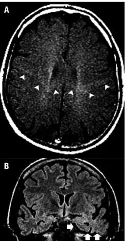
Figure 1. A man of 42 years of age presented with UMN + LMN signs
as-sociated with dementia and language-speech abnormalities (his mother had died at 67 years old with a diagnosis of ALS-FTD). [A] Axial T1 MTC image showed an abnormally selective hyperintensity throughout the corticospinal tracts, predominantly on the left side, assuming a typical ‘W-like appearance’ crossing the corpus callosum (arrowheads). [B] Coronal FLAIR image depicted a marked atrophy in the left temporal pole with blurring of the grey/white matter differentiation (arrows). Note the abnormal hyperintensity in the left amygdala and hippocampus.
and T2/FLAIR images. Our pathological indings also predominated in the same sites depicted by these MRI
sequences.66 While T1 MTC is able to demonstrate motor
and extra-motor involvement in both CST and frontal lobes and is positively coincident to brain injury, as relected in the UI distribution, T2/FLAIR images are useful for demonstrating subcortical gliosis, as conirmed by histopathological analysis (Figure 2).
Structural MRI studies have shown that bvFTD typically presents asymmetrically with a combination of frontal and anterior temporal cortical atrophy. Patterns of brain atrophy are likely to be associated with the dif-ferent pathological substrates of bvFTD.67 On structural
of the anterior temporal and ventromedial frontal lobe in the semantic variant, and left temporoparietal atrophy in logopenic patients.67,68 Conversely, subjects who
pres-ent with prosopagnosia also prespres-ent with predominantly right temporal lobe atrophy. his pattern of atrophy can also be observed in patients who present with predomi-nantly behavioural features and in those who have prom-inent geographic disorientation.69,70
In addition to the morphological alterations outlined, functional studies have encouraged the view of a con-tinuum between ALS and FTD. In a prospective study of cognition in ALS across two time points separated by six months, Strong et al.71 observed a signiicant loss
of neurons (as indicated by a reduction in the NAA/Cr ratio) in the anterior cingulate gyrus that preceded a sig-niicant loss of motor neurons in the precentral gyrus (motor cortex). he loss of anterior cingulate gyrus neu-rons was correlated with impairments in verbal praxis, a feature consistent with previous clinical and functional
neuroimaging studies. In addition, Abrahams et al.72
observed a signiicant impairment in functional MRI (fMRI) activation in the middle and inferior frontal gyri and anterior cingulate gyrus with tasks of letter luency. hese indings were interpreted as suggestive of cerebral
abnormalities in ALS in networks of regions involved in language and executive function.
Frontotemporal hypoperfusion (SPECT) in anterior and inferior regions to the primary cortex and hypome-tabolism (PET), mainly in the thalamo-frontal associa-tion pathways in some non-demented ALS patients, as well as dysfunction of the dorsolateral prefrontal cortex in ALS patients with associated cognitive impairment (i.e., deicits in letter luency, executive and memory dys-function) have been described in cognitively unimpaired patients with ALS. his indicates that an extension of cerebral involvement accompanies cognitive impairment in ALS, with a pattern shared by FTD; however, these
symptoms do not completely overlap.73-76
Difusion tensor imaging (DTI) provides quantitative information about the magnitude and directionality of water difusion in 3D space and has been used to assess patients with isolated forms of ALS (without
demen-tia) and FTD (without motor symptoms).77 Decreased
fractional anisotropy (FA) in ALS patients was found to correlate with several clinical aspects of the disease.78-80
Conversely, in bvFTD, DTI abnormalities involved pref-erentially white matter tracts located in the frontal lobes and those passing through the temporal lobes.
Never-A
B
C
D
Figure 2. A man of 61 years of
Dement Neuropsychol 2015 December;9(4):369-379
377
Rocha et al. MRI in dementia with MND
theless, difusivity changes were also identiied in more posterior white matter regions.67
Using voxel-based morphometry (VBM) and DTI analysis of brain MRI to examine grey and white matter diferences and commonalities across the continuum,
Lillo et al.54 demonstrated that, in comparison to
con-trols, bvFTD showed substantial degeneration in the forceps minor, anterior corpus callosum, anterior infe-rior longitudinal fasciculus and CST. Similarly, ALS-FTD patients showed white matter degeneration in the same tracts as bvFTD but to a lesser degree in the for-ceps minor and anterior corpus callosum. he authors also demonstrated that a more anterior portion of the inferior longitudinal fasciculus was afected, and the CST more substantially degenerated. As expected, ALS patients demonstrated more substantial changes in the CST compared to controls, while only mild abnormalities were observed in the forceps minor, anterior corpus cal-losum and the inferior longitudinal fasciculus.
Advanced MRI techniques hold the promise of cap-turing UMN loss as well as extramotor brain abnormali-ties in MND and as such deliver biomarkers relevant to diagnosis.63 Nevertheless, a correlation between imaging
parameters and clinical metrics has thus far been incon-sistent across studies.80,81
We argue that T1 MTC should be routinely included in the workup of patients with weakness and pyrami-dal signs as a sensitive and accurate imaging acquisi-tion approach useful for depicting CST involvement in ALS suspected patients.58,63,64 Structural MR sequences,
including FLAIR and 3D acquisitions, are also recom-mended when extra-motor involvement is suspected,
considering the MND/FTD spectrum.66,68 Further
investigations using structural and nonconventional
techniques are recommended to identify MR features in
MND-FTD patients and to correlate in vivo
abnormali-ties with neuropathological diagnostic criteria. Future genetic, clinicopathological and biochemical results remain necessary for fuller comprehension of the MND-FTD spectrum, while determining the imaging correla-tion amongst clinical, imaging, and histopathologic fea-tures in this overlapping syndrome is highly desirable.
CONCLUSIONS
Neuroimaging studies have provided consistent evidence for a more difuse metabolic derangement in MND that extends well beyond traditional ‘motor neuron speciic’ domains. hese indings not only support the concept of MND as syndromic but have also conirmed the wide-spread involvement of the disease process.
Despite each MND-FTLD variant being associated with characteristic behavioural and/or linguistic fea-tures, the fact that they harbour diferent underlying pathological processes renders the diagnostic work-up of these patients a highly challenging task. Nevertheless, when associated with motor neuron damage visible on MTI, the detection of distinct patterns of atrophy and/ or subcortical gliosis on combined structural and non-conventional MRI, mainly using T2/FLAIR, 3D-T1WI, DTI and VBM, in addition to functional abnormalities on SPECT and PET scans, have been shown to contribute to support a correct diagnosis of MND-FTD.
Author contributions. Dr. Rocha was responsible for the
study concept, critical revision of the manuscript and for imaging selection. Dr. Maia Jr and Dr. Nunes were responsible for the literature review and writing the manuscript.
REFERENCES
1. Leblond CS, Kaneb HM, Dion PA, Rouleau GA. Dissection of genetic factors associated with amyotrophic lateral sclerosis. Exp Neurol 2014; 262:91-101.
2. Kiernan MC, Vucic S, Cheah BC, et al. Amyotrophic lateral sclerosis. Lancet 2011;377(9769):942-955.
3. Goetz CG. Amyotrophic lateral sclerosis: early contributions of Jean-Martin Charcot. Muscle Nerve 2000;23:336-343.
4. Marie P. Lecons sur les maladies de la moelle. Paris: Masson [English translation Montagu Lubbock. London: The New Sydenham Society, 1895;CLII]. 1892.
5. Logroscino G, Traynor BJ, Hardiman O, et al. Incidence of amyo-trophic lateral sclerosis in Europe. J Neurol Neurosurg Psychiatry 2010; 81:385-390.
6. Chio A, Logroscino G, Traynor BJ, et al. Global epidemiology of amyo-trophic lateral sclerosis: a systematic review of the published literature. Neuroepidemiology 2013;41:118-130.
7. Linden-Junior E, Becker J, Schestatsky P, Rotta FT, Marrone CD, Gomes I. Prevalence of amyotrophic lateral sclerosis in the city of Porto Alegre, in Southern Brazil. Arq Neuropsiquiatr 2013;71:959-962.
8. Neusch C, Bahr M, Schneider-Gold C. Glia cells in amyotrophic lateral sclerosis: new clues to understanding an old disease? Muscle Nerve 2007;35:712-724.
9. Philips T, Rothstein JD. Glial cells in amyotrophic lateral sclerosis. Exp Neurol 2014;262:111-120.
10. Brettschneider J, Del Tredici K, Toledo JB, et al. Stages of pTDP-43 pathology in amyotrophic lateral sclerosis. Ann Neurol 2013;74:20-38. 11. Rosen DR, Siddique T, Patterson D, et al. Mutations in Cu/Zn superoxide
dismutase gene are associated with familial amyotrophic lateral sclerosis. Nature 1993;362(6415):59-62.
12. Brooks BR, Miller RG, Swash M, Munsat TL, World Federation of Neurology Research Group on Motor Neuron D. El Escorial revisited: revised criteria for the diagnosis of amyotrophic lateral sclerosis. Amyo-troph Lateral Scler Other Motor Neuron Disord 2000;1:293-299. 13. Carvalho MD, Swash M. Awaji diagnostic algorithm increases sensitivity
of El Escorial criteria for ALS diagnosis. Amyotroph Lateral Scler 2009; 10:53-57.
15. Pick A. Über die Beziehungen der senilen Hirnatrophie zur Aphasie. Prag Med Wochenschr 1892;17:165-167.
16. Alzheimer A. Über eigenartige Krankheitsfälle des späeren Alters. Z. Für. Gesamte. Neurol. Psychiatr 1911;4:356-385.
17. Harvey RJ, Skelton-Robinson M, Rossor MN. The prevalence and causes of dementia in people under the age of 65 years. J Neurol Neuro-surg Psychiatry 2003;74:1206-1209.
18. Gislason TB, Ostling S, Borjesson-Hanson A, et al. Effect of diag-nostic criteria on prevalence of frontotemporal dementia in the elderly. Alzheimers Dement 2015;11:425-433.
19. Mackenzie IR, Neumann M, Bigio EH, et al. Nomenclature and nosology for neuropathologic subtypes of frontotemporal lobar degeneration: an update. Acta Neuropathol 2010;119:1-4.
20. Hardy J, Rogaeva E. Motor neuron disease and frontotemporal dementia: sometimes related, sometimes not. Exp Neurol 2014;262 Pt B: 75-83.
21. Renton AE, Chio A, Traynor BJ. State of play in amyotrophic lateral sclerosis genetics. Nat Neurosci 2014;17:17-23.
22. Riedl L, Mackenzie IR, Forstl H, Kurz A, Diehl-Schmid J. Frontotemporal lobar degeneration: current perspectives. Neuropsychiatr Dis Treat 2014; 10:297-310.
23. Wright AF. Neurogenetics II: complex disorders. J Neurol Neurosurg Psychiatry 2005;76:623-631.
24. Hudson AJ. Amyotrophic lateral sclerosis/parkinsonism/dementia: clinico-pathological correlations relevant to Guamanian ALS/PD. Can J Neurol Sci 1991;18(3 Suppl):387-389.
25. Mitsuyama Y. Presenile dementia with motor neuron disease. Dementia 1993;4:137-142.
26. Neary D, Snowden JS, Mann DM, Northen B, Goulding PJ, Macdermott N. Frontal lobe dementia and motor neuron disease. J Neurol Neurosurg Psychiatry 1990;53:23-32.
27. Leigh PN, Anderton BH, Dodson A, Gallo JM, Swash M, Power DM. Ubiquitin deposits in anterior horn cells in motor neurone disease. Neurosci Lett 1988;93:197-203.
28. Leigh PN, Whitwell H, Garofalo O, et al. Ubiquitin-immunoreactive intra-neuronal inclusions in amyotrophic lateral sclerosis. Morphology, distribu-tion, and specificity. Brain 1991;114:775-788.
29. Neumann M, Sampathu DM, Kwong LK, et al. Ubiquitinated TDP-43 in frontotemporal lobar degeneration and amyotrophic lateral sclerosis. Science 2006;314(5796):130-133.
30. Vance C, Rogelj B, Hortobagyi T, et al. Mutations in FUS, an RNA processing protein, cause familial amyotrophic lateral sclerosis type 6. Science 2009;323(5918):1208-1211.
31. Renton AE, Majounie E, Waite A, et al. A hexanucleotide repeat expan-sion in C9ORF72 is the cause of chromosome 9p21-linked ALS-FTD. Neuron 2011;72:257-268.
32. DeJesus-Hernandez M, Mackenzie IR, Boeve BF, et al. Expanded GGGGCC hexanucleotide repeat in noncoding region of C9ORF72 causes chromosome 9p-linked FTD and ALS. Neuron 2011;72:245-256. 33. Cooper-Knock J, Shaw PJ, Kirby J. The widening spectrum of
C9ORF72-related disease; genotype/phenotype correlations and poten-tial modifiers of clinical phenotype. Acta Neuropathol 2014;127:333-345. 34. Souza PV, Pinto WB, Oliveira AS. C9orf72-related disorders: expanding
the clinical and genetic spectrum of neurodegenerative diseases. Arq Neuropsiquiatr 2015;73:246-256.
35. Woollacott IO, Mead S. The C9ORF72 expansion mutation: gene structure, phenotypic and diagnostic issues. Acta Neuropathol 2014; 127:319-332.
36. Ling SC, Polymenidou M, Cleveland DW. Converging mechanisms in ALS and FTD: disrupted RNA and protein homeostasis. Neuron 2013;79:416-438.
37. Lomen-Hoerth C, Anderson T, Miller B. The overlap of amyotrophic lateral sclerosis and frontotemporal dementia. Neurology 2002;59:1077-1079. 38. Lomen-Hoerth C, Murphy J, Langmore S, Kramer JH, Olney RK,
Miller B. Are amyotrophic lateral sclerosis patients cognitively normal? Neurology 2003;60:1094-1097.
39. Neary D, Snowden JS, Mann DM. Cognitive change in motor neurone disease/amyotrophic lateral sclerosis (MND/ALS). J Neurol Sci 2000;180: 15-20.
40. Caselli RJ, Windebank AJ, Petersen RC, et al. Rapidly progressive aphasic dementia and motor neuron disease. Ann Neurol 1993;33: 200-207.
41. Murphy JM, Henry RG, Langmore S, Kramer JH, Miller BL,
Lomen-Hoerth C. Continuum of frontal lobe impairment in amyotrophic lateral sclerosis. Arch Neurol 2007;64:530-534.
42. Abrahams S, Goldstein LH, Al-Chalabi A, et al. Relation between cogni-tive dysfunction and pseudobulbar palsy in amyotrophic lateral sclerosis. J Neurol Neurosurg Psychiatry 1997;62:464-472.
43. Hodges JR, Davies R, Xuereb J, Kril J, Halliday G. Survival in frontotem-poral dementia. Neurology 2003;61:349-354.
44. Lomen-Hoerth C. Clinical phenomenology and neuroimaging correlates in ALS-FTD. J Mol Neurosci 2011;45:656-662.
45. Swash M, Carvalho M. Muscle ultrasound detects fasciculations and facilitates diagnosis in ALS. Neurology 2011;77:1508-1509.
46. Tremolizzo L, Susani E, Aliprandi A, Salmaggi A, Ferrarese C, Appol-lonio I. Muscle ultrasonography for detecting fasciculations in fronto-temporal dementia. Amyotroph Lateral Scler Frontofronto-temporal Degener 2014;15:546-550.
47. Strong MJ, Grace GM, Freedman M, et al. Consensus criteria for the diagnosis of frontotemporal cognitive and behavioural syndromes in amyotrophic lateral sclerosis. Amyotroph Lateral Scler 2009;10:131-146. 48. Mitsuyama Y, Inoue T. Clinical entity of frontotemporal dementia with
motor neuron disease. Neuropathology 2009;29:649-654.
49. Boxer AL, Mackenzie IR, Boeve BF, et al. Clinical, neuroimaging and neuropathological features of a new chromosome 9p-linked FTD-ALS family. J Neurol Neurosurg Psychiatr 2011;82:196-203.
50. Mori H, Yagishita A, Takeda T, Mizutani T. Symmetric Temporal Abnor-malities on MR Imaging in Amyotrophic Lateral Sclerosis with Dementia. AJNR Am J Neuroradiol 2007;28:1511-1516.
51. Ambikairajah A, Devenney E, Flanagan E, et al. A visual MRI atrophy rating scale for the amyotrophic lateral sclerosis-frontotemporal dementia continuum. Amyotroph Lateral Scler Frontotemporal Degener 2014; 15:226-234.
52. Kato S, Hayashi H, Yagishita A. Involvement of the frontotemporal lobe and limbic system in amyotrophic lateral sclerosis: as assessed by serial computed tomography and magnetic resonance imaging. J Neurol Sci 1993;116:52-58.
53. Whitwell JL, Jack CR, Senjem ML, Josephs KA. Patterns of atrophy in pathologically confirmed FTLD with and without motor neuron degenera-tion. Neurology 2006; 66:102-104.
54. Lillo P, Mioshi E, Burrell JR, Kiernan MC, Hodges JR, Hornberger M. Grey and white matter changes across the amyotrophic lateral sclerosis-frontotemporal dementia continuum. PLoS ONE 2012;72:e43993. 55. Yokoyama JS, Rosen HJ. Neuroimaging features of C9ORF72
expan-sion. Alzheimers Res Ther 2012;4:45.
56. Avants B, Khan A, McCluskey L, Elman L, Grossman M. Longitudinal Cortical Atrophy in Amyotrophic Lateral Sclerosis With Frontotemporal Dementia. Arch Neurol 2009;66:138-139.
57. Comi G, Rovaris M, Leocani L. Review neuroimaging in amyotrophic lateral sclerosis. Eur J Neurol 1999;6:629-637.
58. da Rocha AJ, Oliveira AS, Fonseca RB, Maia Jr. AC, Buainain RP, Lederman HM. Detection of corticospinal tract compromise in amyo-trophic lateral sclerosis with brain MR imaging: relevance of the T1-weighted spin-echo magnetization transfer contrast sequence. AJNR Am J Neuroradiol 2004;25:1509-1515.
59. da Rocha AJ, Maia AC Jr., Valerio BC. Corticospinal tract MR signal-intensity pseudonormalization on magnetization transfer contrast imaging: a potential pitfall in the interpretation of the advanced compro-mise of upper motor neurons in amyotrophic lateral sclerosis. AJNR Am J Neuroradiol 2012;33:E79-80.
60. Imon Y, Yamaguchi S, Katayama S, et al. A decrease in cerebral cortex intensity on T2-weighted with ageing images of normal subjects. Neuro-radiology 1998;40:76-80.
61. Yoshida M. Amyotrophic lateral sclerosis with dementia: the clinicopatho-logical spectrum. Neuropathology 2004;24:87-102.
62. Turner MR, Kiernan MC, Leigh PN, Talbot K. Biomarkers in amyotrophic lateral sclerosis. Lancet Neurol 2009;8:94-109.
63. Rocha AJ, Maia Junior AC. Is magnetic resonance imaging a plausible biomarker for upper motor neuron degeneration in amyotrophic lateral sclerosis/primary lateral sclerosis or merely a useful paraclinical tool to exclude mimic syndromes? A critical review of imaging applicability in clinical routine. Arq Neuropsiquiatr 2012;70:532-539.
Dement Neuropsychol 2015 December;9(4):369-379
379
Rocha et al. MRI in dementia with MND
65. da Rocha AJ, Nunes RH. A distinct imaging phenotype in amyotrophic lateral sclerosis confidently detected on T1 MTC. BMJ case reports 2014;2014.
66. da Rocha AJ, Valerio BC, Buainain RP, et al. Motor neuron disease associated with non-fluent rapidly progressive aphasia: case report and review of the literature. Eur J Neurol 2007;14:971-975.
67. Agosta F, Canu E, Sarro L, Comi G, Filippi M. Neuroimaging findings in frontotemporal lobar degeneration spectrum of disorders. Cortex 2012;48: 389-413.
68. Gorno-Tempini ML, Dronkers NF, Rankin KP, et al. Cognition and anatomy in three variants of primary progressive aphasia. Ann Neurol 2004;55:335-346.
69. Joubert S, Felician O, Barbeau E, et al. Progressive prosopagnosia: clinical and neuroimaging results. Neurology 2004;63:1962-1965. 70. da Rocha AJ, Braga FT, da Silva CJ, Toyama C, Gama HP, de Oliveira
MA. Asymmetric cortical degenerative syndromes: an integrated approach to clinical and imaging review. Neurologist 2010;16:298-305. 71. Strong MJ, Grace GM, Orange JB, Leeper HA, Menon RS, Aere C.
A prospective study of cognitive impairment in ALS. Neurology 1999; 53:1665-1670.
72. Abrahams S, Goldstein LH, Simmons A, et al. Word retrieval in amyo-trophic lateral sclerosis: a functional magnetic resonance imaging study. Brain 2004;127:1507-1517.
73. Abrahams S, Goldstein LH, Kew JJ, et al. Frontal lobe dysfunction in amyotrophic lateral sclerosis. A PET study. Brain 1996;119:2105-2120. 74. Abrahams S, Leigh PN, Goldstein LH. Cognitive change in ALS: a
prospective study. Neurology 2005;64:1222-1226.
75. Guedj E, Le Ber I, Lacomblez L, et al. Brain spect perfusion of fronto-temporal dementia associated with motor neuron disease. Neurology 2007;69:488-490.
76. Talbot PR, Goulding PJ, Lloyd JJ, Snowden JS, Neary D, Testa HJ. Inter-relation between "classic" motor neuron disease and frontotemporal dementia: neuropsychological and single photon emis-sion computed tomography study. J Neurol Neurosurg Psychiatr 1995; 58:541-547.
77. Canu E, Agosta F, Riva N, et al. The topography of brain microstructural damage in amyotrophic lateral sclerosis assessed using diffusion tensor MR imaging. AJNR Am J Neuroradiol 2011;32:1307-1314.
78. Cosottini M, Giannelli M, Siciliano G, et al. Diffusion-tensor MR imaging of corticospinal tract in amyotrophic lateral sclerosis and progressive muscular atrophy. Radiology 2005;237:258-264.
79. Cosottini M, Giannelli M, Vannozzi F, et al. Evaluation of corticospinal tract impairment in the brain of patients with amyotrophic lateral scle-rosis by using diffusion tensor imaging acquisition schemes with different numbers of diffusion-weighting directions. J Comput Assist Tomogr 2010;34:746-750.
80. Verstraete E, Turner MR, Grosskreutz J, Filippi M, Benatar M, attendees of the 4th Ni Sm. Mind the gap: The mismatch between clinical and imaging metrics in ALS. Amyotroph Lateral Scler Frontotemporal Degener 2015:1-6.
81. Bede P, Hardiman O. Lessons of ALS imaging: Pitfalls and future direc-tions - A critical review. NeuroImage Clin 2014;4:436-443.
82. Ng ASL, Rademakers R, Miller BL. Frontotemporal dementia: a bridge between dementia and neuromuscular disease. Ann NY Acad Sci 2014; 1338:71-93.