R E S E A R C H
Open Access
In vitro characterization of jellyfish venom
fibrin(ogen)olytic enzymes from
Nemopilema nomurai
Seong Kyeong Bae
1†, Hyunkyoung Lee
4†, Yunwi Heo
3, Min Jung Pyo
1, Indu Choudhary
1, Chang Hoon Han
2,
Won Duk Yoon
2, Changkeun Kang
1and Euikyung Kim
1,4*Abstract
Background:Because jellyfish are capable of provoking envenomation in humans, they are considered hazardous organisms. Although the effects of their toxins are a matter of concern, information on the venom components, biological activity and pathological mechanisms are still scarce. Therefore, the aim of the present study was to investigate a serine protease component ofNemopilema nomuraijellyfish venom (NnV) and unveil its characteristics.
Methods:To determine the relationship between fibrinolytic activity of NnV and the serine protease, fibrin zymography was performed using metalloprotease and serine protease inhibitors. The biochemical characterization of serine proteases of NnV were determined by the amidolytic assay. Fractions with fibrinolytic activity were obtained by DEAE cation exchange column.
Results: NnV displayed fibrinolytic activities with molecular masses of approximately 70, 35, 30, and 28 kDa. The fibrinolytic activity of NnV was completely obliterated by phenylmethylsulfonyl fluoride, a prototype serine protease inhibitor. Based on amidolytic assays using chromogenic substrates specific for various kinds of serine proteases, NnV predominantly manifested a chymotrypsin-like feature. Its activity was completely eliminated at low pH (< 6) and high temperatures (> 37 °C). Some metal ions (Co2+, Cu2+, Zn2+and Ni2+) strongly suppressed its fibrinolytic activity, while others (Ca2+and Mg2+) failed to do so. Isolation of a serine protease with fibrionolytic activity from NnV revealed that only p3 showed the fibrinolytic activity, which was completely inhibited by PMSF.
Conclusion:The present study showed thatN. nomuraijellyfish venom has a chymotrypsin-like serine protease with fibrinolytic activity. Such information might be useful for developing clinical management of jellyfish envenomation and pharmacological agents with therapeutic potential for thrombotic diseases in the future.
Keywords:Nemopilema nomurai, Jellyfish venom, Chymotrypsin, Serine protease, Fibrinolytic activity
Background
Over the last decade, there has been a dramatic increase of global jellyfish blooming from many places in the world, including the oceans of Korea, China, and Japan. Jellyfish blooming can cause a number of social, eco-nomic, and public health problems. Power plants may shutdown due to jellyfish clogging the cooling water
system. In addition, jellyfish can damage fishery industries and sting humans [1–3]. Jellyfish stinging is considered a
serious envenomation for sea bathers due to its life-threating effects, including diffused neurotoxicity, cardio-vascular collapse, respiratory failure, hypotension, shock and even death [4–7]. However, only a few toxic
compo-nents have been successfully identified from jellyfish venom. Their molecular mechanisms of actions remain unclear from toxicological/pathological point of view.
Nemopilema nomurai is a giant jellyfish with a bell
size up to 2 m in diameter. This jellyfish is one of the dominant jellyfish species in Korean coast. Its sting acci-dents have been increasing every year [8]. N. nomurai
* Correspondence:[email protected]
†
Equal contributors
1College of Veterinary Medicine, Gyeongsang National University, Jinju 660-701, Korea
4Institute of Animal Medicine, Gyeongsang National University, Jinju, 660-701, Korea
Full list of author information is available at the end of the article
venom causes various symptoms that are mild or severe such as itching, redness, edema, hypotension, shock, and even death [9]. To date, several reports revealed that NnV induces cardiotoxic, cytotoxic, dermonecrotic, hemolytic, myotoxic, and proteolytic effects in in vitro and in vivo studies [10–13]. Our previous study has demonstrated
that NnV has a proteolytic activity which is closely associated with cytotoxicity in skin cells [13]. Co-treatment of NnV with 1, 10-phenanthroline (metallopro-tease inhibitor) can suppress its proteolytic activity on gel-atin, fibrin, and casein as well as its cytotoxicity [13]. Furthermore, it has been demonstrated that the metallo-protease activity of NnV plays an important role in dermal pathology by its envenomation [12]. NnV causes severe damage accompanied by collapse of skin barriers, hemorrhage, and neutrophil infiltration in dermis. Treat-ment with tetracycline (a metalloprotease inhibitor) can alleviate pathological skin lesion. Therefore, like snake venom, the proteolytic activity might play a central role in local pathological alterations caused byN. nomuraisting.
The proteolytic activity of venom is mainly associated with two protease groups: metalloprotease and serine protease. Serine protease is particularly abundant in snake venoms. It can directly affect the coagulation cas-cade [14]. It influences the degradation of coagulation factor, disturbance of platelet aggregation, and fibrinolysis, thus preventing clot formation and causing systemic bleeding, hypovolemia, and hemodynamic shock [14, 15]. Serine protease also influences cell differentiation, im-mune response, and digestion [14, 16]. Transcriptomics and proteomics analysis have shown that serine protease is one toxic component in jellyfish venoms [17–19]. Our
previous study has found a chymotrypsin-like serine pro-tease in NnV and determined its full-length cDNA and gene sequence [20]. However, little information is available for serine protease in NnV. Therefore, the objective of this study was to investigate the serine protease component of NnV and unveil its characteristics using various biochem-ical methods.
Methods
Chemicals and reagents
Fibrinogen (type I-S from bovine plasma), thrombin (from bovine plasma), 1, 10-phenanthroline, phenyl-methanesulfonyl fluoride (PMSF), benzamidine and chromogenic substrates were purchased from Sigma Chemical Co. (USA). Ethylenediaminetetraacetic acid (EDTA) and β-mercaptoethanol were obtained from Amresco Chemical (USA). All other reagents used were of the purest grade available.
Jellyfish nematocyst preparation
Five different species of jellyfish samples were collected from various geographical locations around the coasts of
South Korea as follows: N. nomurai jellyfish from the Korea Strait along the coasts of Geoje in September 2012; Aurelia aurita from Masan in September 2012;
Dactylometra quinquecirrhafrom Tongyoung in August
2011;Physalia physalisfrom the Jeju island in July 2012;
and Carybdea brevipedalia from the South sea near
Samcheonpo in August 2012. Only tentacles were collected and transferred immediately to a laboratory for further preparation. Nematocysts were isolated from the dissected tentacles as described by the method of Bloom with slight modifications [21].
In brief, dissected tentacles were rinsed with cold seawater to remove debris. The tentacles were placed in three volumes of cold seawater for 24 h with gentle swirling for 1 h once a day at 4 °C. After autolysis for 24 h at 4 °C, the supernatant was collected and centri-fuged at 4000 g for 10 min. The settled material was resuspended in fresh seawater and set for autolysis for 24 h. This process was repeated for three days. The sedi-ments were collected and centrifuged 4000 g for 10 min and washed several times with fresh distilled water by centrifugation 100 g at 4 °C for 5 min until debris around nematocysts was almost removed. Finally, the undischarged nematocysts were collected, lyophilized and stored at−70 °C until use.
Venom preparation
Venom was extracted from the freeze-dried nematocysts using the technique described by Carrette and Seymour [22] with a minor modification. In brief, venom was ex-tracted from 60 mg of nematocyst powder using glass beads (approximately 8000 beads; 0.5 mm in diameter) and 1 mL of cold phosphate buffered saline (PBS, pH 7.4). These mixtures were shaken in a mini bead mill at 3000 rpm (40 s) for ten times with intermittent cool-ing on ice. The venom extracts were then transferred to a new microfuge tube and centrifuged (13,000×g) at 4 °C for 30 min. The supernatant was used as venom in the present study. Protein concentration of the venom was determined by using Bradford method and the venom was employed based on its protein concentration [23].
SDS polyacrylamide gel electrophoresis (SDS-PAGE)
Electrophoresis was carried out according to Laemmli method [24] using 12% separating gel and 4% stacking gel. The NnV was prepared in non-reducing sample buffer (4% SDS, 125 mM Tris-HCl, pH 6.8, 20% glycerol, 0.01% bromophenol blue) then stored at −20 °C until use. NnV protein (50 μg) was electrophoresed for 90 min at 100 V at constant voltages using Tris-glycine running buffer. The molecular weight markers, 10–
bands were stained with 0.125% Coomassie blue in 40% methanol, 10% acetic acid.
Examination of fibrinolytic activity using fibrin zymography
Fibrin was used as substrate for the evaluation of fibrinolytic activity in zymography assay. For this experi-ment, fibrinogen (0.6 mg/mL) and thrombin (0.01 unit/ mL) dissolved in 20 mM sodium phosphate buffer (pH 7.4) was copolymerized with 12% polyacrylamide to prepare the respective zymography gel. The various venoms (20 μg) to be analyzed were prepared in non-reducing sample buffer, then run on gels at 100 V at 4 °C. After electrophoresis, SDS was removed by washing the gel twice for 30 min in 2.5% Triton X-100. Then, the gel was incubated with 20 mM Tris (pH 7.4), 0.5 mM calcium chloride, 200 mM sodium chloride at 37 °C for 16 h and the gel was stained with 0.125% Coomassie blue. Clear zones of the gel indicate regions of fibrinolytic activity. To know the effect of metal ions and protease inhibitors on fibrinolytic activity, the metal ions and protease inhibitors were added to incubation buffers [20 mM Tris (pH 7.4), 200 mM sodium chloride], and the gel stained as usual. All metal ions and inhibitors give a final concentration of 2 mM except PMSF (1 mM).
Fibrinogenolytic activity
Fibrinogenolytic activity of NnV was investigated ac-cording to the method by Matsubara et al. [25]. Briefly, 15 μL of bovine fibronogen (20 mg/mL) was incubated with 10 μL NnV and 5 μL of reaction buffer (pH 7.4, 200 mM sodium chloride, 0.5 mM calcium chloride, 20 mM Tris) at 37 °C for indicated times and doses. The digested products were analyzed using 7.5% SDS-PAGE.
Amidolytic activity assay
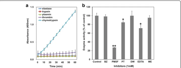
In an attempt to evaluate cleavage specificity, the amidolytic activity of NnV was assessed using various chromogenic substrates: N-Succinyl-Ala-Ala-Ala-ρNA (for elastase), Nα -Benzoyl-DL-Arg-ρNA (for trypsin), N-(p-Tosyl)-Gly-Pro-Lys-4 nitroanilide acetate salt (for plasmin), N-Benzoyl-Phe-Val-Arg-p-nitroanilide hydrochloride (for thrombin), N-Succinyl-Ala-Ala-Pro-Phe-ρNA (for chymotrypsin). For the assays, each substrate was made up to 0.5 mM and then incubated with NnV (0.1 mg/mL) for 1 h at 37 °C. The re-actions were monitored every five minutes using Power-Wave XS microreader (Biotek) at 405 nm.
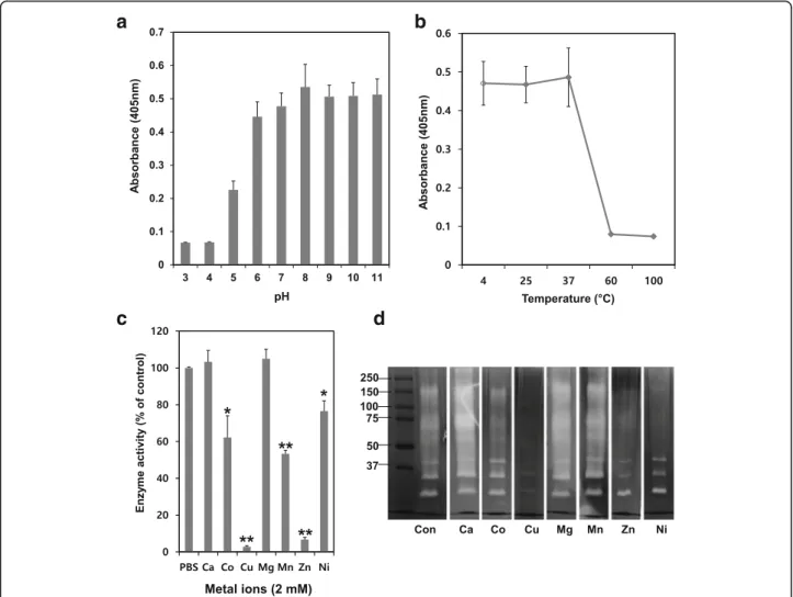
Effects of pH and temperature on amidolytic activity of NnV
The effect of pH on the enzymatic activity of NnV was evaluated under diverse pH conditions. NnV was incu-bated with different pH buffers, including 0.5 M acetate (pH 3 and 4), 0.1 M phosphate (pH 5, 6, 7 and 8) and
0.5 M glycine-NaOH (pH 9, 10 and 11) for 1 h at 4 °C. It reacted against N-Succinyl-Ala-Ala-Pro-Phe-ρNA (for chymotrypsin) substrate and the proteolysis of the sub-strate was detected at 405 nm. Furthermore, the enzy-matic activity was assessed at various temperature conditions at 4, 25, 37, 60 and 100 °C. The residual activity was measured after incubation of NnV in diffe-rent temperatures for 30 min.
Effects of metal ions and protease inhibitors on enzymatic activity of NnV
The substrate of N-Succinyl-Ala-Ala-Pro-Phe-ρNA (for chymotrypsin) was used to measure the effects of metal ions and inhibitors on the enzymatic activity of NnV. For this assay, NnV was incubated with several divalent metal ions or protease inhibitors for 1 h and followed by reacting chymotrypsin substrate for 30 min at 37 °C.
Isolation of fibrinolytic protease fromN. nomuraivenom
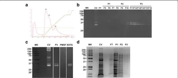
Crude venom of NnV (180 mg) was dissolved in 10 mM Tris-HCl buffer (pH 7.8) and centrifuged at 13000×gfor 30 min. The venom solution was resolved on DEAE column (GE Healthcare), previously dialyzed against extraction buffer at 4 °C. The column was eluted with gradient concentration of NaCl from 0 to 0.8 M at a flow rate of 1 mL per min. Each peak was tested for fibrinolytic activity and SDS-PAGE.
Statistical analysis
The results are expressed as mean ± standard deviation (SD). One-way analysis of variance (ANOVA) was used to evaluate the significance of difference between the two mean values. The values ofpconsidered statistically significant werep< 0.01 andp< 0.05.
Results
SDS-PAGE and fibrinolytic activity of NnV
To compare the fibrinolytic activity between different jelly-fish species, their venom proteins were loaded into fibrin zymogrpahy. The fibrinolytic activity indicated venom of three species, namelyN. nomurai,A. auritaandP. physalis, but notD. quinquecirrhaorC. brevipedalia. Among these,
P. physalisvenom was highly potent for fibrin degradation
and its molecular weight was above 25 kDa. N. nomurai
and A. aurita venoms showed similar banding
patterns, which were distributed in 60–80 kDa and
25–37 kDa (Fig. 1a). The 90 and 70 kDa bands
according to their action mechanisms and site. To deter-mine which kind of protease was associated with the fibrionolytic activity of NnV, broad inhibitors for metal-loprotease [EDTA and 1, 10-Phenanthroline (PT)] and serine protease [PMSF and benzamidine (BZ)] were in-cubated with NnV. Results showed that all inhibitors partially inhibited the fibrinolytic activity of NnV. How-ever, only PMSF at a low concentration of 1 mM showed strong inhibitory effect on its fibrinolytic activity (Fig. 1c). These results suggest that serine proteases play a more important role in the fibrinolytic activity of NnV than metallorproteases.
Fibrinogenolytic activity of NnV
The fibrinogenolytic activity of NnV was analyzed by SDS-PAGE. NnV degraded both α-chain and β-chain of fibrinogen in dose- and time-dependent manners
(Fig. 2a and b). The α-chain of fibrinogen was imme-diately digested after incubation with NnV. Its complete degradation was observed at 360 min after incubation with NnV. The β-chain of fibrinogen began to be degraded at 360 min after incubation with NnV. However, γ-chain of fibrinogen was not affect by NnV.
Amidolytic activity of NnV
NnV failed to degrade elastase, trypsin, thrombin, and plasmin substrates. However, it specifically ex-hibited hydrolytic activity against N-Succinyl-Ala-Ala-Pro-Phe-ρNA substrate (for chymotrypsin) (Fig. 3a). In addition, such enzymatic activity of NnV on chymotrypsin substrate was strongly inhibited by PMSF (Fig. 3b).
a
b
c
Fig. 1SDS-PAGE profile and fibrinolytic activity of NnV.aWe compared the fibrinolytic activity between jellyfish species. Lane 1–N. nomurai; lane 2–A. aurita; lane 3–D. quinquecirrha; lane 4–P. physalis, lane 5–C. brevipedalia.bNnV (50μg) was submitted to SDS electrophoresis under non-reducing conditions. The gels were stained with 0.125% Coomassie blue.c Fibrin zymography of NnV with various protease inhibitors, including PMSF (1 mM), BZ (2 mM), EDTA (2 mM) and PT (2 mM). Clear zones in the fibrin gel indicated regions of proteolytic activity
a
b
Hydrolytic activity of NnV is dependent on pH, temperature, metal ions, and protease inhibitors
The effect of pH on the enzymatic activity of NnV was determined using buffers at various pH values. The en-zymatic activity of NnV on chymotrypsin substrate was stable and higher at neutral (pH 7) and basic (pH 8, 9, 10 and 11) pH conditions (Fig. 4a). However, its activity was unstable and lost under acidic conditions. The optimum temperature condition for its activity was found to be between 4 and 37 °C. However, its activity was lost at high temperature including 60 and 100 °C (Fig. 4b). The effects of various metal ions on its enzym-atic activity were also examined. Ca2+and Mg2+failed to activate or inhibit its enzymatic activity and fibrinolytic activity. However, Co2+, Mn2+, and Ni2+ showed slightly inhibitory effect on its enzymatic and fibrinolytic activity (Fig. 4c and d). Particularly, Cu2+and Zn2+exerted com-pletely inhibitory effects on its activities.
Purification of NnV with fibrinolytic activity
After separating NnV on DEAE column, three peaks were obtained and the fibrionolytic activity of each peak was evaluated using fibrin zymography. Peak 3 (P3, Fr 11–15) showed fibrinolytic activity with two
bands, whereas P1 and P2 failed to show any band (Fig. 5b). To determine whether the fibrinolytic acti-vity of P3 was associated with serine protease, fibrin zymography was performed using typical protease inhibitors EDTA and PMSF. It was found that PMSF abolished the fibrinolytic activity of P3, but EDTA did not (Fig. 5c). However, the protein amount of P3 was small and the matching band with fibrinolytic activity at 35 kDa in P3 was very weak (Fig. 5d).
Discussion
Several reports have demonstrated that jellyfish venom consists of various bioactive substances, including cytoly-sins, neurotoxins, peptides and proteases [13, 20, 26–28].
Unlike other venoms (snake, scorpion and spider), jellyfish venom is not secreted from venom glands or injected through specialized nematocysts that are present all over the cnidarians. Hence, it is very difficult to obtain pure jellyfish venom due to poor yield, besides being a time-consuming and technically challenging process [29]. Al-though it is not easy to isolate and characterize individual jellyfish venom components, their characterization is very important in various aspects, including clinical manage-ment, development of therapeutic agents, and pharma-ceutical application.
Our previous study has revealed that NnV contains abundant proteolytic enzymes responsible for dermone-crosis and cytotoxicity after stings [12, 13]. Among pro-teolytic activities, fibrinolytic activity is an established mediator of toxicity in various animal venoms, especially snake venoms. Fibrinolytic enzymes may interfere with coagulation and fibrin(ogen)olytic systems, leading to systemic bleeding, coagulopathy, hypovolemia, and hemodynamic shock [14, 15, 30]. In our previous study, we reported the fibrinolytic activity of NnV, but it only 1,10-phenathroline was used as an enzymatic inhibitor. Therefore, the relationship between fibrinolytic activity and other proteases could not be excluded. Generally, proteases with fibrinolytic activity are closely associated with metalloproteases and serine proteases.
In order to determine which protease contributed to the fibrionolytic activity of NnV, in this study fibrin zymography was performed using metalloprotease
a
b
Fig. 3The catalytic activity of NnV on several chromogenic substrates.aNnV was preincubated with different substrates
[N-Succinyl-Ala-Ala-Ala-ρNA (for elastase), Nα-Benzoyl-DL-Arg-ρNA (for trypsin), N-(p-Tosyl)-Gly-Pro-Lys-ρNA·acetate salt (for plasmin), N-Benzoyl-Phe-Val-Arg-ρNA·HCl (for
thrombin), N-Succinyl-Ala-Ala-Pro-Phe-ρNA (for chymotrypsin)], 0.5 mM for 1 h. The catalytic activity of NnV was assayed at 405 nm.bNnV was
inhibitors (EDTA and PT) and serine protease inhibitors (PMSF and BZ). Although EDTA and PT moderately inhibited the fibrinolytic activity, only PMSF completely inhibited it at a low concentration of 1 mM. Regarding its amidolytic activity, NnV only cleaved chymotrypsin substrates. Such activity was mostly inhibited by PMSF. Hence, we concluded that the fibrinolytic activity of NnV is mostly due to its serine proteases rather than metalloproteases. It demonstrated specific activity to-ward the chymotrypsin substrate.
The fibrinolytic activity of NnV was dependent on pH, temperature, and metal ion concentration. Its fibrinolytic activity was strong at pH values of 8 to 11. However, such activity was lost at pH values of 3 to 5 and wea-kened at pH values of 6 to 7. To date, fibrinolytic enzymes derived from venoms have been found to be stable at pH values of 5.5 to 8.5. For example, the
fibrinolytic activity of neuwiedase, a metalloprotease
from Bothrops neuwiedi snake, has been found to be
strong at a pH range of 7.4 to 8.0 and Brevilysin L6 can persevere its activity at optimal pH of 8.5 to 9.5 [31, 32]. Interestingly, fibrinolytic activity of NnV was found to be stable at basic conditions in this study, even at pH 11. Its activity was fast at temperature below 37 °C. How-ever, this activity was sharply reduced when temperature was increased to be above 37 °C. Furthermore, the fibrinolytic activity of NnV was strongly inhibited by Zn2+ and Cu2+at 1 mM in fibrin zymography and chromogenic substrate test.
Fibrinolytic enzymes commonly have fibrinogenolytic activities. Therefore, we performed assays to evaluate the fibrinogenolytic activity of NnV and found that NnV ra-pidly degradedα-chain of fibrinogen followed byβ-chain in a dose- and time-dependent manner. However, NnV
c
d
a
b
failed to degradeγ-chain of fibrinogen. Most serine prote-ases in snake venoms preferentially hydrolyze β-chain of fibrin(ogen) with low activity toward α-chain of fibrin(o-gen). However, there are a few exceptions to such charac-teristic feature. For example, Dnase purified from the venom ofDeinagkistrodon acutusis found to be a fibrino-genase that belongs to the serine protease family which preferentially digestsα-chain of fibrinogen in comparison withβ-chain [33]. Despite the fact that fibrinogenase iso-lated fromAgkistrodon halysbrevicaudus is a metallopro-tease, it can rapidly cleave β-chain of fibrinogen [32]. Although the fibrin(ogen)olytic enzymes of NnV belonged to the serine protease group, it preferentially degradedα -chain thanβ-chain of fibrinogen.
To isolate fibrinolytic enzymes from NnV, we per-formed chromatography on DEAE column. Among three peaks, only P3 showed fibrinolytic activity with molecular weight of approximately 35 and 37 kDa. Its fibrinolytic activity was completely inhibited by PMSF, but not by EDTA, indicating that the fibrinolytic enzyme in P3 belongs to the serine protease family. Despite the fibrinolytic activity of P3, its protein profile was very week. Hence, it is difficult to identify or characterize its active sources. Obtaining sufficient amounts of NnV for identification and characterization remains a significant challenge.
Serine proteases in venoms are associated with various biological activities, including hemostatic, cell differenti-ation, prey digestion as well as affecting the complement system [30]. It mainly disturbs the coagulation cascade through activation or inactivation of platelet aggregation,
coagulation, and fibrinolysis [15]. Coagulopathy has not yet been observed in patients stung by jellyfish. There-fore, we speculate the reasons why serine proteases are present in jellyfish venom. Serine proteases can act as spreading factors that may increase their permeability into tissue and promote the spread of venom. They can also form complexes capable of activating the complement cascade through the cleavage of specific components, leading to induction and facilitation of inflammation [34, 35]. Although this study did not investigate the pathological mechanisms of serine pro-teases, it could explain that inflammatory symptoms provoked by jellyfish envenomation may be partially caused by serine proteases.
Conclusions
Jellyfish venom research is a very attractive field due to increasing sting accidents and the presence of various bioactive components. Biochemical characterization of toxins can help us understand the pathological symp-toms associated with envenomation, so that clinical agents can be developed. Although the present study partially purified a serine protease from NnV, our results could aid the development of clinical management. Fur-thermore, fibrinolytic properties can be used to treat thrombosis by preventing clot formation [36, 37]. Based on the results of this study, further research can be con-ducted to isolate serine proteases and investigate the pathological mechanisms involved in inflammatory reac-tions, in addition to their thrombolytic potential.
a
b
d
c
Abbreviations
NnV:Nemopilema nomuraijellyfish venom; PMSF: Phenylmethanesulfonyl fluoride; SD: Standard deviation; SDS-PAGE: SDS polyacrylamide gel electrophoresis
Acknowledgments
The authors would like to thank the National Institute of Fisheries Science and the National Research Foundation of Korea for their support.
Funding
This work was supported by a grant from National Institute of Fisheries Science (R2017046 and15-OE-14) and a National Research Foundation of Korea grant funded by the Korea government (no. NRF-2014R1A2A2A01007245).
Authors’contributions
EK conceived and designed the study. SKB and HL mainly performed the experiments and wrote the manuscript. SKB and HL contributed equally to this work. MJP, YH and IC analyzed the data. CHH, WDY and CK interpreted and discussed the data. All authors have read and approved the final manuscript.
Ethics approval and consent to participate
Not applicable.
Consent for publication
Not applicable.
Competing interests
The authors declare that they have no competing interests.
Publisher’s Note
Springer Nature remains neutral with regard to jurisdictional claims in published maps and institutional affiliations.
Author details
1College of Veterinary Medicine, Gyeongsang National University, Jinju 660-701, Korea.2Headquarters for Marine Environment, National Fisheries Research & Development Institute, Shiran-ri, Gijang-eup, Gijang-gun, Busan 619-705, Korea.3Gyeongnam Department of Environment & Toxicology, Korea Institute of Toxicology, Gyeongnam 52834, Jinju, Korea.4Institute of Animal Medicine, Gyeongsang National University, Jinju, 660-701, Korea.
Received: 15 February 2017 Accepted: 3 July 2017
References
1. Dong Z, Liu D, Keesing JK. Jellyfish blooms in China: dominant species, causes and consequences. Mar Pollut Bull. 2010;60(7):954–63.
2. Fu J, Koo K, Sang AX, Shisler DC. Jellyfish envenomation in an ocean swimmer. Intern Emerg Med. 2014;9(1):103–4.
3. Lynam CP, Gibbons MJ, Axelsen BE, Sparks CA, Coetzee J, Heywood BG, et al. Jellyfish overtake fish in a heavily fished ecosystem. Curr Biol. 2006;16(3): R492–3.
4. Ramasamy S, Isbister GK, Seymour JE, Hodgson WC. The in vivo
cardiovascular effects of box jellyfishChironex fleckerivenom in rats: efficacy of pre-treatment with antivenom, verapamil and magnesium sulphate. Toxicon. 2004;43(6):685–90.
5. Xiao L, He Q, Guo Y, Zhang J, Nie F, Li Y, et al.Cyanea capillata Tentacle-only extract as a potential alternative of nematocyst venom: its cardiovascular toxicity and tolerance to isolation and purification procedures. Toxicon. 2009;53(1):146–52.
6. Kang C, Kim YK, Lee H, Cha M, Sohn ET, Jung ES, et al. Target organ identification of jellyfish envenomation using systemic and integrative analyses in anesthetized dogs. J Pharmacol Toxicol Methods. 2011;64(2):173–9.
7. Bengtson K, Nichols MM, Schnadig V, Ellis MD. Sudden death in a child following jellyfish envenomation byChiropsalmus quadrumanus. Case report and autopsy findings. JAMA. 1991;266(10):1404–6.
8. Kim DH, Seo JN, Yoon WD, Suh YS. Estimating the economic damage caused by jellyfish to fisheries in Korea. Fish Sci. 2012;78:1147–52. 9. Kawahara M, Uye S, Burnett J, Mianzan H. Stings of edible jellyfish
(Rhopilema hispidum,Rhopilema esculentumandNemopilema nomurai) in Japanese waters. Toxicon. 2006;48(6):713–6.
10. Choudhary I, Lee H, Pyo MJ, Heo Y, Bae SK, Kwon YC, et al.Nemopilema nomuraijellyfish venom treatment leads to alterations in rat cardiomyocytes proteome. Data Brief. 2015;5:884–7.
11. Kang C, Munawir A, Cha M, Sohn ET, Lee H, Kim JS, et al. Cytotoxicity and hemolytic activity of jellyfishNemopilema nomurai(Scyphozoa: Rhizostomeae) venom. Comp Biochem Physiol C Toxicol Pharmacol. 2009;150(1):85–90.
12. Kang C, Jin YB, Kwak J, Jung H, Yoon WD, Yoon TJ, et al. Protective effect of tetracycline against dermal toxicity induced by jellyfish venom. PLoS One. 2013;8(3):e57658.
13. Lee H, Jung ES, Kang C, Yoon WD, Kim JS, Kim E. Scyphozoan jellyfish venom metalloproteinases and their role in the cytotoxicity. Toxicon. 2011; 58(3):277–84.
14. Swenson S, Markland FS Jr. Snake venom fibrin(ogen)olytic enzymes. Toxicon. 2005;45(8):1021–39.
15. White J. Snake venoms and coagulopathy. Toxicon. 2005;45(8):951–67.
16. Krem MM, Di Cera E. Evolution of enzyme cascades from embryonic development to blood coagulation. Trends Biochem Sci. 2002;27(2):67–74. 17. Li R, Yu H, Xue W, Yue Y, Liu S, Xing R, et al. Jellyfish venomics and venom
gland transcriptomics analysis ofStomolophus meleagristo reveal the toxins associated with sting. J Proteome. 2014;106:17–29.
18. Li R, Yu H, Yue Y, Liu S, Xing R, Chen X, et al. Combined proteomics and transcriptomics identifies sting-related toxins of jellyfishCyanea nozakii. J Proteome. 2016;148:57–64.
19. Weston AJ, Chung R, Dunlap WC, Morandini AC, Marques AC, Moura-da-Silva AM, et al. Proteomic characterisation of toxins isolated from nematocysts of the South Atlantic jellyfishOlindias sambaquiensis. Toxicon. 2013;71:11–7.
20. Heo Y, Kwon YC, Bae SK, Hwang D, Yang HR, Choudhary I, et al. Cloning a Chymotrypsin-like 1 (CTRL-1) protease cDNA from the jellyfishNemopilema nomurai. Toxins (Basel). 2016;8(7):205.
21. Bloom DA, Burnett JW, Alderslade P. Partial purification of box jellyfish (Chironex fleckeri) nematocyst venom isolated at the beachside. Toxicon. 1998;36(8):1075–85.
22. Carrette T, Seymour J. A rapid and repeatable method for venom extraction from cubozoan nematocysts. Toxicon. 2004;44(2):135–9.
23. Bradford MM. A rapid and sensitive method for the quantitation of microgram quantities of protein utilizing the principle of protein-dye binding. Anal Biochem. 1976;72:248–54.
24. Laemmli UK. Cleavage of structural proteins during the assembly of the head of bacteriophage T4. Nature. 1970;227(5259):680–5.
25. Matsubara K, Hori K, Matsuura Y, Miyazawa K. A fibrinolytic enzyme from a marine green alga,Codium latum. Phytochemistry. 1999;52(6):993–9.
26. Heo Y, Kwon YC, Shin K, Yoon WD, Han CH, Yum S, et al. cDNA and gene structures of two phospholipase A2 isoforms, acidic PLA2 PA4 and PLA2 PA3A/ PA3B/PA5, inNemopilema nomuraijellyfish venom. Toxicon. 2016;122:160–6.
27. Junior VH, Zara F, Marangoni S, Toyama Dde O, de Souza AJ, de Oliveira SC, et al. Identification of two novel cytolysins from the hydrozoanOlindias sambaquiensis(Cnidaria). J Venom Anim Toxins incl Trop Dis. 2014;20(1):10. 28. Lassen S, Wiebring A, Helmholz H, Ruhnau C, Prange A. Isolation of a Nav
channel blocking polypeptide fromCyanea capillataMedusae - a neurotoxin contained in fishing tentacle isorhizas. Toxicon. 2012;59(6):610–6.
29. Yanagihara AA, Shohet RV. Cubozoan venom-induced cardiovascular collapse is caused by hyperkalemia and prevented by zinc gluconate in mice. PLoS One. 2012;7(12):e51368.
30. Kini RM. Serine proteases affecting blood coagulation and fibrinolysis from snake venoms. Pathophysiol Haemost Thromb. 2005;34(4–5):200–4.
31. Rodrigues VM, Soares AM, Guerra-Sá R, Rodrigues V, Fontes MR, Giglio JR. Structural and functional characterization of neuwiedase, a nonhemorrhagic fibrin(ogen)olytic metalloprotease fromBothrops neuwiedisnake venom. Arch Biochem Biophys. 2000;381(2):213–24.
32. Terada S, Hori J, Fujimura S, Kimoto E. Purification and amino acid sequence of brevilysin L6, a non-hemorrhagic metalloprotease fromAgkistrodon halys
Brevicaudus venom. J Biochem. 1999;125(1):64–9.
33. Wang S, Xu X, Gao S, Zhu S, Rong R, Li B. Purification and partial characterization of a novel fibrinogenase from the venom of
Deinagkistrodon acutus: inhibition of platelet aggregation. Protein Expr Purif. 2014;99:99–105.
35. Yamamoto C, Tsuru D, Oda-Ueda N, Ohno M, Hattori S, Kim ST. Flavoxobin, a serine protease fromTrimeresurus flavoviridis(habu snake) venom, independently cleaves Arg726-Ser727 of human C3 and acts as a novel, heterologous C3 convertase. Immunology. 2002;107(1):111–7. 36. Barros LC, Ferreira RS Jr, Barraviera SR, Stolf HO, Thomazini-Santos IA,
Mendes-Giannini MJ, et al. A new fibrin sealant fromCrotalus durissus terrificusvenom: applications in medicine. J Toxicol Environ Health B Crit Rev. 2009;12(8):553–71.
37. Ferreira RS Jr, de Barros LC, Abbade LPF, Barraviera SRCS, Silvares MRC, de Pontes LG, et al. Heterologous fibrin sealant derived from snake venom: from bench to bedside - an overview. J Venom Anim Toxins incl Trop Dis. 2017;23:21.
• We accept pre-submission inquiries
• Our selector tool helps you to find the most relevant journal
• We provide round the clock customer support
• Convenient online submission
• Thorough peer review
• Inclusion in PubMed and all major indexing services
• Maximum visibility for your research
Submit your manuscript at www.biomedcentral.com/submit