Screening, production and biochemical characterization
of a new fibrinolytic enzyme produced by
Streptomyces
sp. (Streptomycetaceae) isolated from Amazonian lichens
Germana Michelle de Medeiros e SILVA1,2, Raquel Pedrosa BEZERRA3, José António TEIXEIRA4,
Flávio Oliveira SILVA3, Juliana Mendes CORREIA2, Tatiana Souza PORTO5, José Luis LIMA-FILHO1,2,3,
Ana Lúcia Figueiredo PORTO1,2,3*.
1 Universidade Estatual do Ceará, Rede Nordeste de Biotecnologia, Av. Paranjana, 1.700, 60740-000, Fortaleza, Brazil
2 Universidade Federal de Pernambuco, Laboratory of Immunopathology Keizo Asami, Av. Prof. Moraes s/n, 50670-901, Recife, Brazil
3 Universidade Federal Rural de Pernambuco, Department of Morphology and Animal Physiology, Av. Dom Manoel de Medeiros s/n, 52171-900 , Recife, Brazil
4 Universidade do Minho, Institute for Biotechnology and Bioengineering, Centre of Biological Engineering, Campus de Gualtar, 4710-057, Braga, Portugal
5 Universidade Federal Rural de Pernambuco, Academic Unit of Garanhuns, Avenida Bom Pastor s/n, 55292-270, Garanhuns, Brazil * Corresponding author: analuporto@yahoo.com.br
ABSTRACT
Thrombosis is a pathophysiological disorder caused by accumulation of fibrin in the blood. Fibrinolytic proteases with potent thrombolytic activity have been produced by diverse microbial sources. Considering the microbial biodiversity of the Amazon region, this study aimed at the screening, production and biochemical characterization of a fibrinolytic enzyme produced by Streptomyces sp. isolated from Amazonian lichens. The strain Streptomyces DPUA1576 showed the highest fibrinolytic activity, which was 283 mm2. Three variables at two levels were used to assess their effects on the fibrinolytic production. The
parameters studied were agitation (0.28 – 1.12 g), temperature (28 – 36 ºC) and pH (6.0 – 8.0); all of them had significant effects on the fibrinolytic production. The maximum fibrinolytic activity (304 mm2) was observed at 1.12 g, 28 ºC, and pH
of 8.0. The crude extract of the fermentation broth was used to assess the biochemical properties of the enzyme. Protease and fibrinolytic activities were stable during 6 h, at a pH ranging from 6.8 to 8.4 and 5.8 to 9.2, respectively. Optimum temperature for protease activity ranged between 35 and 55 °C, while the highest fibrinolytic activity was observed at 45 ºC. Proteolytic activity was inhibited by Cu2+ and Co2+ ions, phenylmethylsulfonyl fluoride (PMSF) and pepstatin A, which suggests that
the enzyme is a serine protease. Enzymatic extract cleaved fibrinogen at the subunits Aα-chain, Aβ-chain, and γ-chain. The results indicated that Streptomyces sp. DPUA 1576 produces enzymes with fibrinolytic and fibrinogenolytic activity, enzymes with an important application in the pharmaceutical industry.
KEYWORDS: Actinomycetes, fibrinolytic protease, inhibitor protease, fibrinogenolytic activity.
Seleção, produção e caracterização bioquímica de uma nova enzima
fibrinolítica produzida por
Streptomyces
sp. (Streptomycetaceae) isolada
de liquens da Amazônia
RESUMO
A trombose é uma doença patofisiológica causada pelo acúmulo de fibrina no sangue. Proteases fibrinolíticas com potente atividade trombolítica são produzidas por diversas fontes microbianas. Considerando a biodiversidade microbiana da região amazônica, o presente estudo teve como objetivo a seleção, produção e caracterização bioquímica da enzima fibrinolítica de Streptomyces sp. isolado de líquens da Amazônia. Streptomyces DPUA1576 foi a melhor produtora com atividade fibrinolítica de 283 mm2. Três
variáveis em dois níveis foram utilizadas para determinar as variáveis mais relevantes na produção da enzima fibrinolítica (FA). Os parâmetros estudados foram agitação (0.28 – 1.12 g), temperatura (28 – 36 ºC) e pH (6.0 – 8.0) e todos obtiveram efeitos significativos na produção fibrinolítica. A maior atividade fibrinolítica (304 mm2) foi obtida a 1.12 g, 28 ºC e pH 8.0. O extrato
bruto da fermentação foi usado para determinar as propriedades bioquímicas da enzima. Atividades proteásica e fibrinolítica foram estáveis durante 6 horas no intervalo de pH entre 6.8 – 8.4 e 5.8 – 9.2, respectivamente. Temperatura ótima para a atividade proteásica foi entre 35 – 55 °C, enquanto que para a atividade fibrinolítica foi de 45 ºC. Atividade proteásica foi inibida por íons Cu2+ e Co2+, fluoreto de fenilmetilsulfonil e pepstatina A, na qual sugere que a enzima é uma serino-protease. O extrato enzimático
degradou o fibrinogênio nas subunidades Aα, Aβ e γ. Os resultados apresentados indicam que Streptomyces sp. DPUA 1576 produz enzimas com atividade fibrinolítica e fibrinogenolítica, enzimas com aplicações importantes na indústria farmacêutica.
INTRODUCTION
Thrombosis is one of the most widely occurring diseases in modern life. It occurs by the accumulation of fibrin when injury on tissue occurs. Normally, the fibrin formed in the blood is dissolved by the action of plasmin (E.C. 3.4.21.7). The unsolved clot by plasmin leads to the formation of thrombus, a serious problem because it induces cerebral and cardiovascular disease (Deepak et al. 2010). There are many drugs used on fibrinolytic therapy such as streptokinase (EC 3.2.1.35), staphylokinase (EC 3.4.21.35), urokinase (EC 3.4.21.31) but all of them have side effects. Thrombolytic therapy today is expensive and has undesirable side effects such as the risk for internal hemorrhage within the intestinal tract when orally administrated (Zhang et al. 2007).
Worldwide, over 29% of the total mortalities are due to thrombosis. By the year 2020, cardiovascular diseases (CVDs) may cause an estimated 25 million deaths per year, thus antithrombotic therapy is of great interest (Kotb 2014). Over the years, thrombolytic therapies via injecting or orally administrating thrombolytic agents to lyses thrombi in blood vessels have been extensively investigated (Peng et al. 2005). Therefore, continuous efforts have been focused in the search of safer and less expensive thrombolytic agents from diverse sources. Until recently, fibrinolytic enzyme with potential thrombolytic application has been purified from various sources such as fermented food, earthworms, mushrooms, snake venom and microbial sources (Kotb 2014). Microbial sources, such as bacteria, actinomycetes, fungi and algae are reported to produce fibrinolytic enzyme with few reports on the use of Streptomyces sp. (Chitte et al. 2011). Streptomyces sp.
produces several extracellular enzymes of commercial interest, such as protease, pectinase, xylanase and cellulase. Proteases constitute two thirds of the total number of enzymes used in industry and this is expected to increase. Some of these proteases are fibrinolytic enzymes capable of digesting fibrin (Silva et al. 2015).
Considering the microbiological potential of the Amazon Region and the growing applicability of fibrinolytic proteases, the screening of Streptomyces sp. appears to be an interesting approach to finding novel fibrinolytic producers to reduce the drawbacks of marketed thrombolytic drugs. Therefore, this study aimed at the screening, production and biochemical characterization of a fibrinolytic enzyme produced by
Streptomyces sp. isolated from Amazonian lichens.
MATERIALS AND METHODS
Screening of isolates with fibrinolytic activity
Fifty Streptomyces spp.isolates obtained from Amazonian lichens were used for fibrinolytic enzyme screening (Table 1). These Streptomyces isolates form part of the Culture Collection
of the Parasitology Department of the Federal University of Amazonas (DPUA/UFAM-Brazil).
The inoculum was produced following the International
Streptomyces Project medium 2 (ISP-2) broth in orbital shaker (BR-300LF, TAITEC Co, Tokyo, Japan), agitation of 1.12 g
for 48 h at 28°C. Enzyme production was carried out with initial cell concentration of 1x10⁶ cells mL -1 (A
600 = 0.1) in
250-mL-Erlenmeyer flasks containing 50 mL of soybean flour medium (Porto et al. 1996), and incubated for 72 h at different conditions of agitation, temperature and pH as described in the Table 2. The samples were clarified by centrifugation (KR-20000T, Kubota, Osaka, Japan) at 8000 g and 4 ºC for 10 min, and submitted to fibrinolytic activity determination.
Experimental design and statistical analysis
To assess the protease and fibrinolytic activity, a factorial arrangement with two levels and three variables (agitation, temperature and pH) was used to determine the most relevant combination. There were assessed agitation at 100, 150 and 200 rpm; these values corresponded to g forces of 0.28, 0.63 and 1.12, respectively. There were also evaluated temperature (ranged between 28 - 36ºC) and pH, between 6 and 8 (Table 2). All statistical and graphical analyses were carried out with the Statistica 8.0 software (StatSoft Inc., Tulsa, OK, USA).
Culture condition
The best fibrinolytic enzyme-producing Streptomyces sp. was cultivated in ISP-2 under the production conditions described in the experimental design (Table 2) during 144 h, in order to study cell growth, protease and fibrinolytic enzyme production. Cell concentration, pH and enzyme activity were analyzed every 12 h.
Effects of temperature and pH on protease and fibrinolytic activity
Effects of metal ions and inhibitors
The enzyme extract can be classified by their sensitivity to various inhibitors for 30 min at room temperature and the residual activity was measured. Effect of metal ion Zn+2, Na+, Mg+2, Fe+2, Cu+2, Ca+2, Mn+2, Co+2 and
inhibitors phenylmethylsulfonyl fluoride (PMSF), ethylenediaminetetraacetic (EDTA), Pepstatin A and iodoacetic acid were determined. The activity of the enzyme assayed in the absence of inhibitors was taken as 100%. Each assay was carried out in triplicate.
Protease activity determination
Protease assay was conducted according to Alencar (2003). That is, a micro centrifuge tube was added of 100 µL of 1% azocasein (w/v, in 0.2 M Tris-HCl, pH 7.2), and 60 µL of crude extract. The reaction runs for 60 min at 25 ºC. Then, 480 µL of 10% (w/v) trichloroacetic acid (TCA) were added to stop the reaction. After 15 min, centrifugation was carried out for 5 min at 8000g. The supernatant (320 µL) was added to 1M NaOH (560 µL) and the absorbance of this mixture was measured at 440 nm (B582 spectrofotometer, Micronal,
São Paulo, Brazil) against a blank similarly prepared except that 0.15M NaCl replaced the crude extract sample. One unit (U) of enzymatic activity was defined as the amount of enzyme capable to produce a 0.001 change in absorbance per minute. The analysis was performed in triplicate. Protein content was determined by after Bradford (1976) using bovine serum albumin as standard.
Fibrinolytic activity determination
Fibrinolytic activity was determined using the fibrin plate method of Astrup and Mullertz (1952), with minor modifications, as follows. The fibrin agarose plate was made by 1% agarose, 0.1% human fibrinogen, and 8 U mL-1 of Human thrombin. The clot
was allowed to stand for 1 h at room temperature. Then, 20 µL of sample solution were placed onto the plate. The plate was incubated at 37°C for 18 h and the diameter of the lytic circle measured. In the fibrin plate method, a clear transparent region is observed in which fibrin is hydrolyzed, and its diameter is directly proportional to the intensity of the fibrinolytic activity. The lysed area was evaluated as the circle area (in square millimeter). Plasmin from human plasma was used as positive control.
Table1. Screening of fibrinolytic activity obtained from different Streptomyces sp.
Sample Halo (mm2) Sample Halo (mm2)
Streptomyces sp. DPUA 1541 113 Streptomyces sp. DPUA 1578 132.67
Streptomyces sp. DPUA 1543 50.24 Streptomyces sp. DPUA 1579 143.07
Streptomyces sp. DPUA 1545 201 Streptomyces sp. DPUA 1580 191.04
Streptomyces sp. DPUA1547 254 Streptomyces sp. DPUA 1581 153.86
Streptomyces sp. DPUA 1549 78.50 Streptomyces sp. DPUA 1582 132.67
Streptomyces sp. DPUA1550 254 Streptomyces sp. DPUA 1583 7.07
Streptomyces sp. DPUA 1551 28.26 Streptomyces sp. DPUA 1584 50.24
Streptomyces sp. DPUA 1553 78.50 Streptomyces sp. DPUA 1586 200.96
Streptomyces sp. DPUA 1554 201 Streptomyces sp. DPUA 1587 63.59
Streptomyces sp. DPUA 1557 0.79 Streptomyces sp. DPUA 1589 153.86
Streptomyces sp. DPUA 1559 113 Streptomyces sp. DPUA 1591 176.63
Streptomyces sp. DPUA 1560 113 Streptomyces sp. DPUA 1595 19.63
Streptomyces sp. DPUA 1561 50.24 Streptomyces sp. DPUA 1597 200.96
Streptomyces sp. DPUA 1563 133 Streptomyces sp. DPUA 1598 132.67
Streptomyces sp. DPUA 1565 113 Streptomyces sp. DPUA 1599 153.86
Streptomyces sp. DPUA 1566 154 Streptomyces sp. DPUA 1600 113.04
Streptomyces sp. DPUA 1567 30.18 Streptomyces sp. DPUA 1602 153.86
Streptomyces sp. DPUA 1568 154 Streptomyces sp. DPUA 1603 200.96
Streptomyces sp. DPUA 1570 133 Streptomyces sp. DPUA 1605 153.86
Streptomyces sp. DPUA 1571 0.00 Streptomyces sp. DPUA 1606 254.34
Streptomyces sp. DPUA 1572 30.18 Streptomyces sp. DPUA 1608 113.04
Streptomyces sp. DPUA 1573 189 Streptomyces sp. DPUA 1609 113.04
Streptomyces sp. DPUA 1575 18.09 Streptomyces sp. DPUA 1610 176.63
Streptomycessp. DPUA 1576 283 Streptomyces sp. DPUA 1611 113.04
Sodium dodecyl sulfate polyacrylamide gel
electrophoresis (SDS PAGE) of the crude extract and zymography
The molecular weights of the proteins of crude extract were determined using SDS-PAGE (12%) according to the method of Laemmli (1970). Electrophoresis was carried out at 20 mA/gel in Tris-glycine buffer, pH 8.3, containing 0.01% SDS. Zymography were performed with gelatin. Gelatin at 0.2% was copolymerized with 12% resolving gel. After electrophoresis, the gel was washed successively with Tris-HCL buffer (pH 7.5) containing 2.5% triton X-100 and glycine-NaOH buffer (pH 9.0).The gel was then incubated in glycine-NaOH buffer overnight at 37oC and
stained with coomassie brilliant blue R-250 for visualization of the clear zones. The molecular-weight standard proteins used were phosphorylase b (97.4 kDa), albumin (66.2 kDa), ovoalbumin (45.0 kDa), carbonic anhydrase (30.1 kDa), trypsin inhibitor (20.5 kDa), and α-lactalbumin (14.4 kDa). The slab gels were stained with Coomassie Blue R-250, 0.25% (w/v), in acetic acid : methanol : water (1 : 4.5 : 4.5, v/v). The molecular weight was estimated by LabImage 1D L340 program.
Fibrinogenolytic activity determination
Fibrinogenolytic activity was determined by modified fibrinogenolytic assay (Gao et al. 1998). The fibrinogen solution (0.05 ml of 2% human fibrinogen in 50mM Tris-HCl buffer, pH 7.6, 0.15 M NaCl) was mixed with 300 µg of crude extract and it was incubated at 37 ºC for 0, 5, 15, 30, 60 min and 5, 12, 24 h. After the indicated time intervals, an aliquot of 0.05 ml reaction mixture was transferred to 0.05 ml denaturing reagent (10 M urea, 4% SDS and 4% 2-mercaptoethanol) and incubated at 37 ºC for 24h. The sample was then analyzed by 12.5% (w/v) SDS-PAGE.
RESULTS
Screening of fibrinolytic activity, cell growth and protease production
Screening on fibrin agar plates showed that forty-nine
Streptomyces sp. isolates from Amazonian lichens were able to display extracellular fibrinolytic activity. Streptomyces
DPUA1576 obtained the highest fibrinolytic activity of 283 mm² (Table 1).
The relationship between protease activity and Streptomyces
sp. DPUA 1576 growth is shown in Figure 1. Protease production started at the stationary growth phase at 72 h and remained constant up to 132 h; then a linear decrease was observed. The highest extracellular protease activity of 11.5 U mL-1 and fibrinolytic activity of 300 mm2 obtained after
72 h of fermentation. Maximum cell concentration was 4.5 g L-1 at 36 h of cultivation. The pH value decreased during cell
growth until reaching stationary growth phase.
Effects of agitation, pH and temperature on protease and fibrinolytic production by multi-factorial
experiments
The obtained values for protease and fibrinolytic activity for the different trials are shown in Table 2. Protease activity varied from 1.76 to 105 U mL-1 and fibrinolytic activity from 0 to 304
mm2 (Table 2). A first order (main effects) model was used to
analyze the data from the factorial design. The main effects of the examined factors on the protease and fibrinolytic activities were calculated and are presented graphically in Figure 2A and 2B, respectively. Agitation, pH, interaction between agitation and pH, agitation and temperature had a positive effect (p˂
0.01) on protease production while the interaction between temperature and pH has a negative effect (p < 0.05; Figure 2A). The main effect of temperature on protease production was not significant (p˃ 0.05) as shown on the Pareto chart (Figure 2A).
Figure 1. The time profile of cell growth of Streptomyces sp. DPUA 1576 and fibrinolytic activity during fermentation: (open square) Biomass (g L-1), (closed
triangles) Protease activity (U mL-1);
Table 2. Protease and fibrinolytic protease production by Streptomyces sp DPUA 1576 under the effect of agitation, temperature and pH treatments.
Runs Agitation (g)
Temperature (ºC) pH
PA (U mL-1)
FA (mm2)
1 0.28 28 6 1.76 0
2 1.12 28 6 1.94 0
3 0.28 36 6 1.94 0
4 1.12 36 6 21.7 123
5 0.28 28 8 20.9 13.6
6 1.12 28 8 105 304
7 0.28 36 8 7.41 0.35
8 1.12 36 8 96.6 268
9 0.63 32 7 3.43 0
10 0.63 32 7 3.61 0
11 0.63 32 7 2.13 0
Figure 2. Pareto Chart showing variables and interactions on (A)protease and (B) fibrinolytic productionby Streptomyces sp DPUA 1576.
Regarding the fibrinolytic enzyme production, only the main effect of agitation was positive and significant (p < 0.05, Figure 2B). As shown in Table 2, the increase in agitation of the flask shaker led to a remarkable enhancement of enzyme production and the highest enzyme activity was 304 mm2 at
1.12 g (200 rpm). The highest fibrinolytic activity (304 mm2)
was obtained under initial culture pH 8.0, agitation of 1.12
g and temperature of 28 ºC (run 6; Table 2).
Effects of temperature and pH on protease and fibrinolytic activity
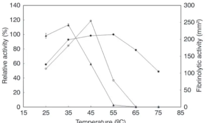
The protease was active at temperatures ranging from 25 to 55 ºC and had an optimum temperature between 35 to 55 ºC, while the activity decreased at a temperature higher than 55 ºC (Figure 3). The relative activities at 25 and 75 ºC were 59 % and 49 %, respectively. Protease activity was stable during 120 min at 25 and 35 ºC and showed 98, 100, and 59% residual activity at 25, 35 and 45 ºC, respectively. The enzyme in the crude extract lost its activity after 60 min between 55 to 75 ºC.The effect of temperature on the
Figure 3. Effect of temperature on the proteolytic activity (closed circles), fibrinolytic activity (open circle) and proteolytic stability (closed triangles) from Streptomyces sp. DPUA1576. Optimum temperature of the enzyme was determined by assaying the activity at various temperatures. Thermal stability of the enzyme was determined by measuring the residual activity after the enzyme was incubated at selected temperatures for 6 h.
fibrinolytic activity was examined. The temperature showing the maximal enzyme activity was 45 ºC (Figure 3).
The pH between 4.0 to 9.2 has effect on the proteolytic activity. The highest proteolytic activities were obtained in the alkaline range and the protease was stable in the pH range from 6.5 to 9.2 during 120 min (Figure 4). The optimum pH for the fibrinolytic activity was 7.5, and the enzyme activity decreased rapidly at levels below pH 7.0 and above pH 8.0. The enzyme was stable between pH 6.0 and 9.5 for 360 min and the stability enzymatic decreased below pH 5.5 (Figure 4).
Effects of metal ions and inhibitors
The effects of metal ions and inhibitors on protease activity were investigated after incubation of the protease with different metal ions and inhibitors for 1 h at 25 oC. The activity of
protease was slightly enhanced by the presence of Na+ ions.
Table 3. Effect of metal ions and protease inhibitors on proteolytic activity of
Streptomyces sp. DPUA1576.
Metal ion or inhibitor Concentration (mM) Residual activity %
None 0 100 ± 1.14
ZnSO4 1 81.4 ± 1.09
NaCl 1 106 ± 0.26
MgSO4 1 94.4 ± 0.60
FeSO4 1 95.3 ± 2.14
CuSO4 1 34.2 ± 0.43
CaCl2 1 88.8 ± 1.67
MnCl2 1 99.8 ± 2.28
CoCl2 1 60.8 ± 2.68
FeCl3 1 95.4 ± 0.81
Pepstatin A 1 14.6 ± 0.26
Phenylmethylsulfonyl fluoride
(PMSF) 1 94.0 ± 0.26
10 91.4 ± 0.31
50 25.4 ± 0.30
EDTA 1 71.8 ± 0.52
10 39.9 ± 0.65
IODO 1 105.7 ± 0.88
10 100.1 ± 0.50
On the other hand, the activity was significantly suppressed by Cu+2 and slightly inhibited by Co+2. No significant effects were
observed by the presence of other metal ions on the protease activity (Table 3). Proteolytic activity was slightly inhibited by 1mM of EDTA and phenylmethylsulfonyl fluoride (PMSF), but significantly inhibited by 50mM of PMSF and 10mM of EDTA showing a residual activity of 25.4 ± 0.30 % and 39.9 ± 0.65%, respectively. Pepstatin A at 1mM also inhibited the protease activity, resulting in a residual activity of 14.6 ± 0.26% (Table 2). Inhibition profile showed in the Table 2 indicates that protease from Streptomyces sp. L5A35 is a serine protease.
SDS-PAGE and fibrinogenolytic activity of crude fermentation extract
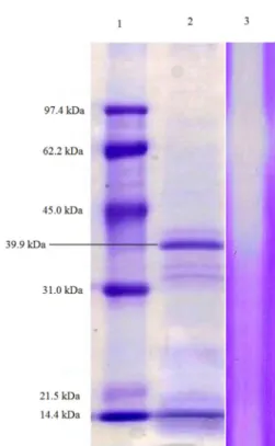
SDS-PAGE analysis of the crude fermentation extract indicates the existence of several proteins in the range of 31 to 45 kDa (line 2, Figure 5). A higher intensity band is observed at 39 kDa suggesting that it corresponds to the fibrinolytic enzyme that correlated with the clear hydrolytic bands observed in the gelatin zymogram (line 3, Figure 5).
The fibrinogenolytic reaction of the extracellular protease produced from Streptomyces sp. DPUA1576 was analyzed with SDS-PAGE (Figure 6). In this case, the subunit chains of fibrinogen were completely hydrolyzed
after 0.5 h of incubation. Figure 5. by Streptomyces Electrophoresissp. DPUA1576. 225 µg of protein was loaded on a 12.5% SDS-PAGE of crude extract with fibrinolytic activity
acrylamide gel. Lane 1 shows the standard molecular weight; lane 2 shows the protein content of crude extract, lane 3 shows zymography.
DISCUSSION
Extracellular fibrinolytic activity showed that Streptomyces
sp. isolated from Amazonian lichens presents clear haloes on fibrin plates indicating the ability of the produced enzymes to degrade fibrin in a direct way. Streptomyces DPUA1576 was selected as the best fibrinolytic producer (283 mm²) with enzyme activity higher than that obtained by Rovati et al.
(2010) in fungal specimens isolated from the sub-tropical Las Yungas Pedemontana forest (Tucuman, Argentina). It reveals the potential of Streptomyces sp. strains from Amazonian lichens as an unconventional and unexplored production alternative to already known thrombolytic agents as well as a reservoir of microorganism species with promising biotechnological value could be also highlighted.
Streptomyces sp. DPUA 1576 showed maximum protease production after 72 h of incubation (stationary phase of cell growth) and the enzyme level declined with further incubation. Protease production during the stationary growth phase is in accordance with the studies of other authors (Miyaji et al. 2006; Gouda 2006). The protease production is directly related to biomass production (Gupta et al. 2002; Puri et al. 2002) and is associated with the stationary phase of cell growth (Puri et al. 2002; Rao 1998). The decline in protease production upon prolonged incubation may be due to autolysis or proteolytic activity of other proteases under starvation conditions (Puri et al. 2002).
At the growth stationary phase (Figure 1), the maximum proteolytic activity was observed after 72 h, and its production was not growth-associated.
The pH value decreased during cell growth until reaching stationary growth phase, after remaining constant between 72 to 132 h. The drop in pH may be attributed to the production of organic acids, which were consumed during later stages of growth resulting in a slight pH increase to 4.0 (Futamura et al. 2001). These results show that alkalinization of the medium is closely related with the degradation of extracellular proteins by proteases produced from Streptomyces
sp DPUA1576, suggesting that alkalinization is adaptive. Our observations with Streptomyces sp. DPUA1576 suggest that this phenomenon may be widespread.
Temperature, agitation and pH are factors that influenced on protease and fibrinolytic productions. The decrease in the temperature caused an increase in protease production by Streptomyces sp DPUA1576. Other studies also showed that temperature is relevant factor for microbial protease production (Thys et al. 2006; Ellaiah et al. 2002) and an enhancement in protease production occurred when the temperature decreased from 50 to 25 ºC (Thys et al. 2006).
An increase in agitation of the flask shaker to 1.12 g
(200 rpm) led to a remarkable enhancement of enzyme
production. Streptomyces sp. is an aerophilic bacteria and increase the agitation providing a greater amount of oxygen for microorganism multiplication and metabolism. Al-Juamily and Al-Zaidy (2012) obtained a maximum fibrinolytic activity of 290 U, at 250 rpm.
The best pH for fibrinolytic enzyme production by
Streptomyces sp DPUA 1576 was 8.0, similar to that reported by Bhavani et al. (2012) using Streptomycesvenezuelae. The effect of pH is in agreement with previous reports on its effect on extracellular enzymatic activity (Caracuel et al. 2003).
In the initial culture, the highest fibrinolytic activity of 304 mm2 was obtained at pH 8.0, agitation of 1.12 g (200
rpm) and temperature of 28 ºC. This result corresponds to a 41% increase in the enzyme activity, when compared to fibrinolytic activity of 215 mm² using Bacillus licheniformis
KJ-31 (Hwang et al. 2007). Silva et al. (2015) showed that
Streptomyces sp. DPUA 1576 produced the highest fibrinolytic activity of 706.5 mm2 at 1.7% soybean flour and 1.0% glucose
concentration, which was 33% higher than plasmin. Optimum temperature for protease was between 35 to 55 ºC and to fibrinolytic was at 45 ºC. Simkhada et al
(2010) reported that the optimum temperature of fibrinolytic protease from Streptomyces sp CS684 was 45 ºC, but it was not stable at this temperature, losing more than 50% of its activity after 90 min. This enzyme was stable below 40 ºC.
The highest proteolytic activities were obtained in the alkaline range. Several other researchers have also described alkaline proteases with broad pH activities and stabilities. A purified alkaline protease from B. subtilis was observed to be stable with activity ≥ 78% in the pH range of 7.0–11.0, with the highest activity at pH 9.0 (Pandee et al. 2008)
The fibrinolytic enzyme was activated at neutral and alkaline pH values, and optimal reaction for fibrinolytic enzyme was obtained at pH 7.5, which is similar to the optimum observed for Streptomyces sp. CS684 (Simkhada et al. 2010), but higher than Streptomyces sp. P3 (Cheng et al. 2015). The fibrinolytic enzyme from Streptomyces DPUA1576 was stable at pH 6.0–9.5, and the activity was greatly decreased below pH 6.0, exhibiting a pH stability comparably wider than that of Streptomyces sp. P3 (Cheng et al. 2015) and similar to Streptomyces sp. CS684 (Simkhada et al. 2010). These results suggest that the enzyme is active over a very narrow range of pH values, which indicates that FSP3 may be suitable for use in the human in vivo environment
Proteolytic activity significantly was inhibited by PMSF, an inhibitor of serine proteases, which indicates that the enzyme’s catalytic site includes a serine residue. These results are in agreement with (Afifah et al. 2014; Mukherjee and Rai 2011). SDS-PAGE of crude fermentation extract indicates the highest intensity band at 39 kDa suggesting it corresponds to the fibrinolytic enzyme, which is similar to that of fibrinolytic enzyme from Streptomyces megaspores SD5 (Chitte and Dey 2000) and Streptomyces sp. CS684 (Simkhada et al. 2010).
Another important activity of the fibrinolytic enzyme is its fibrinogenolytic reaction, by which fibrinogen is degraded. Extracellular protease produced from Streptomyces
sp. DPUA1576 hydrolyzed completely fibrinogen after 0.5 h of incubation. This fibrinogenolytic pattern is completely different from the one obtained with the enzyme produced by Streptomyces sp. CS684, where degraded Bβ-chains and slowly released γ-chains and the Aa-subunit of fibrinogen remained resistant to degradation (Simkhada et al. 2010) and by Streptomyces omiyaensis which degraded the Aα and Bβ chains and γ chains after 3h of incubation (Uesugi et al. 2011).
CONCLUSIONS
Streptomyces sp. DPUA 1576 is a highly promising strain to be used in the production of fibrinolytic enzyme with a maximal enzyme activity of about 304 mm2 and may be a
potential source of new and unexploited fibrinolytic enzymes for different therapeutic purposes.It was also demonstrated that the protease present in the medium broth could act both on fibrinogen and fibrin blocking the activation process of fibrinogen to fibrin.
The obtained results demonstrate that microbial enzymes have the potential to be developed as drugs to prevent or cure cardiovascular diseases, as fibrinolytic enzymes in the treatment of thrombosis.
ACKNOWLEDGMENTS
The authors grateful acknowledge the financial support of Fundação de Amparo a Pesquisa do Estado de Pernambuco (FACEPE, Pernambuco, Brazil, N. 0158-2.12/11), CNPq/ RENORBIO (National Counsel of Technological and Scientific Development, N.55146/2010-3) and National Council for the Improvement of Higher Education (CAPES, Brazil) for the scholarship. The author thanks editor and reviewers for their review and comments.
REFERENCES
Afifah, D.N.; Sulchan, M.; Syah, D.; Yanti, Suhartono, M.T.; Kim, J.H. 2014. Purification and characterization of a fibrinolytic enzyme from Bacillus pumilus 2.g isolated from Gembus, an
Indonesian fermented food. Preventive Nutrition and Food Science, 19: 213–219.
Alencar, R.B.; Biondi, M.M.; Paiva, P.M.G.; Vieira, V.L.A.; Carvalho, L.B.; Bezerra, R.S. 2003. Alkaline proteases from digestive tract of four tropical fishes. Brazilian Journal Food Technology,
6: 279-284.
Al-Juamily, E.; Al-Zaidy, B.H. 2012. Optimization conditions of production fibrinolytic enzyme from Bacillus lichniformis B4 local isolate. British Journal of Pharmacology and Toxicology, 3: 289-295.
Astrup, T.; Müllertz, S. 1952. The fibrin plate method for estimating fibrinolytic activity. Archives of Biochemistry and Biophysics, 40: 346-351.
Bhavani, B.; Naveena, B.; Partha, N. 2012. Strain improvement of Streptomyces venezuelae for enhanced fibrinolytic enzyme production. Advanced Materials Research, 584: 440-444. Bradford, M.M. 1976. A dye binding assay for protein. Analytical
Biochemistry, 72: 248-254.
Caracuel, Z.; Roncero, I.G.; Espeso, E.A.; González-Verdejo, C.I.; García-Maceira, F.I.; Di Pietro A. 2003. The pH signalling transcription factor PacC controls virulence in the plant pathogen Fusarium oxysporum. Molecular Microbiology, 48: 765–779.
Cheng, G.; He, L.; Sun, Z.; Cui, Z.; Du, Y.; Kong, Y. 2015. Purification and biochemical characterization of a novel fibrinolytic enzyme from Streptomyces sp. P3. Journal of Microbiology and Biotechnology, 25: 1449-1459.
Chitte, R.R.; Dey, S. 2000. Potent fibrinolytic enzyme from a thermophilic Streptomyces megaspores strain SD5. Letters Applied of Microbiology, 31: 405–110.
Chitte, R.R.; Deshmukh, S.V.; Kanekar, P.P. 2011. Production, purification, and biochemical characterization of a fibrinolytic enzyme from thermophilic Streptomyces sp. MCMB-379. Applied Biochemistry and Biotechnology, 165: 1406–1413.
Deepak, V.; Ilanvan, S.; Sampathkumar, M.V.; Victoria, M.J.; Pasha, S.P.B.S.; Pandian, S.B.R.K.; Gurunathan, S. 2010. Medium optimization and immobilization of purified fibrinolytic URAK from Bacillus cereus NK1 on PHB nanoparticles. Enzyme Microbiology and Technology, 47: 297–304.
Ellaiah, P.; Srinivasulu, B.; Adinarayana, K. 2002. A review on microbial alkaline proteases. Journal of Scientific and Industrial Research, 61: 690–704
Futamura, T.; Ishihara, H.; Tamura, T.; Yasutake, T.; Huang, G.; Kojima, M.; Okabe, M. 2001. Kojic acid production in an airlift bioreactor using partially hydrolyzed raw corn starch. Journal of Bioscience and Bioengineering, 92: 360–365.
Gao, R.; Zhang, Y.; Meng, Q.X.; Lee, W.H.; Li, D.S.; Xiong, Y.L.; Wang, W.Y. 1998 Characterization of three fibrinogenolytic enzymes from Chinese green tree viper (Trimeresurus stejnegeri) venom. Toxicon, 36: 457-467.
Gupta, R.; Beg, Q.; Lorenz, P. 2002. Bacterial alkaline proteases: molecular approaches and industrial applications. Applied Microbiology and Biotechnology, 59: 15-32.
Hwang, J.K.; Choi, K.H.; Kim, M.J.; Park, C.S.; Cha, J. 2007. Purification and characterization of a new fibrinolytic enzyme of
Bacillus licheniformis KJ-31, isolated from korean traditional jeot-gal. Journal of Microbiology and Biotechnology, 17: 1469-1476 Kotb, E. 2014. The biotechnological potential of fibrinolytic enzymes
in the dissolution of endogenous blood thrombi. Biotechnology Progress, 30: 656-672.
Laemmli, U.K. 1970. Cleavage of structural proteins during the assembly of the head of bacteriophage T4. Nature, 227: 680-685. Miyaji, T.; Otta, Y.; Nakagawa, T.; Watanabe, T.; Niimura, Y.; Tomizuka, N. 2006 Purification and molecular characterization of subtilisin like alkaline protease. Letters in Applied Microbiology,
42: 242-247.
Mukherjee, A.K.; Rai, S.K. 2011. A statistical approach for the enhanced production of alkaline protease showing fibrinolytic activity from a newly isolated Gram-negative Bacillus sp. strain AS-S20-I. New Biotechnology, 28: 182-189.
Pandee, P.; H-Kittikul, A.; Masahiro, O.; Dissara, Y. 2008. Production and properties of a fibrinolytic enzyme by
Schizophyllum commune BL23 Songklanakarin. Journal of Science and Technology, 30: 447-453.
Peng, Y.; Xiaojuan, Y.; Yizheng, Z. 2005. Microbial fibrinolytic enzymes: an overview of source, production, properties, and thrombolytic activity in vivo. Applied Microbiology and Biotechnology, 69: 126-132.
Porto, A.L.F.; Campos-Takaki, G.M.; Lima-Filho, J.L. 1996. Effects of cultural conditions on protease production by Streptomyces clavuligerus.Applied Biochemistry and Biotechnology, 60: 115-121. Puri, S.; Beg, Q.K.; Gupta, R.G. 2002. Optimization of alkaline protease production from Bacillus sp. using response surface methodology. Current Microbiology, 44: 286–290.
Rao, M.B.; Tanksale, A.M.; Ghatge, M.S.; Deshpande, V.V. 1998. Molecular and biotechnological aspects of microbial proteases.
Microbiology and Molecular Biology Reviews, 62: 597-635. Rovati, J.I.; Delgado, O.D.; Figueroa, I.L.C.; Farina, J.I. 2010.
A novel source of fibrinolytic activity: Bionectria sp., an unconventional enzyme-producing fungus isolated form Yungas rainforest (Tucumán, Argentina). World Journal Microbiology and Biotechnology, 26: 55-62.
Silva, G.M.M.; Bezerra, R.P.; Teixeira, J.A.; Porto, T.S.; Lima-Silva, J.L.; Porto, A.L.F. 2015. Fibrinolytic protease production by new
Streptomyces sp. DPUA 1576 from Amazon lichens. Electronic Journal of Biotechnology, 18: 16–19.
Simkhada, J.R.; Mander, P.; Cho, S.S.; Yoo, J.C. 2010. A novel fibrinolytic protease from Streptomyces sp. CS684. Process Biochemistry, 45: 88-93.
Thys, R.C.S.; Guzzon, S.O.; Cladera-Olivera, F.; Brandelli, A. 2006. Optimization of protease production by Microbacterium
sp. in feather meal using response surface methodology. Process Biochemistry, 42: 67-73.
Uesugi, Y.; Usuki, H.; Iwabuchi, M.; Hatanaka, T. 2011. Highly potent fibrinolytic serine protease from Streptomyces. Enzyme Microbiology and Technology, 48: 7-12.
Vjayaraghavan, P.; Vincent, S.G.P. 2015. A low cost fermentation medium for potential fibrinolytic enzyme production by a newly isolated marine bacterium, Shewanella sp. IND20. Biotechnology Reports,7: 135-142.
Zhang, L.; Zhang, Z.G.; Liu, X.S.; Solgot, A.; Chopp, M. 2007. The PI3K/Akt Pathway Mediates the Neuroprotective Effect of Atorvastatin in Extending Thrombolytic Therapy After Embolic Stroke in the Rat. Arteriosclerosis, Thrombosis and Vascular Biology, 27: 2470–2475.